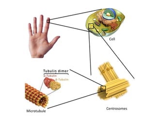
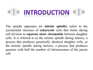
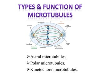
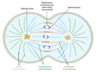
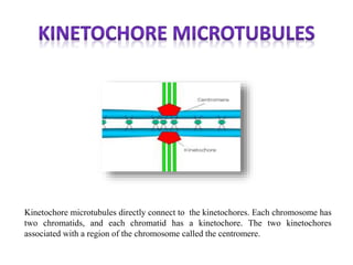
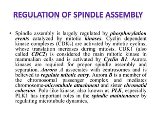
Seminar - 01
Presented by: Jaineel Dharod
M.Pharm Sem-1
Cell
Centrosomes
Microtubule
The spindle apparatus (or mitotic spindle) refers to the
cytoskeletal structure of eukaryotic cells that forms during
cell division to separate sister chromatids between daughter
cells. It is referred to as the mitotic spindle during mitosis, a
process that produces genetically identical daughter cells, or
the meiotic spindle during meiosis, a process that produces
gametes with half the number of chromosomes of the parent
cell.
In 1986, Marc Kirschner and Tim Mitchison
proposed that microtubules use their dynamic
properties of growth and shrinkage at their plus
ends to probe the three dimensional space of the
cell. Plus ends that encounter kinetochores or sites
of polarity become captured and no longer display
growth or shrinkage. In contrast to normal dynamic
microtubules, which have a half-life of 5–10
minutes, the captured microtubules can last for
hours.
Self-organization of molecular components
provides a variety of possible spatial
structures. This model proposes that
microtubules are nucleated acentrosomally
near chromosomes and spontaneously
assemble into anti-parallel bundles and
adopt a spindle-like structure
Astral microtubules.
Polar microtubules.
Kinetochore microtubules.
Astral microtubules develop in the actin skeleton and interact
with the cell cortex to aid in spindle orientation. They are
organized into radial arrays around the centrosomes. The
turn-over rate of this population of microtubules is higher
than any other population.
The role of astral microtubules is assisted by dyneins specific
to this role. These dyneins have their light chains (static
portion) attached to the cell membrane, and their globular
parts (dynamic portions) attached to the microtubules. The
globular chains attempt to move towards the centrosome, but
as they are bound to the cell membrane, this results in pulling
the centrosomes towards the membrane, thus assisting
cytokinesis.
Polar microtubules
interdigitate at the
spindle midzone and
push the spindle poles
apart via motor
proteins.
Kinetochore microtubules directly connect to the kinetochores. Each chromosome has
two chromatids, and each chromatid has a kinetochore. The two kinetochores
associated with a region of the chromosome called the centromere.
• Spindle assembly is largely regulated by phosphorylation
events catalyzed by mitotic kinases. Cyclin dependent
kinase complexes (CDKs) are activated by mitotic cyclins,
whose translation increases during mitosis. CDK1 (also
called CDC2) is considered the main mitotic kinase in
mammalian cells and is activated by Cyclin B1. Aurora
kinases are required for proper spindle assembly and
separation. Aurora A associates with centrosomes and is
believed to regulate mitotic entry. Aurora B is a member of
the chromosomal passenger complex and mediates
chromosome-microtubule attachment and sister chromatid
cohesion. Polo-like kinase, also known as PLK, especially
PLK1 has important roles in the spindle maintenance by
regulating microtubule dynamics.
1. Principles of anatomy and physiology by Gerard J Tortora/
Bryan Derrickson, pg-92
2. https://www.nature.com/scitable/content/types-of-
microtubules-involved-in-mitosis-14752887
3. https://en.wikipedia.org/wiki/Microtubule
4. https://en.wikipedia.org/wiki/Spindle_apparatus
5. https://en.wikipedia.org/wiki/Tubulin#Eukaryotic
Mitotic spindle

Mitotic spindle

  • 1.
    Seminar - 01 Presentedby: Jaineel Dharod M.Pharm Sem-1
  • 2.
  • 3.
    The spindle apparatus(or mitotic spindle) refers to the cytoskeletal structure of eukaryotic cells that forms during cell division to separate sister chromatids between daughter cells. It is referred to as the mitotic spindle during mitosis, a process that produces genetically identical daughter cells, or the meiotic spindle during meiosis, a process that produces gametes with half the number of chromosomes of the parent cell.
  • 4.
    In 1986, MarcKirschner and Tim Mitchison proposed that microtubules use their dynamic properties of growth and shrinkage at their plus ends to probe the three dimensional space of the cell. Plus ends that encounter kinetochores or sites of polarity become captured and no longer display growth or shrinkage. In contrast to normal dynamic microtubules, which have a half-life of 5–10 minutes, the captured microtubules can last for hours.
  • 5.
    Self-organization of molecularcomponents provides a variety of possible spatial structures. This model proposes that microtubules are nucleated acentrosomally near chromosomes and spontaneously assemble into anti-parallel bundles and adopt a spindle-like structure
  • 8.
  • 10.
    Astral microtubules developin the actin skeleton and interact with the cell cortex to aid in spindle orientation. They are organized into radial arrays around the centrosomes. The turn-over rate of this population of microtubules is higher than any other population. The role of astral microtubules is assisted by dyneins specific to this role. These dyneins have their light chains (static portion) attached to the cell membrane, and their globular parts (dynamic portions) attached to the microtubules. The globular chains attempt to move towards the centrosome, but as they are bound to the cell membrane, this results in pulling the centrosomes towards the membrane, thus assisting cytokinesis.
  • 11.
    Polar microtubules interdigitate atthe spindle midzone and push the spindle poles apart via motor proteins.
  • 12.
    Kinetochore microtubules directlyconnect to the kinetochores. Each chromosome has two chromatids, and each chromatid has a kinetochore. The two kinetochores associated with a region of the chromosome called the centromere.
  • 13.
    • Spindle assemblyis largely regulated by phosphorylation events catalyzed by mitotic kinases. Cyclin dependent kinase complexes (CDKs) are activated by mitotic cyclins, whose translation increases during mitosis. CDK1 (also called CDC2) is considered the main mitotic kinase in mammalian cells and is activated by Cyclin B1. Aurora kinases are required for proper spindle assembly and separation. Aurora A associates with centrosomes and is believed to regulate mitotic entry. Aurora B is a member of the chromosomal passenger complex and mediates chromosome-microtubule attachment and sister chromatid cohesion. Polo-like kinase, also known as PLK, especially PLK1 has important roles in the spindle maintenance by regulating microtubule dynamics.
  • 15.
    1. Principles ofanatomy and physiology by Gerard J Tortora/ Bryan Derrickson, pg-92 2. https://www.nature.com/scitable/content/types-of- microtubules-involved-in-mitosis-14752887 3. https://en.wikipedia.org/wiki/Microtubule 4. https://en.wikipedia.org/wiki/Spindle_apparatus 5. https://en.wikipedia.org/wiki/Tubulin#Eukaryotic