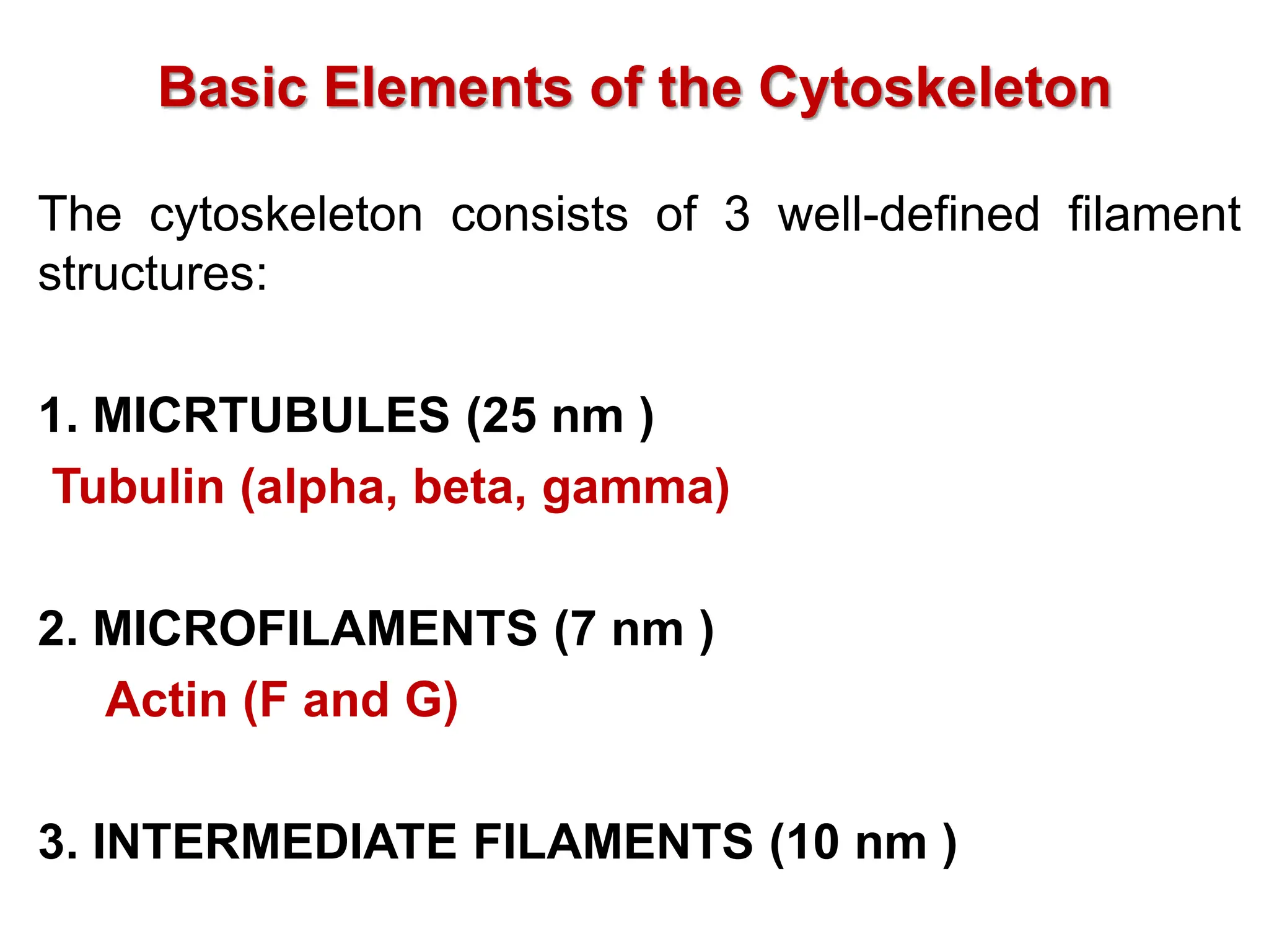
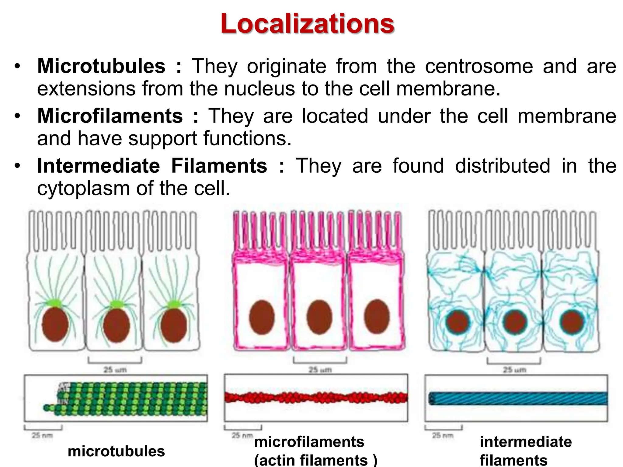
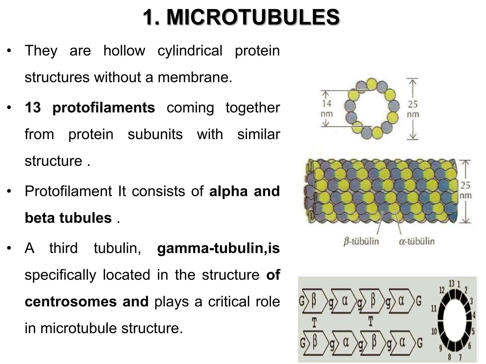
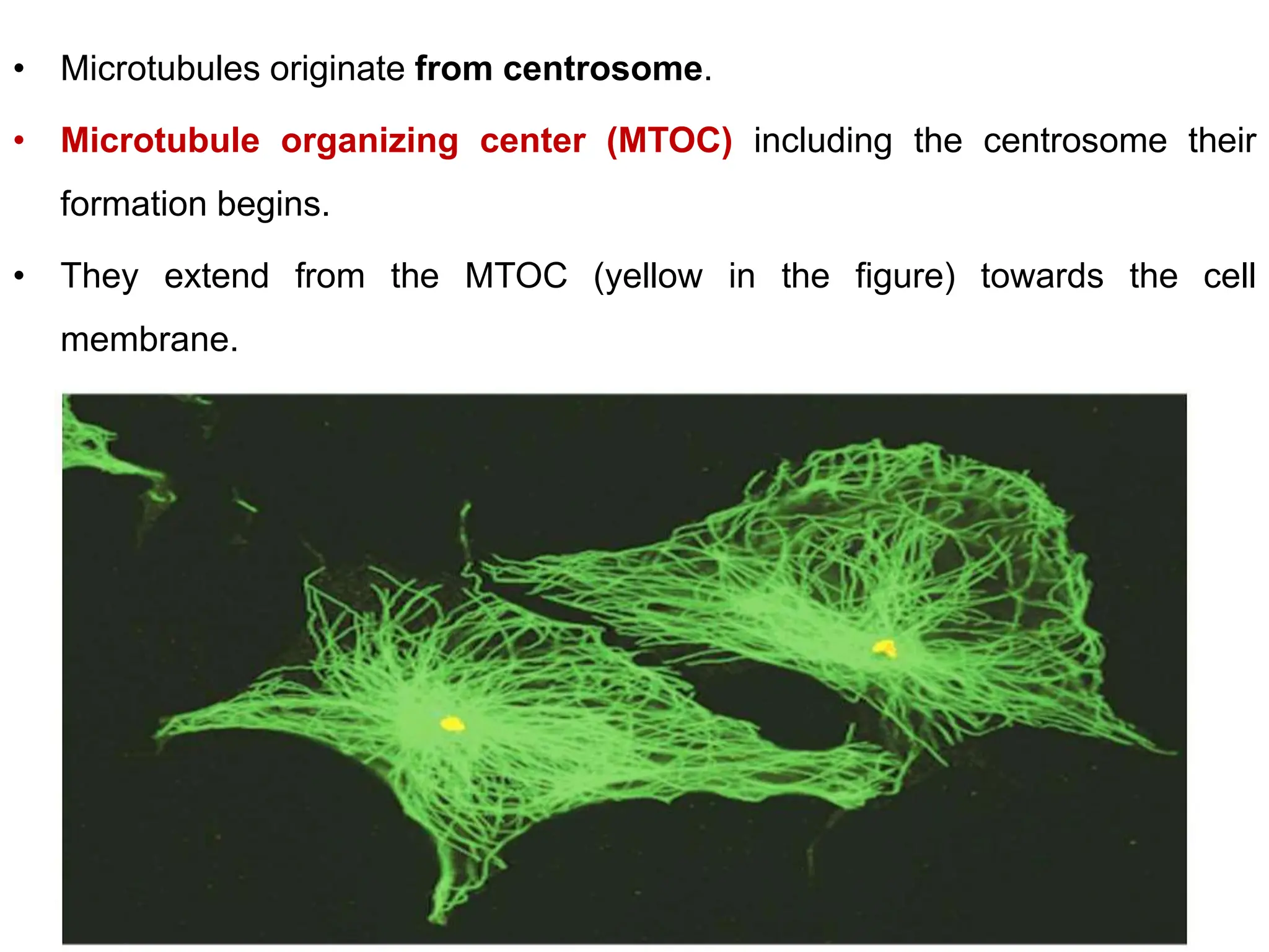
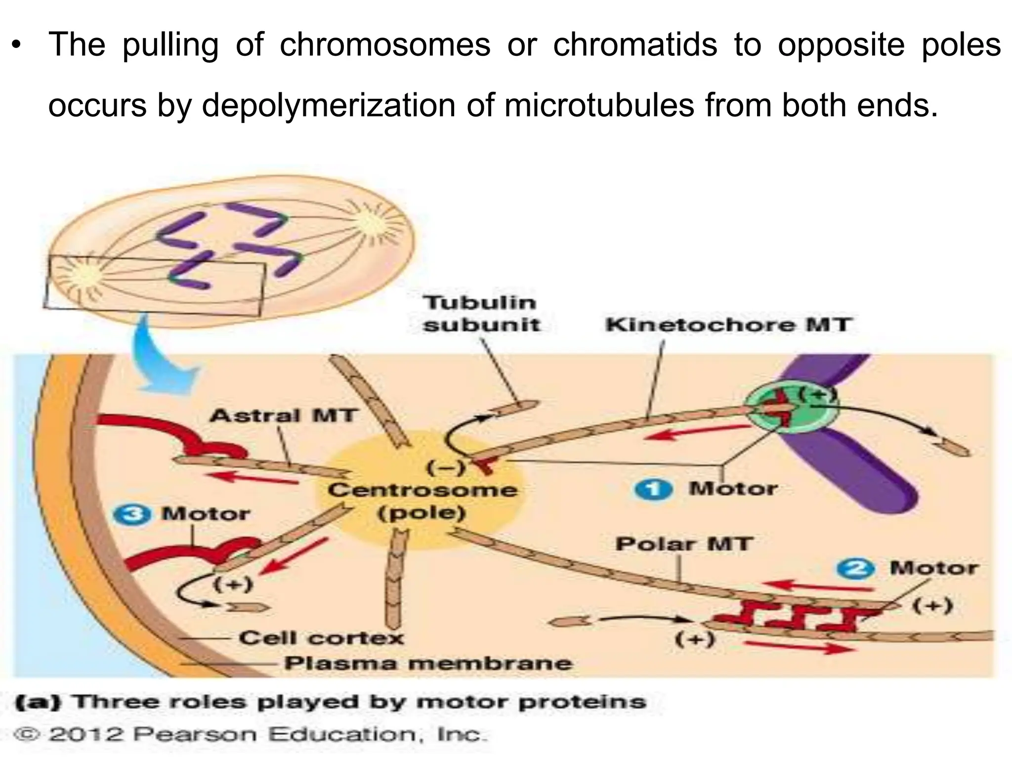
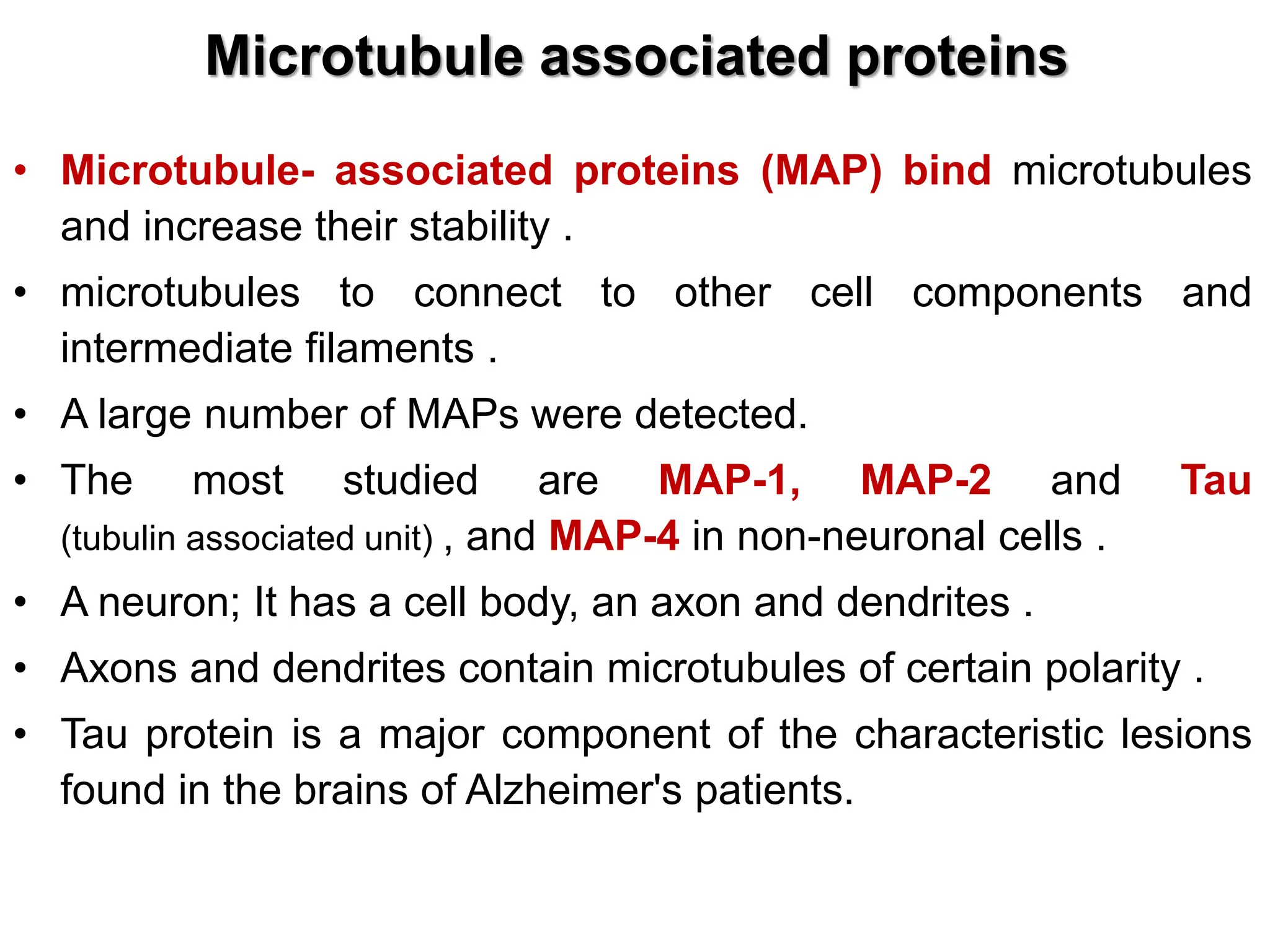
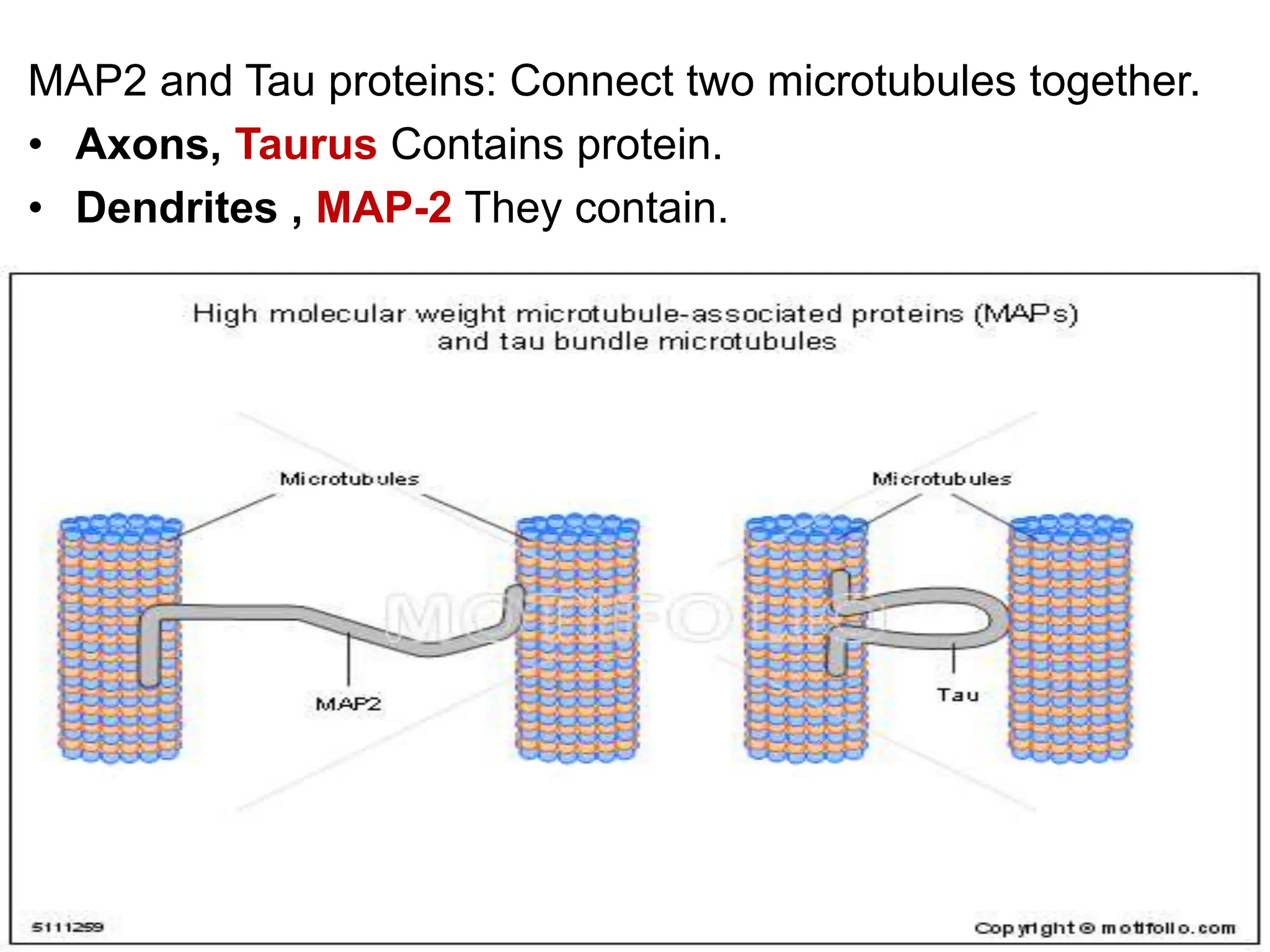
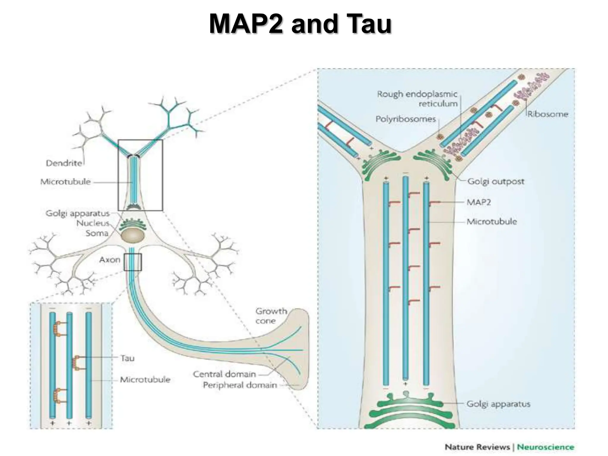
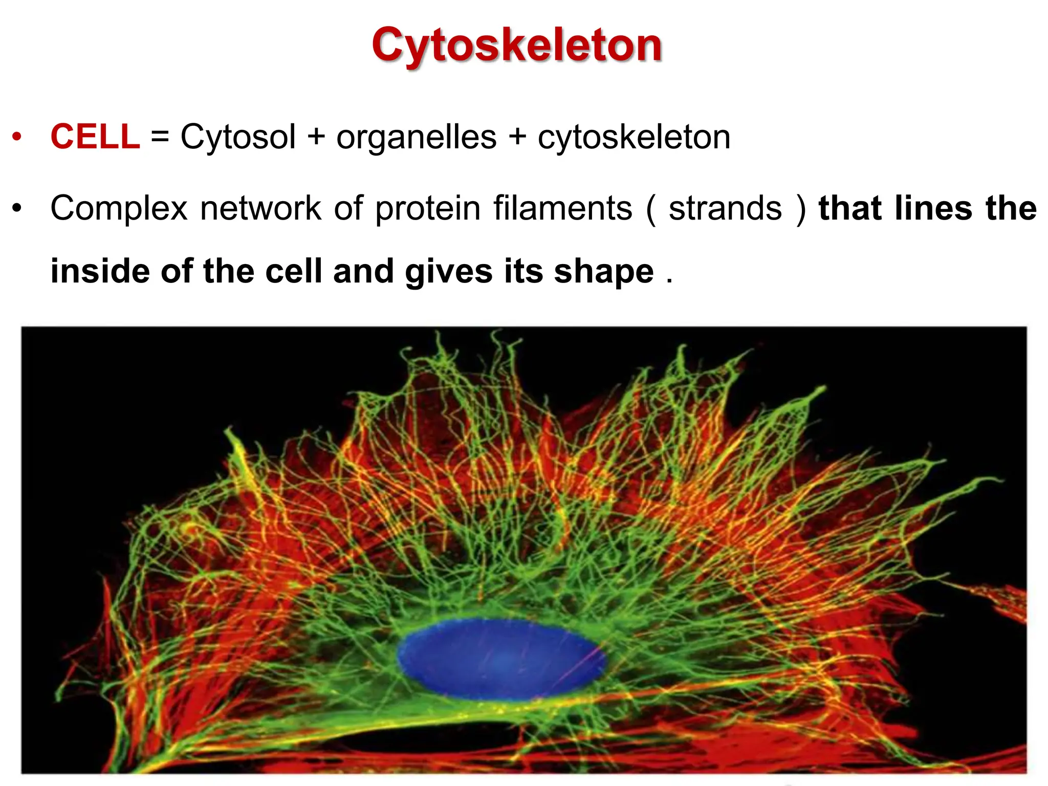
The cytoskeleton is composed of microtubules, microfilaments, and intermediate filaments that give cells their shape and allow cellular movement. Microtubules originate from the microtubule organizing center and extend towards the cell membrane. They are involved in many cellular functions like intracellular transport, cell division, and maintaining organelle structure. Microtubule associated proteins help stabilize microtubules and connect them to other cell components.
![Funtions
• Determination and preservation of Cell/Nucleus shape and
placement (elasticity, strength)
• Movement of the cell (migration, diapedesis [leakage of
blood cells from the vessel walls into tissues], etc.),
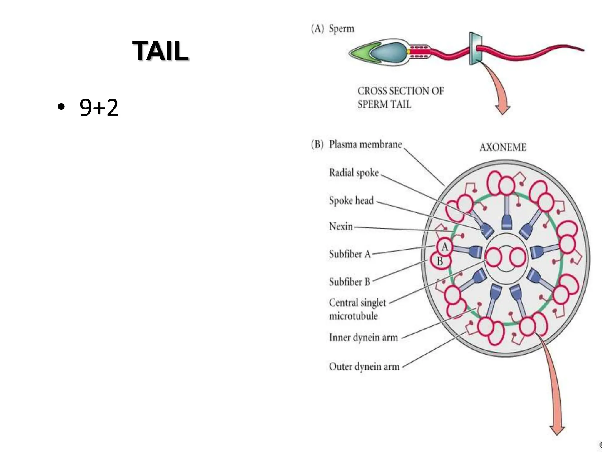
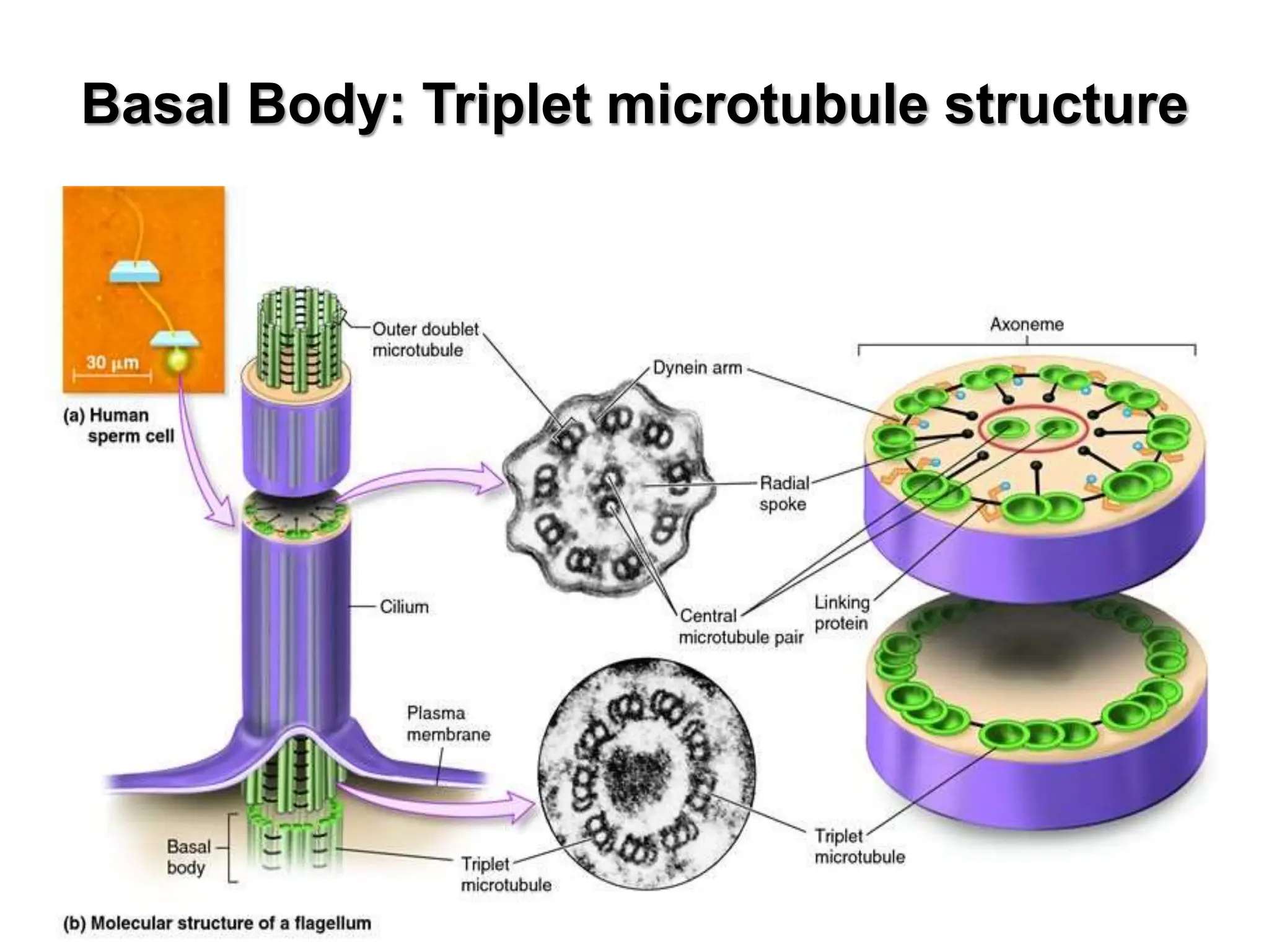
movement of a part of the cell ( cilia , tail, etc.)
• Phagocytosis, endocytosis , exocytosis
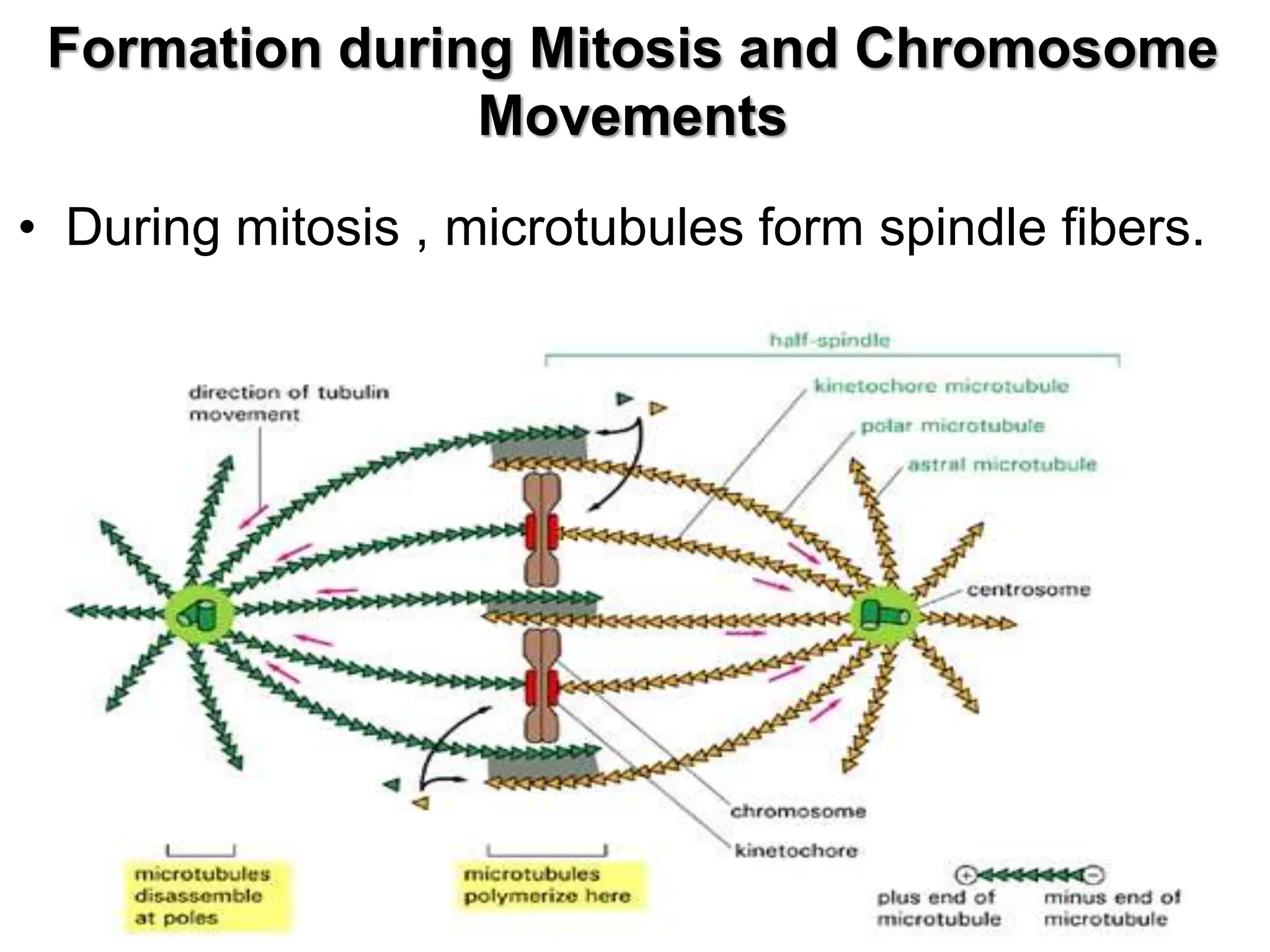
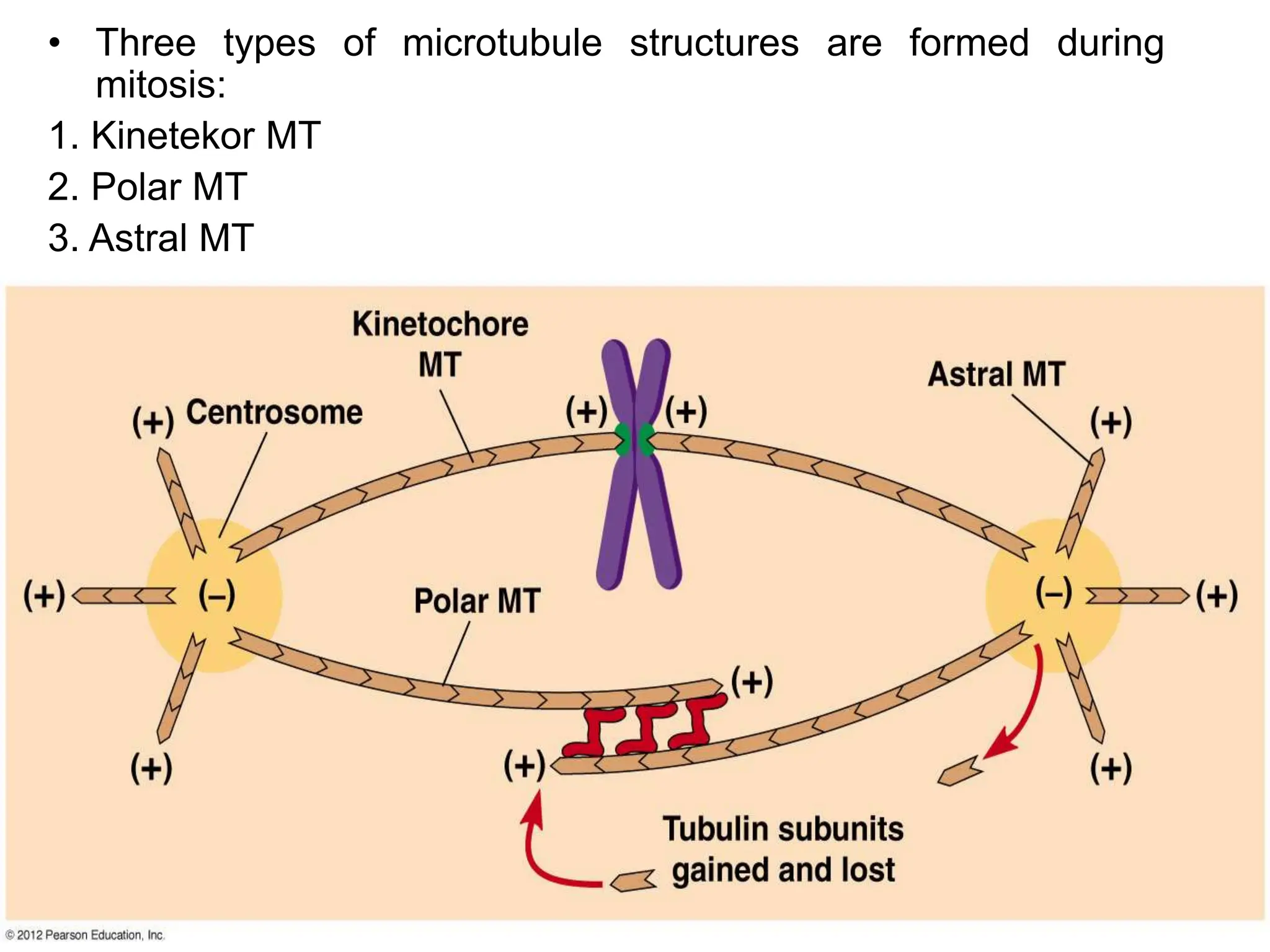
• Cytokinesis
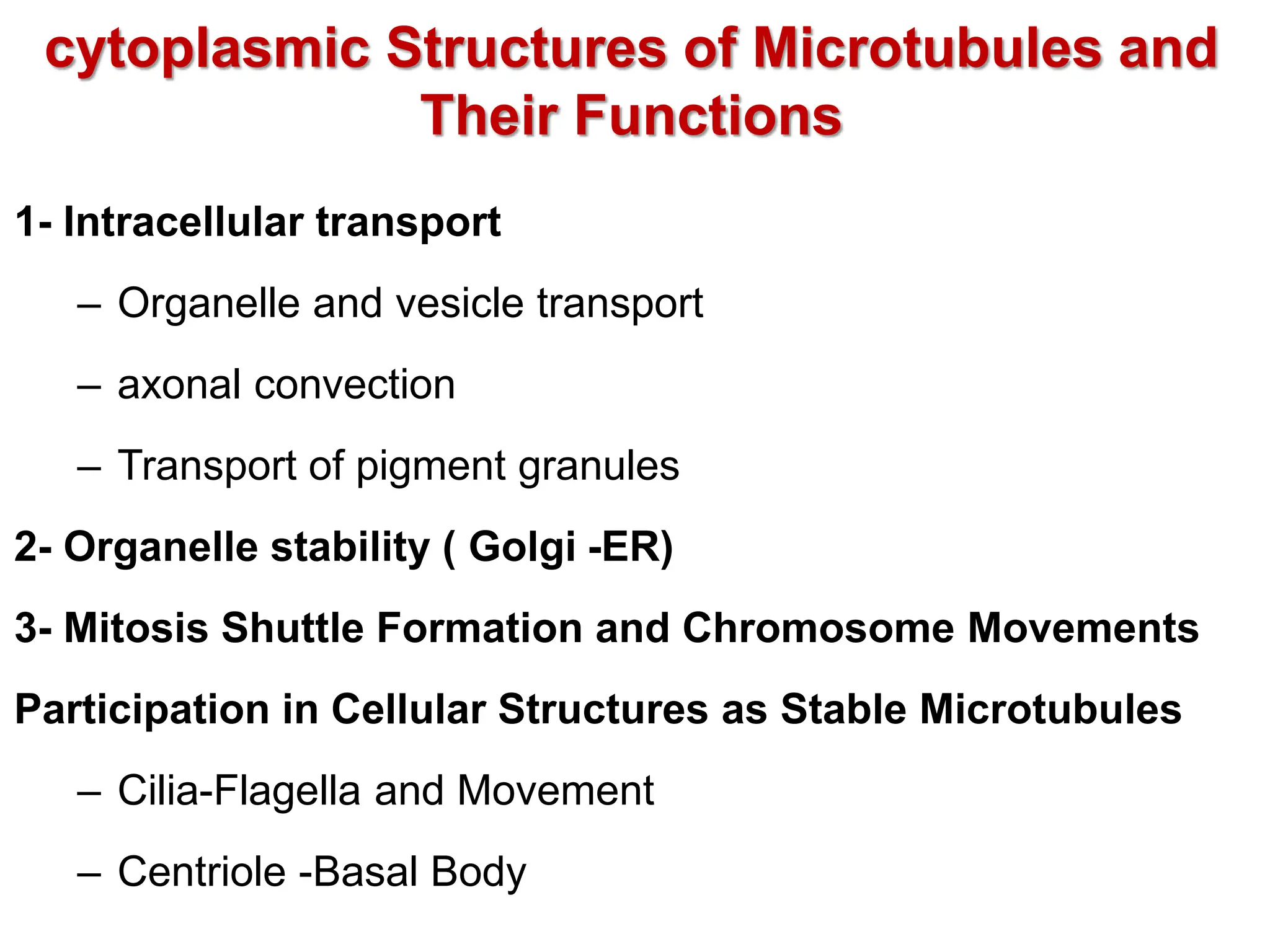
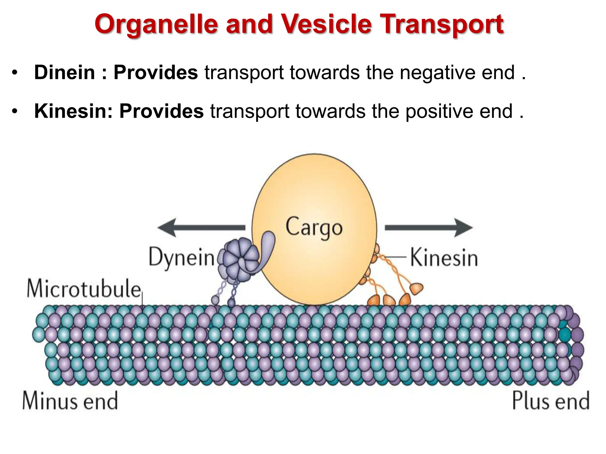
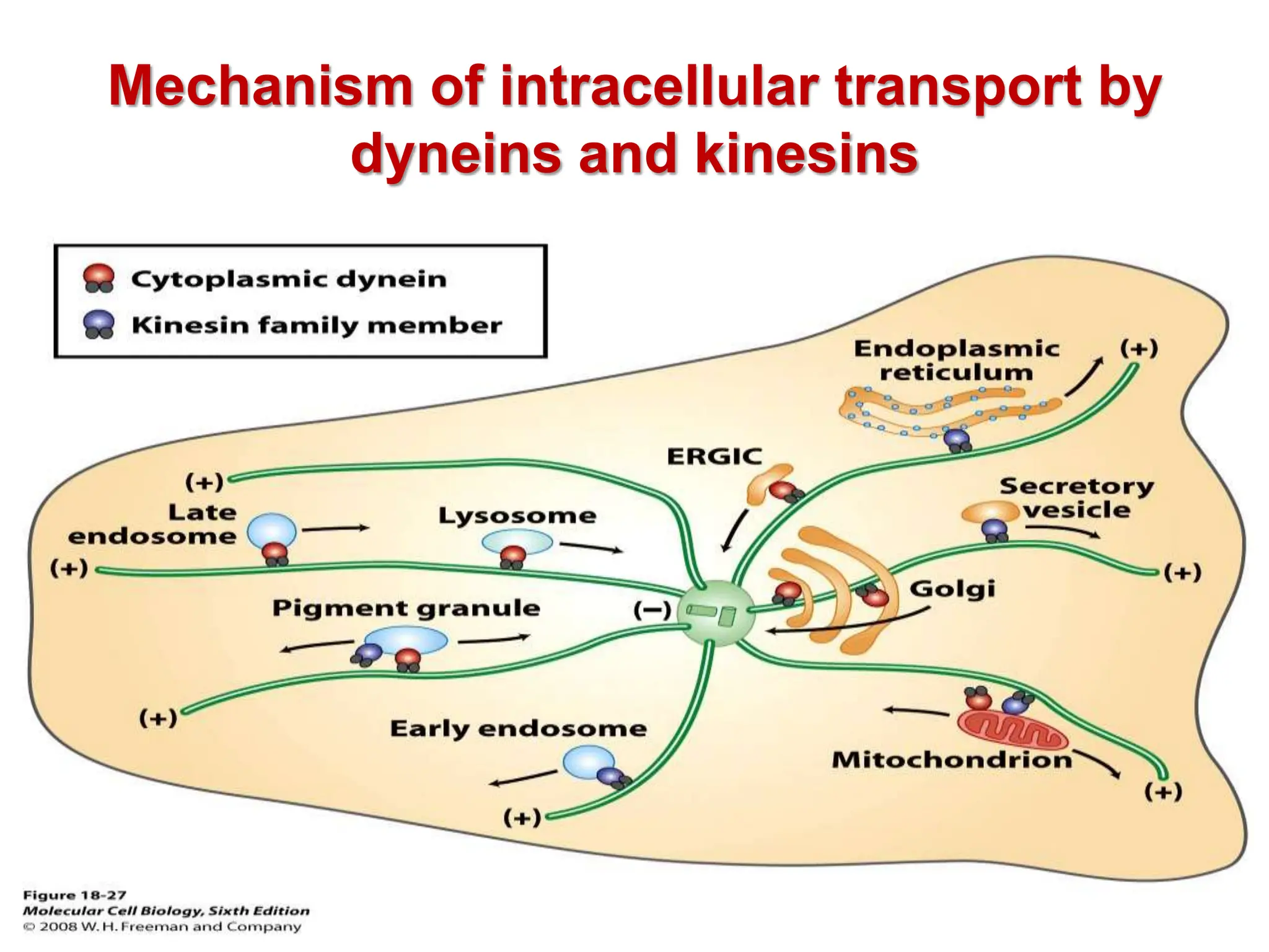
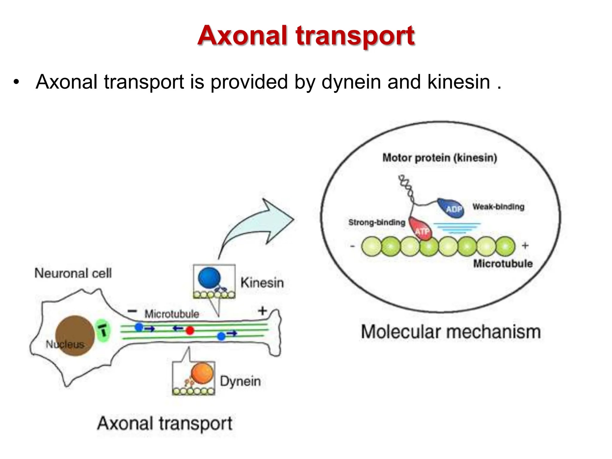
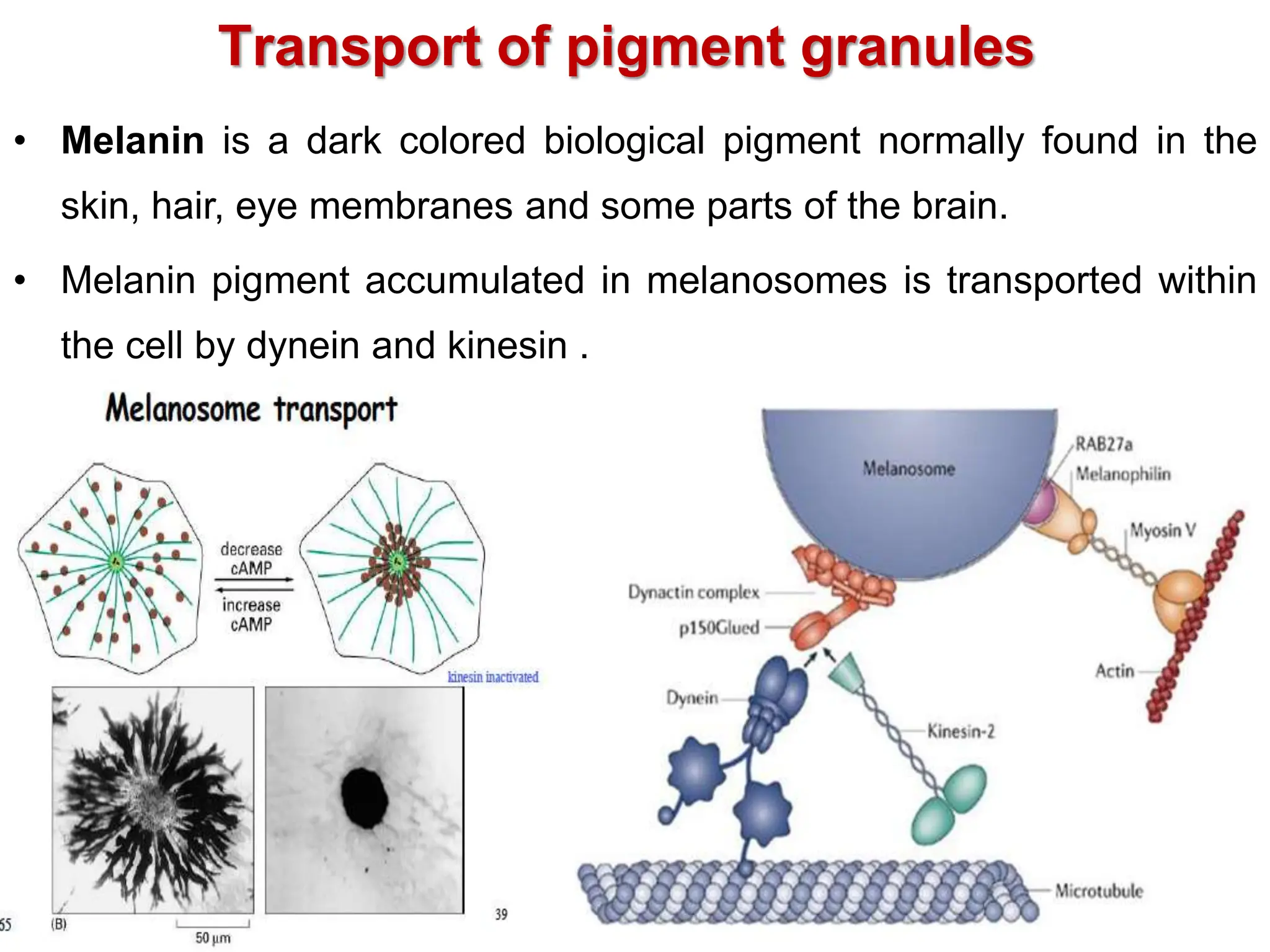
• Intracellular transport
• Contraction
• Supporting cell-cell and cell-extracellular media relationships
• Creating a structural framework within the cytoplasm
• Effective regulation of cell cytoplasm
• Stabilization of intercellular connections](https://image.slidesharecdn.com/cytoskeleton1-240416225044-01d0a7ff/75/structure-and-functions-of-cytoskeletons-3-2048.jpg)