SlideShare a Scribd company logo
1 of 4
Download to read offline
International Research Journal of Engineering and Technology (IRJET) e-ISSN: 2395 -0056
Volume: 04 Issue: 01 | Jan -2017 www.irjet.net p-ISSN: 2395-0072
© 2017, IRJET | Impact Factor value: 5.181 | ISO 9001:2008 Certified Journal | Page 335
An Ameliorate Technique for Brain Lumps Detection Using Fuzzy C-
Means Clustering
Pritam R. Dungarwal1, Prof. Dinesh D. Patil
1Research Scholar Student, M.E (CSE), SSGBCOET,Bhusawal,Maharashtra,India
2HOD, Professor, Dept. of CSE, SSGBCOET, Bhusawal,Maharashtra,India
---------------------------------------------------------------------***--------------------------------------------------------------------
ABSTRACT-Clustering is commonly used in biomedical
applications particularly for brain lumps detection in
Abnormal magnetic resonance images (MRI). In terms of
segmentation efficiency Fuzzy clustering using fuzzy local
information C-means algorithm proved to be greater over the
other clustering methodologies. Most Research in developed
countries show that the number of people who have brain
cancer were died due to the fact of inaccurate detection and
not in time. Generally, CT scan or MRI that is directed into
intracranial cavity produces a complete image of brain. This
image is visually examined by the physician for detection &
diagnosis of brain cancer. However this method of detection
resists the accurate determination of size of lump. In addition,
it also reduces the time for analysis. At the end of the process
the lump is extracted from the MR image and itsexactposition
and the shape also determined. The graphbasedonpixel value
is drawn taking the various points from the swelledcellsliesin
the original position from the affected region. Here the
affected region is considered as ellipsoid shape and the
volumes have been calculated from it. A fuzzy level set
algorithm is proposed in this paper to facilitatemedical image
segmentation & on this performance of evaluation of the
proposed algorithm was carried.
Index Terms— introduction, clustering, Fuzzy clustering,
image segmentation, Fuzzy C-Mean, result
1 INTRODUCTION
Now a day’s brain abnormality mainly brain lumps are
one of the most common brain diseases, so detection
and quantification of lumps in MRI are important in
medical diagnosis. Through past many researchers have
prepared important research in the field of brain
abnormality segmentation but still now it is very
important research fields due to the large number of
variation of MRI of brain. The accurate segmentation
of internal structures of the brain is of great interest
for the study and very helpful for the treatment of lumps. It
aims at reducing the mortality and improving the surgical or
radio therapeutic management of lumps. The most
important aim of medical image analysis in general and
brain MRI analysis in particular, is to extract clinical
information that would improve diagnosis and treatment of
disease. The aim is to provide information associated to
anatomical structures as well as potential abnormal
tissues necessary to treatment planning and patient follow-
up. There are different brain lumps detection and
segmentation methods to detect and segment a brain
lumps from MRI images. The measurement of brain
lumps volume can assist lumps staging for brain lumps
volume measurements is developed which overcome
the problem of inter-operator variance, besides partial
volume effects and shows satisfactory performance
for segmentation, thus segmentation is very important for
volume calculation. Different segmentation techniques
such as thresholding based segmentation methodology,
Region Growing based segmentation, K-nearest
neighbours (KNN), Bayesian approach, Markov Random
Field Models , Expectation maximization (EM),
Support vector machine (SVM), Fuzzy c-means
algorithms, K-means algorithms, Morphology-based
segmentation, Atlas-guided based segmentation,
Knowledge based segmentation, Texture-based
segmentation, Artificial neural networks (ANNs), Fusion-
based, Fuzzy connectedness, Watershed Methods, Level
set based segmentation, Hybrid Self Organizing Map
(SOM), SOM, Graph Cut based segmentation, Fractal-
based segmentation, Parametric deformable models
(snakes), Boundary based methods, Geometric
deformable model, The Combination of Watershed and
Level Set segmentation, Spatio-Temporal Model, Hidden
Markov Model, Genetic algorithms based segmentation,
Kohon-en Self Organizing Map (SOM) with a common
phase pre-processing and segmentation and its steps are
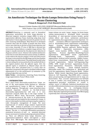
shown below. Thus accurate segmentation over full field
of view is another very much problem but during the
segmentation procedure verification of results is
another source of difficulty. Statistical classification may
not allow differentiation between non-enhancinglumps and
normal tissue due to overlapping intensity
distributions of healthy tissue with lumps and surrounding
edema. Manually segmenting brain lumps from MR imaging
is generally time consuming and difficult.
Fig: 1 Segmentation process
Brain MRI Images
Pre-processing
Detection Phase
Post-Processing Segmentation
Volume Calculation
Diagnosis
International Research Journal of Engineering and Technology (IRJET) e-ISSN: 2395 -0056
Volume: 04 Issue: 01 | Jan -2017 www.irjet.net p-ISSN: 2395-0072
© 2017, IRJET | Impact Factor value: 5.181 | ISO 9001:2008 Certified Journal | Page 336
An automate segmentation method is desirable because it
reduces the load on the operator and generates satisfactory
results.
The region growing segmentation is used to segment the
brain lumps due to its wide range of applications and
automatic features. After taking the image of the lumps
brain there is a need to process it. The image clearly shows
the place of the lumps portion of the brain. The image
does not give the information about the numerical
parameters such as area and volume of the lumps portion
of the brain. After segmentation the desired lumps area is
selected from the segmented image. This selected region
is used to calculate the area and volumeofthelumpspresent
in the MR image.
2. LITERATURE SURVEY
The previous methods for brain lumps segmentation are
thresholding, region growing & clustering. Thresholding is
the simplest method of image segmentation. From a
grayscale image, thresholding can be used to create binary
images. During the thresholding process, individual
pixels in an image are marked as "object" pixels if
their value is greater than some threshold value
(assuming an object to be brighter than the background)
and as "background" pixels otherwise. This convention is
known as threshold above. Variants include threshold
below, which is opposite of threshold above; threshold
inside, where a pixel is labelled "object" if its value is
between two thresholds; and threshold outside, which is
the opposite of threshold inside. Typically, an object pixel
is given a value of “1” while a background pixel is given
a value of “0.” Finally, a binary image is created by
colouring each pixel white or black, depending on a
pixel's labels.
The major drawback to threshold-based approaches is that
they often lack the sensitivity and specificity needed for
accurate classification. Brain cancer cells have high
proteinaceous fluid which has very high density and hence
very high intensity, therefore watershedsegmentationisthe
best tool to classify cancers and high intensity tissues of
brain [6]. Watershed segmentation can classify the
intensities with very small difference also, which is not
possible with snake and level set method. A similar method
for tumor detection is proposed by Malhotra [2], but multi-
parameter extraction was not used. Hossam and P Vasuda
[10] have proposed a method for brain tumor detection and
segmentation using histogram Thresholding detects the
tumor but the result shown crops excessive area ofbrain. An
efficient and improved brain tumor detectionalgorithm was
developed by Rajeev Ratan, Sanjay Sharma and S. K. Sharma
[8] which makes use of multi-parameter MRI analysis and
the lumps cannot be segmented in 3-D unless and until we
have 3-D MRI image data set. So, a relatively simple method
for detection of brain lumps is presentedwhichmakesuseof
marker basedwatershedsegmentationwithimprovementto
avoid over & under segmentation. The Segmentation of an
image entails the division or separation of the image into
regions of similar attribute. The ultimate aim in a large
number of image processing applications is to extract
important features from the image data, from which a
description, interpretation, or understanding of the scene
can be provided by the machine. The segmentation of brain
lumps from magnetic resonance images is an important but
time consuming task performed by medical experts.[5] The
digital image processing community has developed several
segmentation methods, many of them ad hoc. Suchendra et
al. (1997) suggested a multi scale image segmentation using
a hierarchical self-organizing map; a high speed parall fuzzy
c-mean algorithm for brain lumps segmentation; an
improved implementation of brain lumps detection using
segmentation based on neuron fuzzy technique designed a
method on 3D variational segmentation forprocessesdueto
the high diversity in appearance of lumps tissue from
various patients. When experts work on lumps images then
they use three different types of algorithms. Some of the
techniques based on pixel based, some based on texture of
images and some of them based on structure of images.
3. AN AMELIORATE TECHNIQUE FORBRAINLUMPS
DETECTION
Content based image retrieval methods areoneofthem with
widely used in retrieving medical imagesfromMRIdata sets.
In this research paper we are presenting An Ameliorate
Technique, based on modified classifiers&feedback method
for lumps image retrieval from MRI images. This technique
uses new concepts of texture and shape properties of the
images by using fuzzy c-means clustering and modified SVM
classifier. It also uses subjective feedback method for
efficient and accurate results. This method uses following
phases –
Fig2: Phases of Brain Lumps Detection
Algorithm for an Ameliorate Technique for Brain Lumps
Detection
Input- Set of MRI images
Output- Detect Brain Lumps accurately
1- Select input image from image data set
2- Apply pre-processing on image
o Noise removal- by using Median filter
International Research Journal of Engineering and Technology (IRJET) e-ISSN: 2395 -0056
Volume: 04 Issue: 01 | Jan -2017 www.irjet.net p-ISSN: 2395-0072
© 2017, IRJET | Impact Factor value: 5.181 | ISO 9001:2008 Certified Journal | Page 337
 Select a noisy MRI image and separate each plane and
each scalar component is treated in dependently.
 Generate zero arrays around an image based on image
mask size using pad array command.
 Select 3 * 3 masks from an image and the mask should
be odd sized
 For each component of each point under the mask a
single median component is determined.
 Contrast enhancement
3- Feature Extraction
 Boundary detection
 The Intensity Gradient and Texture map are calculated
 Extra features extraction such as texture and shape
properties
 Measure SVM classifiers
4- Modified Subjective feedback phase
 Apply Fussy C-Means clustering
4. CLUSTERING TECHNIQUES
Clustering is the processes of collection of objects which are
similar between them and are dissimilar objects belong to
the other clusters. The clustering is suitable in biomedical
image segmentation when the number of cluster is known
for particular clustering of human anatomy. Clustering
(pixel) is belong to the one cluster then it couldnotbelong to
another cluster. K-mean is the example of exclusive
clustering algorithm. In overlapping clustering, one data
(pixel) is belonging to the two or more clusters. Fuzzy C-
mean is example of the overlapping clustering algorithm.
[10]
K-MEANS CLUSTERING
K-mean clustering is unsupervised algorithms that solve
clustering problem. The procedure for k mean clustering
algorithm is the simple and easy way to segment the image
using basic knowledge of the clustering value. In k-mean
clustering initially randomly define k centroids. Selection of
this k centroid is placed in thecunningwaybecausedifferent
location makes the different clustering. So that, the better is
to place centroid value will be as much as far away from the
each other. Secondly calculate the distance between each
pixel to selected cluster centroid. Each pixel compares with
the k clusters centroids and finding the distance using the
distance formula. If pixel has theshortestdistanceamong all,
than it is move to particular cluster. Repeatthisprocessuntil
all pixel compare to the cluster centroids. The process
continues until some convergence criteria are the met [10]
FUZZY C-MEANS CLUSTERING
Fuzzy C-means clustering is the overlapping clustering
technique. One pixel value depending upon the two or more
clusters centers. It is also known as soft clustering method.
Most widely used the fuzzy clustering algorithm is theFuzzy
C-means (FCM) algorithm (Bezdek 1981). The FCM
algorithm is the partition of the n element X={x1,...,xn} intoa
collection of the c fuzzy clustering with respect to the below
given criteria [9][10][4] It is based on the minimization of
the following objective function:
Where, m = level of the fuzziness and real number
greater than 1. uij= degree of the membership of xi in the
cluster cj x = data value Fuzzy C-means is the popular
method for medical image segmentation but only consider
the image intensity thereby producingunsatisfactoryresults
in noisy images. A bunch of the algorithms are proposed to
make the FCM robust against noise and in homogeneity but
it’s still not perfect. In 2012, J. Selvakumar, A,LakshmiandT.
Arivoli [7] proposed a technique for brain lumps
segmentation using the k-means and the fuzzy c-means
algorithm. Its use the pre-processing steps for filtering the
noise and other artefacts in image and apply the K-means
and fuzzy c-mean algorithm. This purposed algorithm,fuzzy
c-mean is slower than the K-means in efficiencybutgivesthe
accurate prediction of lumps cellswhicharenotpredicted by
the K-means algorithm.
5. EXPERIMENTAL RESULT
The proposed algorithm and Fuzzy C-Means algorithm is
implemented using MATLAB software and tested on the
brain MRI images to explore the segmentation accuracy of
the proposed approach. The comparison is made between
the Fuzzy C-Means and proposed algorithm. The quality of
the segmentation of the proposed algorithm can be
calculated by segmentation accuracy which is given as.
The input image and the corresponding segmented image is
shown below.
Fig: 3 Experimental Results on different brain images
Following table & graph shows result comparison of k-
means and FCM algorithm
Table 1. Results
Parameters In %
Existing K-Means Implemented FCM
Precision 91 94.73
Recall 90.54 92.52
Specificity 88.70 96.77
International Research Journal of Engineering and Technology (IRJET) e-ISSN: 2395 -0056
Volume: 04 Issue: 01 | Jan -2017 www.irjet.net p-ISSN: 2395-0072
© 2017, IRJET | Impact Factor value: 5.181 | ISO 9001:2008 Certified Journal | Page 338
Accuracy 89.90 92.51
Fig 4. Graphical comparison of Results
6. CONCLUSION
Recently, the applications of the image processing can be
found in the areas like electronics, remote sensing, natural
scene detection, biometrics, bio-medical etc. If we
concentration the bio-medical applications, image
processing is commonly used for the diagnosis of different
tissues purpose. By use of suitable image segmentation
method and the use of accurate input image we get accurate
results.
REFERENCES
[1] B.Jyothia, Y.MadhaveeLathab, P.G.Krishna Mohanc
,V.S.K.Reddy,” Integrated MultipleFeaturesforTumorImage
Retrieval Using Classifier and Feedback Methods”,
International Conference on Computational Modeling and
Security (CMS 2016), Science direct, 2016, PP 141-148.
[2] L.A. Khoo, P. Taylor, and R.M. Given-Wilson, “Computer-
Aided Detection in the United Kingdom National Breast
Screening Programme: Prospective Study,” Radiology, vol.
237, pp. 444-449, 2005.
[3] Pritam Dungarwal and Prof. Dinesh Patil, “Literature
Survey on Detection of lumps in brain” International Journal
of Modern Trends in Engineering and Research (IJMTER)
Volume 03, Issue 12, December – 2016,pp 312-316
[4] Krit Somkantha, Nipon Theera-Umpon, “Boundary
Detection in Medical ImagesUsingEdgeFollowingAlgorithm
Based on Intensity Gradient and TextureGradientFeatures,”
in Proc. IEEE transactionson biomedical engineering,vol.58,
no. 3, march 2011,pp. 567–573.
[5] B.Jyothi, Y.MadhaveeLatha, P.G.Krishna Mohan,”
Multidimentional Feature Vector Space for an Effective
Content Based Medical Image Retrieval 5th IEEE
International Advance Conputing Conference (IACC-
2015),BMS College of engineering Bangalore,June 12 to 13
,2015.
[6] Nitin Jain & Dr. S. S. Salankar,” Color & Texture Feature
Extraction for Content Based Image Retrieval”, IOSR Journal
of Electrical andElectronicsEngineering(IOSR-JEEE)e-ISSN:
2278-1676, p-ISSN: 2320-3331 PP 53-58.
[7] J. Selvakumar, A. Lakshmi, T. Arivoli, “Brain tumor
segmentation and its Area calculation in Brain MR images
using K-Mean clustering and Fuzzy C-Mean Algorithm”,
IEEE-International conference on advances in engineering
science and management, March-2012.
[8] Cs Pillai, “A Survey of Shape Descriptors forDigital Image
Processing”, IRACST - International Journal of Computer
Science and Information Technology & Security (IJCSITS),
ISSN: 2249-9555 Vol. 3, No.1, February 2013
[9] Prashant Aher and Umesh Lilhore, “An improved cbmir
architecture, based on modified classifiers & feedback
method for tumor image retrieval from mri images”
International Journal of Modern Trends in Engineering and
Research (IJMTER) Volume 03, Issue 12,Dec – 2016,pp-157-
160
[10] Ramakrishna Reddy, Eamani and G.V Hari Prasad,”
Content-Based Image Retrieval Using Support Vector
Machine in digital image processing techniques”,
International Journal of Engineering and Technology
(IJEST),IIN:0977-5462 vol.04 No04 April 2012

More Related Content

What's hot

3D Segmentation of Brain Tumor Imaging
3D Segmentation of Brain Tumor Imaging3D Segmentation of Brain Tumor Imaging
3D Segmentation of Brain Tumor ImagingIJAEMSJORNAL
 
IRJET - Machine Learning Applications on Cancer Prognosis and Prediction
IRJET - Machine Learning Applications on Cancer Prognosis and PredictionIRJET - Machine Learning Applications on Cancer Prognosis and Prediction
IRJET - Machine Learning Applications on Cancer Prognosis and PredictionIRJET Journal
 
Mri brain tumour detection by histogram and segmentation
Mri brain tumour detection by histogram and segmentationMri brain tumour detection by histogram and segmentation
Mri brain tumour detection by histogram and segmentationiaemedu
 
Brain Tumor Detection and Classification using Adaptive Boosting
Brain Tumor Detection and Classification using Adaptive BoostingBrain Tumor Detection and Classification using Adaptive Boosting
Brain Tumor Detection and Classification using Adaptive BoostingIRJET Journal
 
Neuroendoscopy Adapter Module Development for Better Brain Tumor Image Visual...
Neuroendoscopy Adapter Module Development for Better Brain Tumor Image Visual...Neuroendoscopy Adapter Module Development for Better Brain Tumor Image Visual...
Neuroendoscopy Adapter Module Development for Better Brain Tumor Image Visual...IJECEIAES
 
IRJET - Detection of Heamorrhage in Brain using Deep Learning
IRJET - Detection of Heamorrhage in Brain using Deep LearningIRJET - Detection of Heamorrhage in Brain using Deep Learning
IRJET - Detection of Heamorrhage in Brain using Deep LearningIRJET Journal
 
IRJET- Brain Tumor Detection using Convolutional Neural Network
IRJET- Brain Tumor Detection using Convolutional Neural NetworkIRJET- Brain Tumor Detection using Convolutional Neural Network
IRJET- Brain Tumor Detection using Convolutional Neural NetworkIRJET Journal
 
Techniques of Brain Cancer Detection from MRI using Machine Learning
Techniques of Brain Cancer Detection from MRI using Machine LearningTechniques of Brain Cancer Detection from MRI using Machine Learning
Techniques of Brain Cancer Detection from MRI using Machine LearningIRJET Journal
 
Brain Image Fusion using DWT and Laplacian Pyramid Approach and Tumor Detecti...
Brain Image Fusion using DWT and Laplacian Pyramid Approach and Tumor Detecti...Brain Image Fusion using DWT and Laplacian Pyramid Approach and Tumor Detecti...
Brain Image Fusion using DWT and Laplacian Pyramid Approach and Tumor Detecti...INFOGAIN PUBLICATION
 
IRJET-A Review on Brain Tumor Detection using BFCFCM Algorithm
IRJET-A Review on Brain Tumor Detection using BFCFCM   AlgorithmIRJET-A Review on Brain Tumor Detection using BFCFCM   Algorithm
IRJET-A Review on Brain Tumor Detection using BFCFCM AlgorithmIRJET Journal
 
Automated brain tumor detection and segmentation from mri images using adapti...
Automated brain tumor detection and segmentation from mri images using adapti...Automated brain tumor detection and segmentation from mri images using adapti...
Automated brain tumor detection and segmentation from mri images using adapti...Tamilarasan N
 
Brain Tumor Detection using Neural Network
Brain Tumor Detection using Neural NetworkBrain Tumor Detection using Neural Network
Brain Tumor Detection using Neural Networkijtsrd
 
Classification of Abnormalities in Brain MRI Images Using PCA and SVM
Classification of Abnormalities in Brain MRI Images Using PCA and SVMClassification of Abnormalities in Brain MRI Images Using PCA and SVM
Classification of Abnormalities in Brain MRI Images Using PCA and SVMIJERA Editor
 
A Wavelet Based Automatic Segmentation of Brain Tumor in CT Images Using Opti...
A Wavelet Based Automatic Segmentation of Brain Tumor in CT Images Using Opti...A Wavelet Based Automatic Segmentation of Brain Tumor in CT Images Using Opti...
A Wavelet Based Automatic Segmentation of Brain Tumor in CT Images Using Opti...CSCJournals
 
Multistage Classification of Alzheimer’s Disease
Multistage Classification of Alzheimer’s DiseaseMultistage Classification of Alzheimer’s Disease
Multistage Classification of Alzheimer’s DiseaseIJLT EMAS
 
Paper id 25201482
Paper id 25201482Paper id 25201482
Paper id 25201482IJRAT
 

What's hot (20)

E0413024026
E0413024026E0413024026
E0413024026
 
3D Segmentation of Brain Tumor Imaging
3D Segmentation of Brain Tumor Imaging3D Segmentation of Brain Tumor Imaging
3D Segmentation of Brain Tumor Imaging
 
IRJET - Machine Learning Applications on Cancer Prognosis and Prediction
IRJET - Machine Learning Applications on Cancer Prognosis and PredictionIRJET - Machine Learning Applications on Cancer Prognosis and Prediction
IRJET - Machine Learning Applications on Cancer Prognosis and Prediction
 
Mri brain tumour detection by histogram and segmentation
Mri brain tumour detection by histogram and segmentationMri brain tumour detection by histogram and segmentation
Mri brain tumour detection by histogram and segmentation
 
C1103041623
C1103041623C1103041623
C1103041623
 
Brain Tumor Detection and Classification using Adaptive Boosting
Brain Tumor Detection and Classification using Adaptive BoostingBrain Tumor Detection and Classification using Adaptive Boosting
Brain Tumor Detection and Classification using Adaptive Boosting
 
Neuroendoscopy Adapter Module Development for Better Brain Tumor Image Visual...
Neuroendoscopy Adapter Module Development for Better Brain Tumor Image Visual...Neuroendoscopy Adapter Module Development for Better Brain Tumor Image Visual...
Neuroendoscopy Adapter Module Development for Better Brain Tumor Image Visual...
 
IRJET - Detection of Heamorrhage in Brain using Deep Learning
IRJET - Detection of Heamorrhage in Brain using Deep LearningIRJET - Detection of Heamorrhage in Brain using Deep Learning
IRJET - Detection of Heamorrhage in Brain using Deep Learning
 
IRJET- Brain Tumor Detection using Convolutional Neural Network
IRJET- Brain Tumor Detection using Convolutional Neural NetworkIRJET- Brain Tumor Detection using Convolutional Neural Network
IRJET- Brain Tumor Detection using Convolutional Neural Network
 
Techniques of Brain Cancer Detection from MRI using Machine Learning
Techniques of Brain Cancer Detection from MRI using Machine LearningTechniques of Brain Cancer Detection from MRI using Machine Learning
Techniques of Brain Cancer Detection from MRI using Machine Learning
 
Brain Image Fusion using DWT and Laplacian Pyramid Approach and Tumor Detecti...
Brain Image Fusion using DWT and Laplacian Pyramid Approach and Tumor Detecti...Brain Image Fusion using DWT and Laplacian Pyramid Approach and Tumor Detecti...
Brain Image Fusion using DWT and Laplacian Pyramid Approach and Tumor Detecti...
 
Bt36430432
Bt36430432Bt36430432
Bt36430432
 
IRJET-A Review on Brain Tumor Detection using BFCFCM Algorithm
IRJET-A Review on Brain Tumor Detection using BFCFCM   AlgorithmIRJET-A Review on Brain Tumor Detection using BFCFCM   Algorithm
IRJET-A Review on Brain Tumor Detection using BFCFCM Algorithm
 
MH_Report
MH_ReportMH_Report
MH_Report
 
Automated brain tumor detection and segmentation from mri images using adapti...
Automated brain tumor detection and segmentation from mri images using adapti...Automated brain tumor detection and segmentation from mri images using adapti...
Automated brain tumor detection and segmentation from mri images using adapti...
 
Brain Tumor Detection using Neural Network
Brain Tumor Detection using Neural NetworkBrain Tumor Detection using Neural Network
Brain Tumor Detection using Neural Network
 
Classification of Abnormalities in Brain MRI Images Using PCA and SVM
Classification of Abnormalities in Brain MRI Images Using PCA and SVMClassification of Abnormalities in Brain MRI Images Using PCA and SVM
Classification of Abnormalities in Brain MRI Images Using PCA and SVM
 
A Wavelet Based Automatic Segmentation of Brain Tumor in CT Images Using Opti...
A Wavelet Based Automatic Segmentation of Brain Tumor in CT Images Using Opti...A Wavelet Based Automatic Segmentation of Brain Tumor in CT Images Using Opti...
A Wavelet Based Automatic Segmentation of Brain Tumor in CT Images Using Opti...
 
Multistage Classification of Alzheimer’s Disease
Multistage Classification of Alzheimer’s DiseaseMultistage Classification of Alzheimer’s Disease
Multistage Classification of Alzheimer’s Disease
 
Paper id 25201482
Paper id 25201482Paper id 25201482
Paper id 25201482
 

Similar to An Ameliorate Technique for Brain Lumps Detection Using Fuzzy C-Means Clustering

DIRECTIONAL CLASSIFICATION OF BRAIN TUMOR IMAGES FROM MRI USING CNN-BASED DEE...
DIRECTIONAL CLASSIFICATION OF BRAIN TUMOR IMAGES FROM MRI USING CNN-BASED DEE...DIRECTIONAL CLASSIFICATION OF BRAIN TUMOR IMAGES FROM MRI USING CNN-BASED DEE...
DIRECTIONAL CLASSIFICATION OF BRAIN TUMOR IMAGES FROM MRI USING CNN-BASED DEE...IRJET Journal
 
Exploring Deep Learning-based Segmentation Techniques for Brain Structures in...
Exploring Deep Learning-based Segmentation Techniques for Brain Structures in...Exploring Deep Learning-based Segmentation Techniques for Brain Structures in...
Exploring Deep Learning-based Segmentation Techniques for Brain Structures in...IRJET Journal
 
Brain Tumor Detection and Segmentation using UNET
Brain Tumor Detection and Segmentation using UNETBrain Tumor Detection and Segmentation using UNET
Brain Tumor Detection and Segmentation using UNETIRJET Journal
 
IRJET - An Efficient Approach for Multi-Modal Brain Tumor Classification usin...
IRJET - An Efficient Approach for Multi-Modal Brain Tumor Classification usin...IRJET - An Efficient Approach for Multi-Modal Brain Tumor Classification usin...
IRJET - An Efficient Approach for Multi-Modal Brain Tumor Classification usin...IRJET Journal
 
An Investigation into Brain Tumor Segmentation Techniques
An Investigation into Brain Tumor Segmentation Techniques An Investigation into Brain Tumor Segmentation Techniques
An Investigation into Brain Tumor Segmentation Techniques IIRindia
 
IRJET - Fusion of CT and MRI for the Detection of Brain Tumor by SWT and Prob...
IRJET - Fusion of CT and MRI for the Detection of Brain Tumor by SWT and Prob...IRJET - Fusion of CT and MRI for the Detection of Brain Tumor by SWT and Prob...
IRJET - Fusion of CT and MRI for the Detection of Brain Tumor by SWT and Prob...IRJET Journal
 
Brain Tumor Diagnosis using Image De Noising with Scale Invariant Feature Tra...
Brain Tumor Diagnosis using Image De Noising with Scale Invariant Feature Tra...Brain Tumor Diagnosis using Image De Noising with Scale Invariant Feature Tra...
Brain Tumor Diagnosis using Image De Noising with Scale Invariant Feature Tra...ijtsrd
 
IMAGE SEGMENTATION USING FCM ALGORITM | J4RV3I12021
IMAGE SEGMENTATION USING FCM ALGORITM | J4RV3I12021IMAGE SEGMENTATION USING FCM ALGORITM | J4RV3I12021
IMAGE SEGMENTATION USING FCM ALGORITM | J4RV3I12021Journal For Research
 
A Review on Brain Disorder Segmentation in MR Images
A Review on Brain Disorder Segmentation in MR ImagesA Review on Brain Disorder Segmentation in MR Images
A Review on Brain Disorder Segmentation in MR ImagesIJMER
 
IRJET- Analysis of Brain Tumor Classification by using Multiple Clustering Al...
IRJET- Analysis of Brain Tumor Classification by using Multiple Clustering Al...IRJET- Analysis of Brain Tumor Classification by using Multiple Clustering Al...
IRJET- Analysis of Brain Tumor Classification by using Multiple Clustering Al...IRJET Journal
 
IRJET- Diversified Segmentation and Classification Techniques on Brain Tu...
IRJET-  	  Diversified Segmentation and Classification Techniques on Brain Tu...IRJET-  	  Diversified Segmentation and Classification Techniques on Brain Tu...
IRJET- Diversified Segmentation and Classification Techniques on Brain Tu...IRJET Journal
 
Survey of various methods used for integrating machine learning into brain tu...
Survey of various methods used for integrating machine learning into brain tu...Survey of various methods used for integrating machine learning into brain tu...
Survey of various methods used for integrating machine learning into brain tu...Drjabez
 
Survey on “Brain Tumor Detection Using Deep Learning
Survey on “Brain Tumor Detection Using Deep LearningSurvey on “Brain Tumor Detection Using Deep Learning
Survey on “Brain Tumor Detection Using Deep LearningIRJET Journal
 
Description of Different Phases of Brain Tumor Classification
Description of Different Phases of Brain Tumor ClassificationDescription of Different Phases of Brain Tumor Classification
Description of Different Phases of Brain Tumor Classificationasclepiuspdfs
 
Brain Tumor Detection Using Deep Learning
Brain Tumor Detection Using Deep LearningBrain Tumor Detection Using Deep Learning
Brain Tumor Detection Using Deep LearningIRJET Journal
 
Automatic brain tumor detection using adaptive region growing with thresholdi...
Automatic brain tumor detection using adaptive region growing with thresholdi...Automatic brain tumor detection using adaptive region growing with thresholdi...
Automatic brain tumor detection using adaptive region growing with thresholdi...IAESIJAI
 
Performance Analysis of SVM Classifier for Classification of MRI Image
Performance Analysis of SVM Classifier for Classification of MRI ImagePerformance Analysis of SVM Classifier for Classification of MRI Image
Performance Analysis of SVM Classifier for Classification of MRI ImageIRJET Journal
 
IRJET- Brain Tumor Detection using Hybrid Model of DCT DWT and Thresholding
IRJET- Brain Tumor Detection using Hybrid Model of DCT DWT and ThresholdingIRJET- Brain Tumor Detection using Hybrid Model of DCT DWT and Thresholding
IRJET- Brain Tumor Detection using Hybrid Model of DCT DWT and ThresholdingIRJET Journal
 
Brain Tumor Detection From MRI Image Using Deep Learning
Brain Tumor Detection From MRI Image Using Deep LearningBrain Tumor Detection From MRI Image Using Deep Learning
Brain Tumor Detection From MRI Image Using Deep LearningIRJET Journal
 

Similar to An Ameliorate Technique for Brain Lumps Detection Using Fuzzy C-Means Clustering (20)

DIRECTIONAL CLASSIFICATION OF BRAIN TUMOR IMAGES FROM MRI USING CNN-BASED DEE...
DIRECTIONAL CLASSIFICATION OF BRAIN TUMOR IMAGES FROM MRI USING CNN-BASED DEE...DIRECTIONAL CLASSIFICATION OF BRAIN TUMOR IMAGES FROM MRI USING CNN-BASED DEE...
DIRECTIONAL CLASSIFICATION OF BRAIN TUMOR IMAGES FROM MRI USING CNN-BASED DEE...
 
Exploring Deep Learning-based Segmentation Techniques for Brain Structures in...
Exploring Deep Learning-based Segmentation Techniques for Brain Structures in...Exploring Deep Learning-based Segmentation Techniques for Brain Structures in...
Exploring Deep Learning-based Segmentation Techniques for Brain Structures in...
 
Brain Tumor Detection and Segmentation using UNET
Brain Tumor Detection and Segmentation using UNETBrain Tumor Detection and Segmentation using UNET
Brain Tumor Detection and Segmentation using UNET
 
IRJET - An Efficient Approach for Multi-Modal Brain Tumor Classification usin...
IRJET - An Efficient Approach for Multi-Modal Brain Tumor Classification usin...IRJET - An Efficient Approach for Multi-Modal Brain Tumor Classification usin...
IRJET - An Efficient Approach for Multi-Modal Brain Tumor Classification usin...
 
An Investigation into Brain Tumor Segmentation Techniques
An Investigation into Brain Tumor Segmentation Techniques An Investigation into Brain Tumor Segmentation Techniques
An Investigation into Brain Tumor Segmentation Techniques
 
IRJET - Fusion of CT and MRI for the Detection of Brain Tumor by SWT and Prob...
IRJET - Fusion of CT and MRI for the Detection of Brain Tumor by SWT and Prob...IRJET - Fusion of CT and MRI for the Detection of Brain Tumor by SWT and Prob...
IRJET - Fusion of CT and MRI for the Detection of Brain Tumor by SWT and Prob...
 
Brain Tumor Diagnosis using Image De Noising with Scale Invariant Feature Tra...
Brain Tumor Diagnosis using Image De Noising with Scale Invariant Feature Tra...Brain Tumor Diagnosis using Image De Noising with Scale Invariant Feature Tra...
Brain Tumor Diagnosis using Image De Noising with Scale Invariant Feature Tra...
 
IMAGE SEGMENTATION USING FCM ALGORITM | J4RV3I12021
IMAGE SEGMENTATION USING FCM ALGORITM | J4RV3I12021IMAGE SEGMENTATION USING FCM ALGORITM | J4RV3I12021
IMAGE SEGMENTATION USING FCM ALGORITM | J4RV3I12021
 
A Review on Brain Disorder Segmentation in MR Images
A Review on Brain Disorder Segmentation in MR ImagesA Review on Brain Disorder Segmentation in MR Images
A Review on Brain Disorder Segmentation in MR Images
 
IRJET- Analysis of Brain Tumor Classification by using Multiple Clustering Al...
IRJET- Analysis of Brain Tumor Classification by using Multiple Clustering Al...IRJET- Analysis of Brain Tumor Classification by using Multiple Clustering Al...
IRJET- Analysis of Brain Tumor Classification by using Multiple Clustering Al...
 
IRJET- Diversified Segmentation and Classification Techniques on Brain Tu...
IRJET-  	  Diversified Segmentation and Classification Techniques on Brain Tu...IRJET-  	  Diversified Segmentation and Classification Techniques on Brain Tu...
IRJET- Diversified Segmentation and Classification Techniques on Brain Tu...
 
Survey of various methods used for integrating machine learning into brain tu...
Survey of various methods used for integrating machine learning into brain tu...Survey of various methods used for integrating machine learning into brain tu...
Survey of various methods used for integrating machine learning into brain tu...
 
Survey on “Brain Tumor Detection Using Deep Learning
Survey on “Brain Tumor Detection Using Deep LearningSurvey on “Brain Tumor Detection Using Deep Learning
Survey on “Brain Tumor Detection Using Deep Learning
 
L045047880
L045047880L045047880
L045047880
 
Description of Different Phases of Brain Tumor Classification
Description of Different Phases of Brain Tumor ClassificationDescription of Different Phases of Brain Tumor Classification
Description of Different Phases of Brain Tumor Classification
 
Brain Tumor Detection Using Deep Learning
Brain Tumor Detection Using Deep LearningBrain Tumor Detection Using Deep Learning
Brain Tumor Detection Using Deep Learning
 
Automatic brain tumor detection using adaptive region growing with thresholdi...
Automatic brain tumor detection using adaptive region growing with thresholdi...Automatic brain tumor detection using adaptive region growing with thresholdi...
Automatic brain tumor detection using adaptive region growing with thresholdi...
 
Performance Analysis of SVM Classifier for Classification of MRI Image
Performance Analysis of SVM Classifier for Classification of MRI ImagePerformance Analysis of SVM Classifier for Classification of MRI Image
Performance Analysis of SVM Classifier for Classification of MRI Image
 
IRJET- Brain Tumor Detection using Hybrid Model of DCT DWT and Thresholding
IRJET- Brain Tumor Detection using Hybrid Model of DCT DWT and ThresholdingIRJET- Brain Tumor Detection using Hybrid Model of DCT DWT and Thresholding
IRJET- Brain Tumor Detection using Hybrid Model of DCT DWT and Thresholding
 
Brain Tumor Detection From MRI Image Using Deep Learning
Brain Tumor Detection From MRI Image Using Deep LearningBrain Tumor Detection From MRI Image Using Deep Learning
Brain Tumor Detection From MRI Image Using Deep Learning
 

More from IRJET Journal

TUNNELING IN HIMALAYAS WITH NATM METHOD: A SPECIAL REFERENCES TO SUNGAL TUNNE...
TUNNELING IN HIMALAYAS WITH NATM METHOD: A SPECIAL REFERENCES TO SUNGAL TUNNE...TUNNELING IN HIMALAYAS WITH NATM METHOD: A SPECIAL REFERENCES TO SUNGAL TUNNE...
TUNNELING IN HIMALAYAS WITH NATM METHOD: A SPECIAL REFERENCES TO SUNGAL TUNNE...IRJET Journal
 
STUDY THE EFFECT OF RESPONSE REDUCTION FACTOR ON RC FRAMED STRUCTURE
STUDY THE EFFECT OF RESPONSE REDUCTION FACTOR ON RC FRAMED STRUCTURESTUDY THE EFFECT OF RESPONSE REDUCTION FACTOR ON RC FRAMED STRUCTURE
STUDY THE EFFECT OF RESPONSE REDUCTION FACTOR ON RC FRAMED STRUCTUREIRJET Journal
 
A COMPARATIVE ANALYSIS OF RCC ELEMENT OF SLAB WITH STARK STEEL (HYSD STEEL) A...
A COMPARATIVE ANALYSIS OF RCC ELEMENT OF SLAB WITH STARK STEEL (HYSD STEEL) A...A COMPARATIVE ANALYSIS OF RCC ELEMENT OF SLAB WITH STARK STEEL (HYSD STEEL) A...
A COMPARATIVE ANALYSIS OF RCC ELEMENT OF SLAB WITH STARK STEEL (HYSD STEEL) A...IRJET Journal
 
Effect of Camber and Angles of Attack on Airfoil Characteristics
Effect of Camber and Angles of Attack on Airfoil CharacteristicsEffect of Camber and Angles of Attack on Airfoil Characteristics
Effect of Camber and Angles of Attack on Airfoil CharacteristicsIRJET Journal
 
A Review on the Progress and Challenges of Aluminum-Based Metal Matrix Compos...
A Review on the Progress and Challenges of Aluminum-Based Metal Matrix Compos...A Review on the Progress and Challenges of Aluminum-Based Metal Matrix Compos...
A Review on the Progress and Challenges of Aluminum-Based Metal Matrix Compos...IRJET Journal
 
Dynamic Urban Transit Optimization: A Graph Neural Network Approach for Real-...
Dynamic Urban Transit Optimization: A Graph Neural Network Approach for Real-...Dynamic Urban Transit Optimization: A Graph Neural Network Approach for Real-...
Dynamic Urban Transit Optimization: A Graph Neural Network Approach for Real-...IRJET Journal
 
Structural Analysis and Design of Multi-Storey Symmetric and Asymmetric Shape...
Structural Analysis and Design of Multi-Storey Symmetric and Asymmetric Shape...Structural Analysis and Design of Multi-Storey Symmetric and Asymmetric Shape...
Structural Analysis and Design of Multi-Storey Symmetric and Asymmetric Shape...IRJET Journal
 
A Review of “Seismic Response of RC Structures Having Plan and Vertical Irreg...
A Review of “Seismic Response of RC Structures Having Plan and Vertical Irreg...A Review of “Seismic Response of RC Structures Having Plan and Vertical Irreg...
A Review of “Seismic Response of RC Structures Having Plan and Vertical Irreg...IRJET Journal
 
A REVIEW ON MACHINE LEARNING IN ADAS
A REVIEW ON MACHINE LEARNING IN ADASA REVIEW ON MACHINE LEARNING IN ADAS
A REVIEW ON MACHINE LEARNING IN ADASIRJET Journal
 
Long Term Trend Analysis of Precipitation and Temperature for Asosa district,...
Long Term Trend Analysis of Precipitation and Temperature for Asosa district,...Long Term Trend Analysis of Precipitation and Temperature for Asosa district,...
Long Term Trend Analysis of Precipitation and Temperature for Asosa district,...IRJET Journal
 
P.E.B. Framed Structure Design and Analysis Using STAAD Pro
P.E.B. Framed Structure Design and Analysis Using STAAD ProP.E.B. Framed Structure Design and Analysis Using STAAD Pro
P.E.B. Framed Structure Design and Analysis Using STAAD ProIRJET Journal
 
A Review on Innovative Fiber Integration for Enhanced Reinforcement of Concre...
A Review on Innovative Fiber Integration for Enhanced Reinforcement of Concre...A Review on Innovative Fiber Integration for Enhanced Reinforcement of Concre...
A Review on Innovative Fiber Integration for Enhanced Reinforcement of Concre...IRJET Journal
 
Survey Paper on Cloud-Based Secured Healthcare System
Survey Paper on Cloud-Based Secured Healthcare SystemSurvey Paper on Cloud-Based Secured Healthcare System
Survey Paper on Cloud-Based Secured Healthcare SystemIRJET Journal
 
Review on studies and research on widening of existing concrete bridges
Review on studies and research on widening of existing concrete bridgesReview on studies and research on widening of existing concrete bridges
Review on studies and research on widening of existing concrete bridgesIRJET Journal
 
React based fullstack edtech web application
React based fullstack edtech web applicationReact based fullstack edtech web application
React based fullstack edtech web applicationIRJET Journal
 
A Comprehensive Review of Integrating IoT and Blockchain Technologies in the ...
A Comprehensive Review of Integrating IoT and Blockchain Technologies in the ...A Comprehensive Review of Integrating IoT and Blockchain Technologies in the ...
A Comprehensive Review of Integrating IoT and Blockchain Technologies in the ...IRJET Journal
 
A REVIEW ON THE PERFORMANCE OF COCONUT FIBRE REINFORCED CONCRETE.
A REVIEW ON THE PERFORMANCE OF COCONUT FIBRE REINFORCED CONCRETE.A REVIEW ON THE PERFORMANCE OF COCONUT FIBRE REINFORCED CONCRETE.
A REVIEW ON THE PERFORMANCE OF COCONUT FIBRE REINFORCED CONCRETE.IRJET Journal
 
Optimizing Business Management Process Workflows: The Dynamic Influence of Mi...
Optimizing Business Management Process Workflows: The Dynamic Influence of Mi...Optimizing Business Management Process Workflows: The Dynamic Influence of Mi...
Optimizing Business Management Process Workflows: The Dynamic Influence of Mi...IRJET Journal
 
Multistoried and Multi Bay Steel Building Frame by using Seismic Design
Multistoried and Multi Bay Steel Building Frame by using Seismic DesignMultistoried and Multi Bay Steel Building Frame by using Seismic Design
Multistoried and Multi Bay Steel Building Frame by using Seismic DesignIRJET Journal
 
Cost Optimization of Construction Using Plastic Waste as a Sustainable Constr...
Cost Optimization of Construction Using Plastic Waste as a Sustainable Constr...Cost Optimization of Construction Using Plastic Waste as a Sustainable Constr...
Cost Optimization of Construction Using Plastic Waste as a Sustainable Constr...IRJET Journal
 

More from IRJET Journal (20)

TUNNELING IN HIMALAYAS WITH NATM METHOD: A SPECIAL REFERENCES TO SUNGAL TUNNE...
TUNNELING IN HIMALAYAS WITH NATM METHOD: A SPECIAL REFERENCES TO SUNGAL TUNNE...TUNNELING IN HIMALAYAS WITH NATM METHOD: A SPECIAL REFERENCES TO SUNGAL TUNNE...
TUNNELING IN HIMALAYAS WITH NATM METHOD: A SPECIAL REFERENCES TO SUNGAL TUNNE...
 
STUDY THE EFFECT OF RESPONSE REDUCTION FACTOR ON RC FRAMED STRUCTURE
STUDY THE EFFECT OF RESPONSE REDUCTION FACTOR ON RC FRAMED STRUCTURESTUDY THE EFFECT OF RESPONSE REDUCTION FACTOR ON RC FRAMED STRUCTURE
STUDY THE EFFECT OF RESPONSE REDUCTION FACTOR ON RC FRAMED STRUCTURE
 
A COMPARATIVE ANALYSIS OF RCC ELEMENT OF SLAB WITH STARK STEEL (HYSD STEEL) A...
A COMPARATIVE ANALYSIS OF RCC ELEMENT OF SLAB WITH STARK STEEL (HYSD STEEL) A...A COMPARATIVE ANALYSIS OF RCC ELEMENT OF SLAB WITH STARK STEEL (HYSD STEEL) A...
A COMPARATIVE ANALYSIS OF RCC ELEMENT OF SLAB WITH STARK STEEL (HYSD STEEL) A...
 
Effect of Camber and Angles of Attack on Airfoil Characteristics
Effect of Camber and Angles of Attack on Airfoil CharacteristicsEffect of Camber and Angles of Attack on Airfoil Characteristics
Effect of Camber and Angles of Attack on Airfoil Characteristics
 
A Review on the Progress and Challenges of Aluminum-Based Metal Matrix Compos...
A Review on the Progress and Challenges of Aluminum-Based Metal Matrix Compos...A Review on the Progress and Challenges of Aluminum-Based Metal Matrix Compos...
A Review on the Progress and Challenges of Aluminum-Based Metal Matrix Compos...
 
Dynamic Urban Transit Optimization: A Graph Neural Network Approach for Real-...
Dynamic Urban Transit Optimization: A Graph Neural Network Approach for Real-...Dynamic Urban Transit Optimization: A Graph Neural Network Approach for Real-...
Dynamic Urban Transit Optimization: A Graph Neural Network Approach for Real-...
 
Structural Analysis and Design of Multi-Storey Symmetric and Asymmetric Shape...
Structural Analysis and Design of Multi-Storey Symmetric and Asymmetric Shape...Structural Analysis and Design of Multi-Storey Symmetric and Asymmetric Shape...
Structural Analysis and Design of Multi-Storey Symmetric and Asymmetric Shape...
 
A Review of “Seismic Response of RC Structures Having Plan and Vertical Irreg...
A Review of “Seismic Response of RC Structures Having Plan and Vertical Irreg...A Review of “Seismic Response of RC Structures Having Plan and Vertical Irreg...
A Review of “Seismic Response of RC Structures Having Plan and Vertical Irreg...
 
A REVIEW ON MACHINE LEARNING IN ADAS
A REVIEW ON MACHINE LEARNING IN ADASA REVIEW ON MACHINE LEARNING IN ADAS
A REVIEW ON MACHINE LEARNING IN ADAS
 
Long Term Trend Analysis of Precipitation and Temperature for Asosa district,...
Long Term Trend Analysis of Precipitation and Temperature for Asosa district,...Long Term Trend Analysis of Precipitation and Temperature for Asosa district,...
Long Term Trend Analysis of Precipitation and Temperature for Asosa district,...
 
P.E.B. Framed Structure Design and Analysis Using STAAD Pro
P.E.B. Framed Structure Design and Analysis Using STAAD ProP.E.B. Framed Structure Design and Analysis Using STAAD Pro
P.E.B. Framed Structure Design and Analysis Using STAAD Pro
 
A Review on Innovative Fiber Integration for Enhanced Reinforcement of Concre...
A Review on Innovative Fiber Integration for Enhanced Reinforcement of Concre...A Review on Innovative Fiber Integration for Enhanced Reinforcement of Concre...
A Review on Innovative Fiber Integration for Enhanced Reinforcement of Concre...
 
Survey Paper on Cloud-Based Secured Healthcare System
Survey Paper on Cloud-Based Secured Healthcare SystemSurvey Paper on Cloud-Based Secured Healthcare System
Survey Paper on Cloud-Based Secured Healthcare System
 
Review on studies and research on widening of existing concrete bridges
Review on studies and research on widening of existing concrete bridgesReview on studies and research on widening of existing concrete bridges
Review on studies and research on widening of existing concrete bridges
 
React based fullstack edtech web application
React based fullstack edtech web applicationReact based fullstack edtech web application
React based fullstack edtech web application
 
A Comprehensive Review of Integrating IoT and Blockchain Technologies in the ...
A Comprehensive Review of Integrating IoT and Blockchain Technologies in the ...A Comprehensive Review of Integrating IoT and Blockchain Technologies in the ...
A Comprehensive Review of Integrating IoT and Blockchain Technologies in the ...
 
A REVIEW ON THE PERFORMANCE OF COCONUT FIBRE REINFORCED CONCRETE.
A REVIEW ON THE PERFORMANCE OF COCONUT FIBRE REINFORCED CONCRETE.A REVIEW ON THE PERFORMANCE OF COCONUT FIBRE REINFORCED CONCRETE.
A REVIEW ON THE PERFORMANCE OF COCONUT FIBRE REINFORCED CONCRETE.
 
Optimizing Business Management Process Workflows: The Dynamic Influence of Mi...
Optimizing Business Management Process Workflows: The Dynamic Influence of Mi...Optimizing Business Management Process Workflows: The Dynamic Influence of Mi...
Optimizing Business Management Process Workflows: The Dynamic Influence of Mi...
 
Multistoried and Multi Bay Steel Building Frame by using Seismic Design
Multistoried and Multi Bay Steel Building Frame by using Seismic DesignMultistoried and Multi Bay Steel Building Frame by using Seismic Design
Multistoried and Multi Bay Steel Building Frame by using Seismic Design
 
Cost Optimization of Construction Using Plastic Waste as a Sustainable Constr...
Cost Optimization of Construction Using Plastic Waste as a Sustainable Constr...Cost Optimization of Construction Using Plastic Waste as a Sustainable Constr...
Cost Optimization of Construction Using Plastic Waste as a Sustainable Constr...
 

Recently uploaded

Call Girls Delhi {Jodhpur} 9711199012 high profile service
Call Girls Delhi {Jodhpur} 9711199012 high profile serviceCall Girls Delhi {Jodhpur} 9711199012 high profile service
Call Girls Delhi {Jodhpur} 9711199012 high profile servicerehmti665
 
IVE Industry Focused Event - Defence Sector 2024
IVE Industry Focused Event - Defence Sector 2024IVE Industry Focused Event - Defence Sector 2024
IVE Industry Focused Event - Defence Sector 2024Mark Billinghurst
 
APPLICATIONS-AC/DC DRIVES-OPERATING CHARACTERISTICS
APPLICATIONS-AC/DC DRIVES-OPERATING CHARACTERISTICSAPPLICATIONS-AC/DC DRIVES-OPERATING CHARACTERISTICS
APPLICATIONS-AC/DC DRIVES-OPERATING CHARACTERISTICSKurinjimalarL3
 
Biology for Computer Engineers Course Handout.pptx
Biology for Computer Engineers Course Handout.pptxBiology for Computer Engineers Course Handout.pptx
Biology for Computer Engineers Course Handout.pptxDeepakSakkari2
 
Call Girls Narol 7397865700 Independent Call Girls
Call Girls Narol 7397865700 Independent Call GirlsCall Girls Narol 7397865700 Independent Call Girls
Call Girls Narol 7397865700 Independent Call Girlsssuser7cb4ff
 
VIP Call Girls Service Hitech City Hyderabad Call +91-8250192130
VIP Call Girls Service Hitech City Hyderabad Call +91-8250192130VIP Call Girls Service Hitech City Hyderabad Call +91-8250192130
VIP Call Girls Service Hitech City Hyderabad Call +91-8250192130Suhani Kapoor
 
CCS355 Neural Network & Deep Learning UNIT III notes and Question bank .pdf
CCS355 Neural Network & Deep Learning UNIT III notes and Question bank .pdfCCS355 Neural Network & Deep Learning UNIT III notes and Question bank .pdf
CCS355 Neural Network & Deep Learning UNIT III notes and Question bank .pdfAsst.prof M.Gokilavani
 
Introduction to Microprocesso programming and interfacing.pptx
Introduction to Microprocesso programming and interfacing.pptxIntroduction to Microprocesso programming and interfacing.pptx
Introduction to Microprocesso programming and interfacing.pptxvipinkmenon1
 
Application of Residue Theorem to evaluate real integrations.pptx
Application of Residue Theorem to evaluate real integrations.pptxApplication of Residue Theorem to evaluate real integrations.pptx
Application of Residue Theorem to evaluate real integrations.pptx959SahilShah
 
Study on Air-Water & Water-Water Heat Exchange in a Finned Tube Exchanger
Study on Air-Water & Water-Water Heat Exchange in a Finned Tube ExchangerStudy on Air-Water & Water-Water Heat Exchange in a Finned Tube Exchanger
Study on Air-Water & Water-Water Heat Exchange in a Finned Tube ExchangerAnamika Sarkar
 
ZXCTN 5804 / ZTE PTN / ZTE POTN / ZTE 5804 PTN / ZTE POTN 5804 ( 100/200 GE Z...
ZXCTN 5804 / ZTE PTN / ZTE POTN / ZTE 5804 PTN / ZTE POTN 5804 ( 100/200 GE Z...ZXCTN 5804 / ZTE PTN / ZTE POTN / ZTE 5804 PTN / ZTE POTN 5804 ( 100/200 GE Z...
ZXCTN 5804 / ZTE PTN / ZTE POTN / ZTE 5804 PTN / ZTE POTN 5804 ( 100/200 GE Z...ZTE
 
Artificial-Intelligence-in-Electronics (K).pptx
Artificial-Intelligence-in-Electronics (K).pptxArtificial-Intelligence-in-Electronics (K).pptx
Artificial-Intelligence-in-Electronics (K).pptxbritheesh05
 
HARMONY IN THE NATURE AND EXISTENCE - Unit-IV
HARMONY IN THE NATURE AND EXISTENCE - Unit-IVHARMONY IN THE NATURE AND EXISTENCE - Unit-IV
HARMONY IN THE NATURE AND EXISTENCE - Unit-IVRajaP95
 
Decoding Kotlin - Your guide to solving the mysterious in Kotlin.pptx
Decoding Kotlin - Your guide to solving the mysterious in Kotlin.pptxDecoding Kotlin - Your guide to solving the mysterious in Kotlin.pptx
Decoding Kotlin - Your guide to solving the mysterious in Kotlin.pptxJoão Esperancinha
 
Current Transformer Drawing and GTP for MSETCL
Current Transformer Drawing and GTP for MSETCLCurrent Transformer Drawing and GTP for MSETCL
Current Transformer Drawing and GTP for MSETCLDeelipZope
 
Internship report on mechanical engineering
Internship report on mechanical engineeringInternship report on mechanical engineering
Internship report on mechanical engineeringmalavadedarshan25
 
HARMONY IN THE HUMAN BEING - Unit-II UHV-2
HARMONY IN THE HUMAN BEING - Unit-II UHV-2HARMONY IN THE HUMAN BEING - Unit-II UHV-2
HARMONY IN THE HUMAN BEING - Unit-II UHV-2RajaP95
 
Sheet Pile Wall Design and Construction: A Practical Guide for Civil Engineer...
Sheet Pile Wall Design and Construction: A Practical Guide for Civil Engineer...Sheet Pile Wall Design and Construction: A Practical Guide for Civil Engineer...
Sheet Pile Wall Design and Construction: A Practical Guide for Civil Engineer...Dr.Costas Sachpazis
 
SPICE PARK APR2024 ( 6,793 SPICE Models )
SPICE PARK APR2024 ( 6,793 SPICE Models )SPICE PARK APR2024 ( 6,793 SPICE Models )
SPICE PARK APR2024 ( 6,793 SPICE Models )Tsuyoshi Horigome
 

Recently uploaded (20)

Call Girls Delhi {Jodhpur} 9711199012 high profile service
Call Girls Delhi {Jodhpur} 9711199012 high profile serviceCall Girls Delhi {Jodhpur} 9711199012 high profile service
Call Girls Delhi {Jodhpur} 9711199012 high profile service
 
IVE Industry Focused Event - Defence Sector 2024
IVE Industry Focused Event - Defence Sector 2024IVE Industry Focused Event - Defence Sector 2024
IVE Industry Focused Event - Defence Sector 2024
 
APPLICATIONS-AC/DC DRIVES-OPERATING CHARACTERISTICS
APPLICATIONS-AC/DC DRIVES-OPERATING CHARACTERISTICSAPPLICATIONS-AC/DC DRIVES-OPERATING CHARACTERISTICS
APPLICATIONS-AC/DC DRIVES-OPERATING CHARACTERISTICS
 
Biology for Computer Engineers Course Handout.pptx
Biology for Computer Engineers Course Handout.pptxBiology for Computer Engineers Course Handout.pptx
Biology for Computer Engineers Course Handout.pptx
 
Exploring_Network_Security_with_JA3_by_Rakesh Seal.pptx
Exploring_Network_Security_with_JA3_by_Rakesh Seal.pptxExploring_Network_Security_with_JA3_by_Rakesh Seal.pptx
Exploring_Network_Security_with_JA3_by_Rakesh Seal.pptx
 
Call Girls Narol 7397865700 Independent Call Girls
Call Girls Narol 7397865700 Independent Call GirlsCall Girls Narol 7397865700 Independent Call Girls
Call Girls Narol 7397865700 Independent Call Girls
 
VIP Call Girls Service Hitech City Hyderabad Call +91-8250192130
VIP Call Girls Service Hitech City Hyderabad Call +91-8250192130VIP Call Girls Service Hitech City Hyderabad Call +91-8250192130
VIP Call Girls Service Hitech City Hyderabad Call +91-8250192130
 
CCS355 Neural Network & Deep Learning UNIT III notes and Question bank .pdf
CCS355 Neural Network & Deep Learning UNIT III notes and Question bank .pdfCCS355 Neural Network & Deep Learning UNIT III notes and Question bank .pdf
CCS355 Neural Network & Deep Learning UNIT III notes and Question bank .pdf
 
Introduction to Microprocesso programming and interfacing.pptx
Introduction to Microprocesso programming and interfacing.pptxIntroduction to Microprocesso programming and interfacing.pptx
Introduction to Microprocesso programming and interfacing.pptx
 
Application of Residue Theorem to evaluate real integrations.pptx
Application of Residue Theorem to evaluate real integrations.pptxApplication of Residue Theorem to evaluate real integrations.pptx
Application of Residue Theorem to evaluate real integrations.pptx
 
Study on Air-Water & Water-Water Heat Exchange in a Finned Tube Exchanger
Study on Air-Water & Water-Water Heat Exchange in a Finned Tube ExchangerStudy on Air-Water & Water-Water Heat Exchange in a Finned Tube Exchanger
Study on Air-Water & Water-Water Heat Exchange in a Finned Tube Exchanger
 
ZXCTN 5804 / ZTE PTN / ZTE POTN / ZTE 5804 PTN / ZTE POTN 5804 ( 100/200 GE Z...
ZXCTN 5804 / ZTE PTN / ZTE POTN / ZTE 5804 PTN / ZTE POTN 5804 ( 100/200 GE Z...ZXCTN 5804 / ZTE PTN / ZTE POTN / ZTE 5804 PTN / ZTE POTN 5804 ( 100/200 GE Z...
ZXCTN 5804 / ZTE PTN / ZTE POTN / ZTE 5804 PTN / ZTE POTN 5804 ( 100/200 GE Z...
 
Artificial-Intelligence-in-Electronics (K).pptx
Artificial-Intelligence-in-Electronics (K).pptxArtificial-Intelligence-in-Electronics (K).pptx
Artificial-Intelligence-in-Electronics (K).pptx
 
HARMONY IN THE NATURE AND EXISTENCE - Unit-IV
HARMONY IN THE NATURE AND EXISTENCE - Unit-IVHARMONY IN THE NATURE AND EXISTENCE - Unit-IV
HARMONY IN THE NATURE AND EXISTENCE - Unit-IV
 
Decoding Kotlin - Your guide to solving the mysterious in Kotlin.pptx
Decoding Kotlin - Your guide to solving the mysterious in Kotlin.pptxDecoding Kotlin - Your guide to solving the mysterious in Kotlin.pptx
Decoding Kotlin - Your guide to solving the mysterious in Kotlin.pptx
 
Current Transformer Drawing and GTP for MSETCL
Current Transformer Drawing and GTP for MSETCLCurrent Transformer Drawing and GTP for MSETCL
Current Transformer Drawing and GTP for MSETCL
 
Internship report on mechanical engineering
Internship report on mechanical engineeringInternship report on mechanical engineering
Internship report on mechanical engineering
 
HARMONY IN THE HUMAN BEING - Unit-II UHV-2
HARMONY IN THE HUMAN BEING - Unit-II UHV-2HARMONY IN THE HUMAN BEING - Unit-II UHV-2
HARMONY IN THE HUMAN BEING - Unit-II UHV-2
 
Sheet Pile Wall Design and Construction: A Practical Guide for Civil Engineer...
Sheet Pile Wall Design and Construction: A Practical Guide for Civil Engineer...Sheet Pile Wall Design and Construction: A Practical Guide for Civil Engineer...
Sheet Pile Wall Design and Construction: A Practical Guide for Civil Engineer...
 
SPICE PARK APR2024 ( 6,793 SPICE Models )
SPICE PARK APR2024 ( 6,793 SPICE Models )SPICE PARK APR2024 ( 6,793 SPICE Models )
SPICE PARK APR2024 ( 6,793 SPICE Models )
 

An Ameliorate Technique for Brain Lumps Detection Using Fuzzy C-Means Clustering

  • 1. International Research Journal of Engineering and Technology (IRJET) e-ISSN: 2395 -0056 Volume: 04 Issue: 01 | Jan -2017 www.irjet.net p-ISSN: 2395-0072 © 2017, IRJET | Impact Factor value: 5.181 | ISO 9001:2008 Certified Journal | Page 335 An Ameliorate Technique for Brain Lumps Detection Using Fuzzy C- Means Clustering Pritam R. Dungarwal1, Prof. Dinesh D. Patil 1Research Scholar Student, M.E (CSE), SSGBCOET,Bhusawal,Maharashtra,India 2HOD, Professor, Dept. of CSE, SSGBCOET, Bhusawal,Maharashtra,India ---------------------------------------------------------------------***-------------------------------------------------------------------- ABSTRACT-Clustering is commonly used in biomedical applications particularly for brain lumps detection in Abnormal magnetic resonance images (MRI). In terms of segmentation efficiency Fuzzy clustering using fuzzy local information C-means algorithm proved to be greater over the other clustering methodologies. Most Research in developed countries show that the number of people who have brain cancer were died due to the fact of inaccurate detection and not in time. Generally, CT scan or MRI that is directed into intracranial cavity produces a complete image of brain. This image is visually examined by the physician for detection & diagnosis of brain cancer. However this method of detection resists the accurate determination of size of lump. In addition, it also reduces the time for analysis. At the end of the process the lump is extracted from the MR image and itsexactposition and the shape also determined. The graphbasedonpixel value is drawn taking the various points from the swelledcellsliesin the original position from the affected region. Here the affected region is considered as ellipsoid shape and the volumes have been calculated from it. A fuzzy level set algorithm is proposed in this paper to facilitatemedical image segmentation & on this performance of evaluation of the proposed algorithm was carried. Index Terms— introduction, clustering, Fuzzy clustering, image segmentation, Fuzzy C-Mean, result 1 INTRODUCTION Now a day’s brain abnormality mainly brain lumps are one of the most common brain diseases, so detection and quantification of lumps in MRI are important in medical diagnosis. Through past many researchers have prepared important research in the field of brain abnormality segmentation but still now it is very important research fields due to the large number of variation of MRI of brain. The accurate segmentation of internal structures of the brain is of great interest for the study and very helpful for the treatment of lumps. It aims at reducing the mortality and improving the surgical or radio therapeutic management of lumps. The most important aim of medical image analysis in general and brain MRI analysis in particular, is to extract clinical information that would improve diagnosis and treatment of disease. The aim is to provide information associated to anatomical structures as well as potential abnormal tissues necessary to treatment planning and patient follow- up. There are different brain lumps detection and segmentation methods to detect and segment a brain lumps from MRI images. The measurement of brain lumps volume can assist lumps staging for brain lumps volume measurements is developed which overcome the problem of inter-operator variance, besides partial volume effects and shows satisfactory performance for segmentation, thus segmentation is very important for volume calculation. Different segmentation techniques such as thresholding based segmentation methodology, Region Growing based segmentation, K-nearest neighbours (KNN), Bayesian approach, Markov Random Field Models , Expectation maximization (EM), Support vector machine (SVM), Fuzzy c-means algorithms, K-means algorithms, Morphology-based segmentation, Atlas-guided based segmentation, Knowledge based segmentation, Texture-based segmentation, Artificial neural networks (ANNs), Fusion- based, Fuzzy connectedness, Watershed Methods, Level set based segmentation, Hybrid Self Organizing Map (SOM), SOM, Graph Cut based segmentation, Fractal- based segmentation, Parametric deformable models (snakes), Boundary based methods, Geometric deformable model, The Combination of Watershed and Level Set segmentation, Spatio-Temporal Model, Hidden Markov Model, Genetic algorithms based segmentation, Kohon-en Self Organizing Map (SOM) with a common phase pre-processing and segmentation and its steps are shown below. Thus accurate segmentation over full field of view is another very much problem but during the segmentation procedure verification of results is another source of difficulty. Statistical classification may not allow differentiation between non-enhancinglumps and normal tissue due to overlapping intensity distributions of healthy tissue with lumps and surrounding edema. Manually segmenting brain lumps from MR imaging is generally time consuming and difficult. Fig: 1 Segmentation process Brain MRI Images Pre-processing Detection Phase Post-Processing Segmentation Volume Calculation Diagnosis
  • 2. International Research Journal of Engineering and Technology (IRJET) e-ISSN: 2395 -0056 Volume: 04 Issue: 01 | Jan -2017 www.irjet.net p-ISSN: 2395-0072 © 2017, IRJET | Impact Factor value: 5.181 | ISO 9001:2008 Certified Journal | Page 336 An automate segmentation method is desirable because it reduces the load on the operator and generates satisfactory results. The region growing segmentation is used to segment the brain lumps due to its wide range of applications and automatic features. After taking the image of the lumps brain there is a need to process it. The image clearly shows the place of the lumps portion of the brain. The image does not give the information about the numerical parameters such as area and volume of the lumps portion of the brain. After segmentation the desired lumps area is selected from the segmented image. This selected region is used to calculate the area and volumeofthelumpspresent in the MR image. 2. LITERATURE SURVEY The previous methods for brain lumps segmentation are thresholding, region growing & clustering. Thresholding is the simplest method of image segmentation. From a grayscale image, thresholding can be used to create binary images. During the thresholding process, individual pixels in an image are marked as "object" pixels if their value is greater than some threshold value (assuming an object to be brighter than the background) and as "background" pixels otherwise. This convention is known as threshold above. Variants include threshold below, which is opposite of threshold above; threshold inside, where a pixel is labelled "object" if its value is between two thresholds; and threshold outside, which is the opposite of threshold inside. Typically, an object pixel is given a value of “1” while a background pixel is given a value of “0.” Finally, a binary image is created by colouring each pixel white or black, depending on a pixel's labels. The major drawback to threshold-based approaches is that they often lack the sensitivity and specificity needed for accurate classification. Brain cancer cells have high proteinaceous fluid which has very high density and hence very high intensity, therefore watershedsegmentationisthe best tool to classify cancers and high intensity tissues of brain [6]. Watershed segmentation can classify the intensities with very small difference also, which is not possible with snake and level set method. A similar method for tumor detection is proposed by Malhotra [2], but multi- parameter extraction was not used. Hossam and P Vasuda [10] have proposed a method for brain tumor detection and segmentation using histogram Thresholding detects the tumor but the result shown crops excessive area ofbrain. An efficient and improved brain tumor detectionalgorithm was developed by Rajeev Ratan, Sanjay Sharma and S. K. Sharma [8] which makes use of multi-parameter MRI analysis and the lumps cannot be segmented in 3-D unless and until we have 3-D MRI image data set. So, a relatively simple method for detection of brain lumps is presentedwhichmakesuseof marker basedwatershedsegmentationwithimprovementto avoid over & under segmentation. The Segmentation of an image entails the division or separation of the image into regions of similar attribute. The ultimate aim in a large number of image processing applications is to extract important features from the image data, from which a description, interpretation, or understanding of the scene can be provided by the machine. The segmentation of brain lumps from magnetic resonance images is an important but time consuming task performed by medical experts.[5] The digital image processing community has developed several segmentation methods, many of them ad hoc. Suchendra et al. (1997) suggested a multi scale image segmentation using a hierarchical self-organizing map; a high speed parall fuzzy c-mean algorithm for brain lumps segmentation; an improved implementation of brain lumps detection using segmentation based on neuron fuzzy technique designed a method on 3D variational segmentation forprocessesdueto the high diversity in appearance of lumps tissue from various patients. When experts work on lumps images then they use three different types of algorithms. Some of the techniques based on pixel based, some based on texture of images and some of them based on structure of images. 3. AN AMELIORATE TECHNIQUE FORBRAINLUMPS DETECTION Content based image retrieval methods areoneofthem with widely used in retrieving medical imagesfromMRIdata sets. In this research paper we are presenting An Ameliorate Technique, based on modified classifiers&feedback method for lumps image retrieval from MRI images. This technique uses new concepts of texture and shape properties of the images by using fuzzy c-means clustering and modified SVM classifier. It also uses subjective feedback method for efficient and accurate results. This method uses following phases – Fig2: Phases of Brain Lumps Detection Algorithm for an Ameliorate Technique for Brain Lumps Detection Input- Set of MRI images Output- Detect Brain Lumps accurately 1- Select input image from image data set 2- Apply pre-processing on image o Noise removal- by using Median filter
  • 3. International Research Journal of Engineering and Technology (IRJET) e-ISSN: 2395 -0056 Volume: 04 Issue: 01 | Jan -2017 www.irjet.net p-ISSN: 2395-0072 © 2017, IRJET | Impact Factor value: 5.181 | ISO 9001:2008 Certified Journal | Page 337  Select a noisy MRI image and separate each plane and each scalar component is treated in dependently.  Generate zero arrays around an image based on image mask size using pad array command.  Select 3 * 3 masks from an image and the mask should be odd sized  For each component of each point under the mask a single median component is determined.  Contrast enhancement 3- Feature Extraction  Boundary detection  The Intensity Gradient and Texture map are calculated  Extra features extraction such as texture and shape properties  Measure SVM classifiers 4- Modified Subjective feedback phase  Apply Fussy C-Means clustering 4. CLUSTERING TECHNIQUES Clustering is the processes of collection of objects which are similar between them and are dissimilar objects belong to the other clusters. The clustering is suitable in biomedical image segmentation when the number of cluster is known for particular clustering of human anatomy. Clustering (pixel) is belong to the one cluster then it couldnotbelong to another cluster. K-mean is the example of exclusive clustering algorithm. In overlapping clustering, one data (pixel) is belonging to the two or more clusters. Fuzzy C- mean is example of the overlapping clustering algorithm. [10] K-MEANS CLUSTERING K-mean clustering is unsupervised algorithms that solve clustering problem. The procedure for k mean clustering algorithm is the simple and easy way to segment the image using basic knowledge of the clustering value. In k-mean clustering initially randomly define k centroids. Selection of this k centroid is placed in thecunningwaybecausedifferent location makes the different clustering. So that, the better is to place centroid value will be as much as far away from the each other. Secondly calculate the distance between each pixel to selected cluster centroid. Each pixel compares with the k clusters centroids and finding the distance using the distance formula. If pixel has theshortestdistanceamong all, than it is move to particular cluster. Repeatthisprocessuntil all pixel compare to the cluster centroids. The process continues until some convergence criteria are the met [10] FUZZY C-MEANS CLUSTERING Fuzzy C-means clustering is the overlapping clustering technique. One pixel value depending upon the two or more clusters centers. It is also known as soft clustering method. Most widely used the fuzzy clustering algorithm is theFuzzy C-means (FCM) algorithm (Bezdek 1981). The FCM algorithm is the partition of the n element X={x1,...,xn} intoa collection of the c fuzzy clustering with respect to the below given criteria [9][10][4] It is based on the minimization of the following objective function: Where, m = level of the fuzziness and real number greater than 1. uij= degree of the membership of xi in the cluster cj x = data value Fuzzy C-means is the popular method for medical image segmentation but only consider the image intensity thereby producingunsatisfactoryresults in noisy images. A bunch of the algorithms are proposed to make the FCM robust against noise and in homogeneity but it’s still not perfect. In 2012, J. Selvakumar, A,LakshmiandT. Arivoli [7] proposed a technique for brain lumps segmentation using the k-means and the fuzzy c-means algorithm. Its use the pre-processing steps for filtering the noise and other artefacts in image and apply the K-means and fuzzy c-mean algorithm. This purposed algorithm,fuzzy c-mean is slower than the K-means in efficiencybutgivesthe accurate prediction of lumps cellswhicharenotpredicted by the K-means algorithm. 5. EXPERIMENTAL RESULT The proposed algorithm and Fuzzy C-Means algorithm is implemented using MATLAB software and tested on the brain MRI images to explore the segmentation accuracy of the proposed approach. The comparison is made between the Fuzzy C-Means and proposed algorithm. The quality of the segmentation of the proposed algorithm can be calculated by segmentation accuracy which is given as. The input image and the corresponding segmented image is shown below. Fig: 3 Experimental Results on different brain images Following table & graph shows result comparison of k- means and FCM algorithm Table 1. Results Parameters In % Existing K-Means Implemented FCM Precision 91 94.73 Recall 90.54 92.52 Specificity 88.70 96.77
  • 4. International Research Journal of Engineering and Technology (IRJET) e-ISSN: 2395 -0056 Volume: 04 Issue: 01 | Jan -2017 www.irjet.net p-ISSN: 2395-0072 © 2017, IRJET | Impact Factor value: 5.181 | ISO 9001:2008 Certified Journal | Page 338 Accuracy 89.90 92.51 Fig 4. Graphical comparison of Results 6. CONCLUSION Recently, the applications of the image processing can be found in the areas like electronics, remote sensing, natural scene detection, biometrics, bio-medical etc. If we concentration the bio-medical applications, image processing is commonly used for the diagnosis of different tissues purpose. By use of suitable image segmentation method and the use of accurate input image we get accurate results. REFERENCES [1] B.Jyothia, Y.MadhaveeLathab, P.G.Krishna Mohanc ,V.S.K.Reddy,” Integrated MultipleFeaturesforTumorImage Retrieval Using Classifier and Feedback Methods”, International Conference on Computational Modeling and Security (CMS 2016), Science direct, 2016, PP 141-148. [2] L.A. Khoo, P. Taylor, and R.M. Given-Wilson, “Computer- Aided Detection in the United Kingdom National Breast Screening Programme: Prospective Study,” Radiology, vol. 237, pp. 444-449, 2005. [3] Pritam Dungarwal and Prof. Dinesh Patil, “Literature Survey on Detection of lumps in brain” International Journal of Modern Trends in Engineering and Research (IJMTER) Volume 03, Issue 12, December – 2016,pp 312-316 [4] Krit Somkantha, Nipon Theera-Umpon, “Boundary Detection in Medical ImagesUsingEdgeFollowingAlgorithm Based on Intensity Gradient and TextureGradientFeatures,” in Proc. IEEE transactionson biomedical engineering,vol.58, no. 3, march 2011,pp. 567–573. [5] B.Jyothi, Y.MadhaveeLatha, P.G.Krishna Mohan,” Multidimentional Feature Vector Space for an Effective Content Based Medical Image Retrieval 5th IEEE International Advance Conputing Conference (IACC- 2015),BMS College of engineering Bangalore,June 12 to 13 ,2015. [6] Nitin Jain & Dr. S. S. Salankar,” Color & Texture Feature Extraction for Content Based Image Retrieval”, IOSR Journal of Electrical andElectronicsEngineering(IOSR-JEEE)e-ISSN: 2278-1676, p-ISSN: 2320-3331 PP 53-58. [7] J. Selvakumar, A. Lakshmi, T. Arivoli, “Brain tumor segmentation and its Area calculation in Brain MR images using K-Mean clustering and Fuzzy C-Mean Algorithm”, IEEE-International conference on advances in engineering science and management, March-2012. [8] Cs Pillai, “A Survey of Shape Descriptors forDigital Image Processing”, IRACST - International Journal of Computer Science and Information Technology & Security (IJCSITS), ISSN: 2249-9555 Vol. 3, No.1, February 2013 [9] Prashant Aher and Umesh Lilhore, “An improved cbmir architecture, based on modified classifiers & feedback method for tumor image retrieval from mri images” International Journal of Modern Trends in Engineering and Research (IJMTER) Volume 03, Issue 12,Dec – 2016,pp-157- 160 [10] Ramakrishna Reddy, Eamani and G.V Hari Prasad,” Content-Based Image Retrieval Using Support Vector Machine in digital image processing techniques”, International Journal of Engineering and Technology (IJEST),IIN:0977-5462 vol.04 No04 April 2012