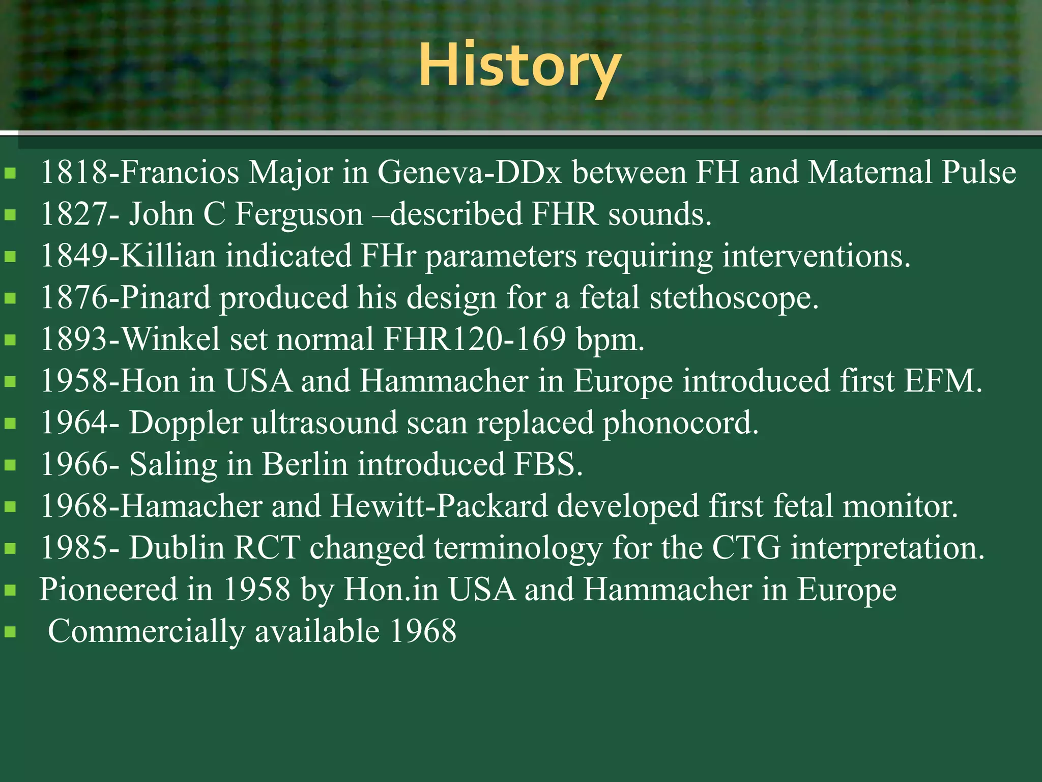
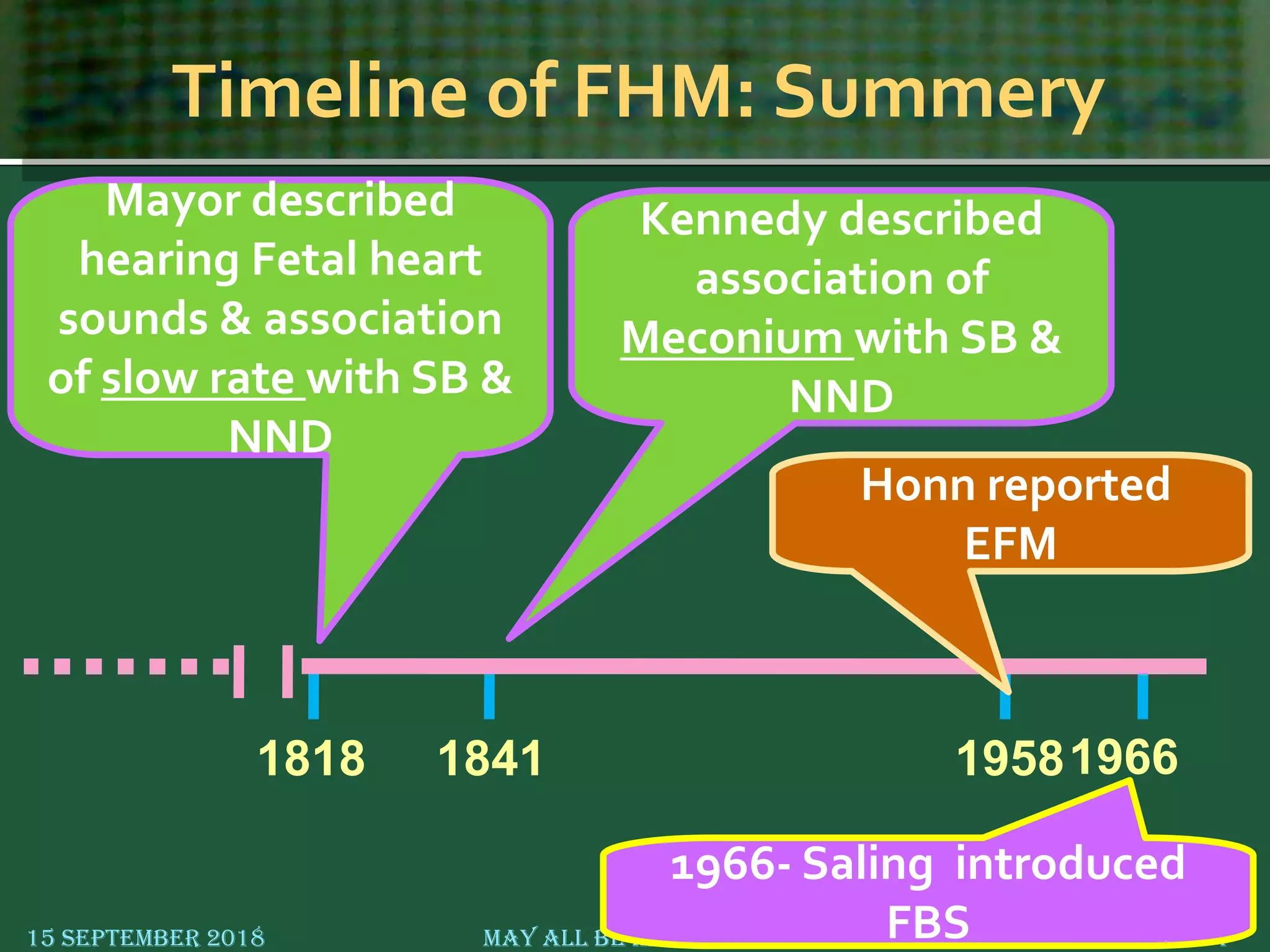
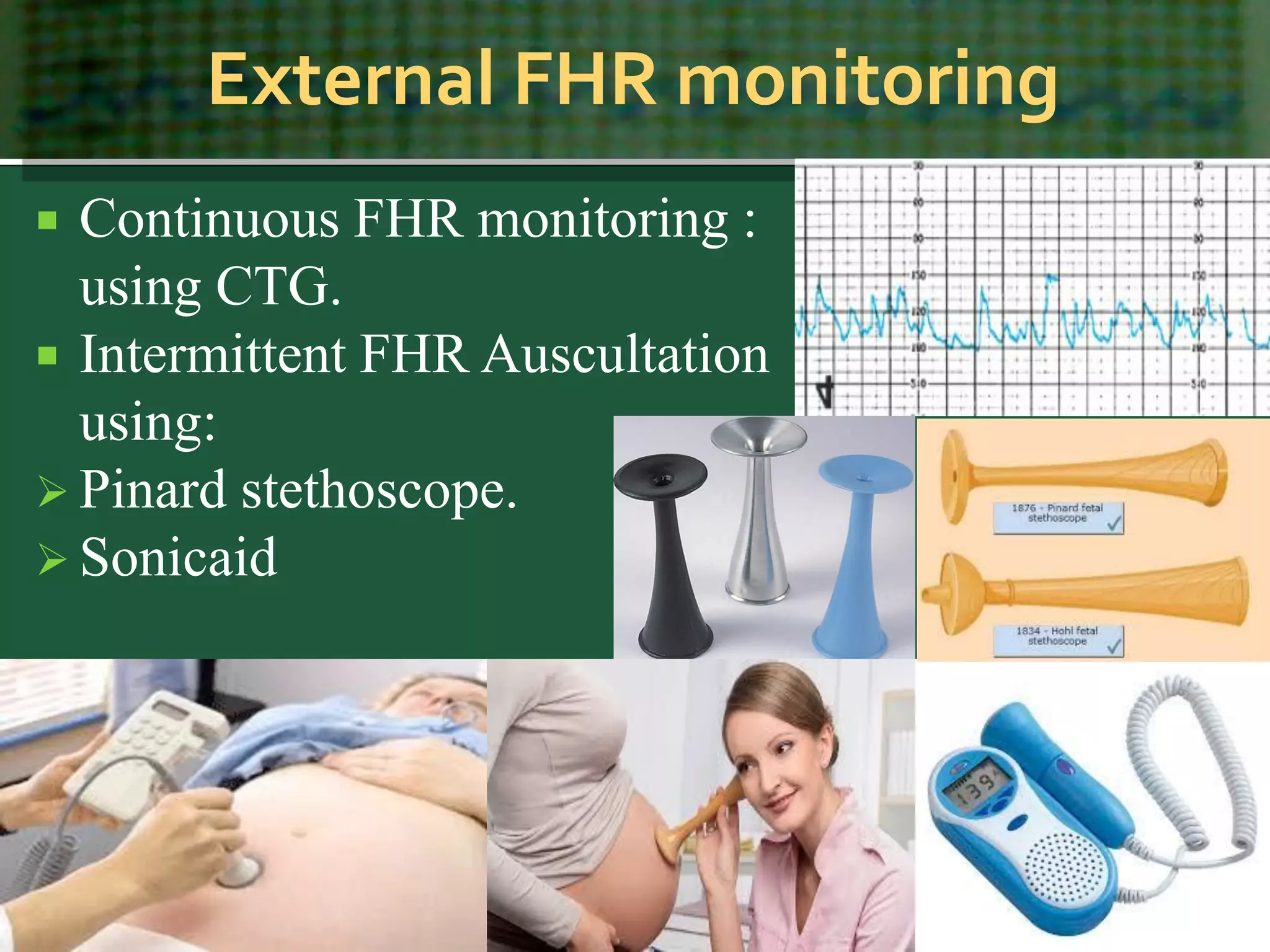
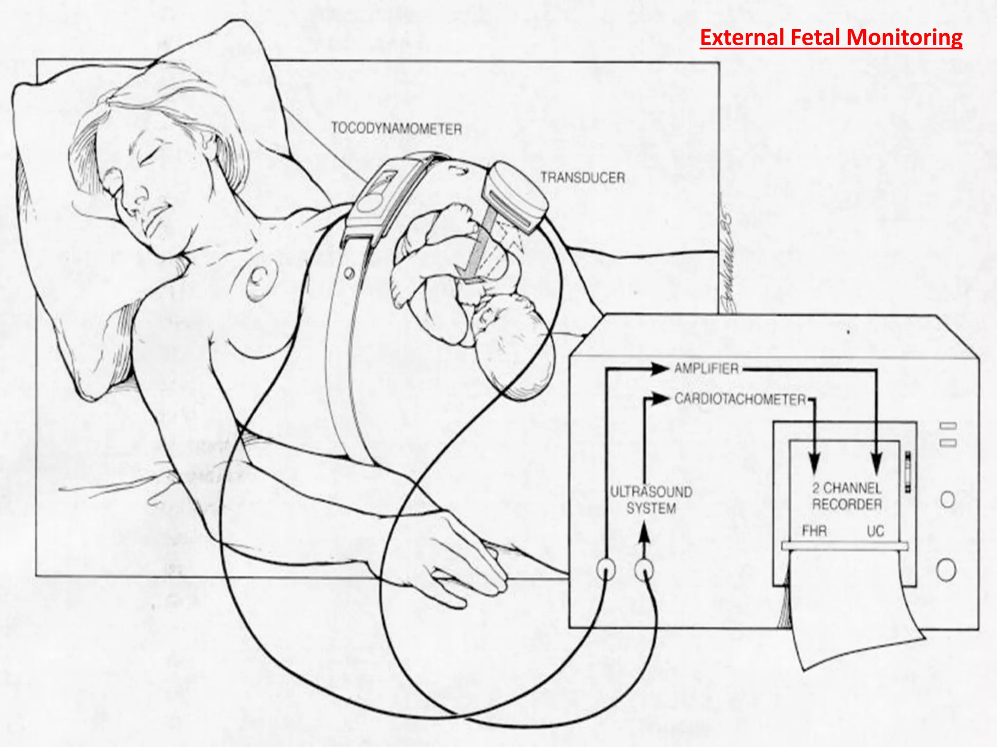
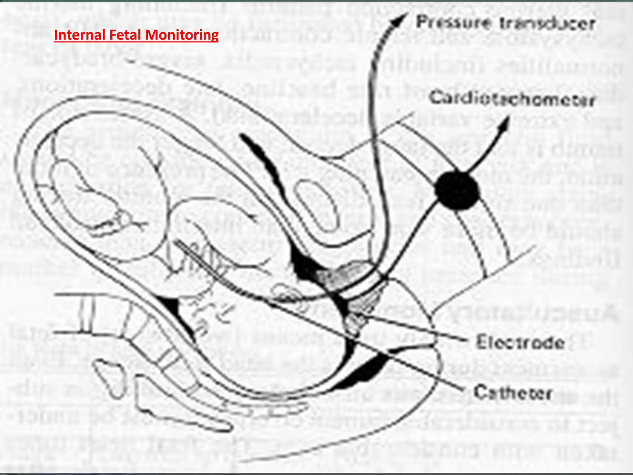
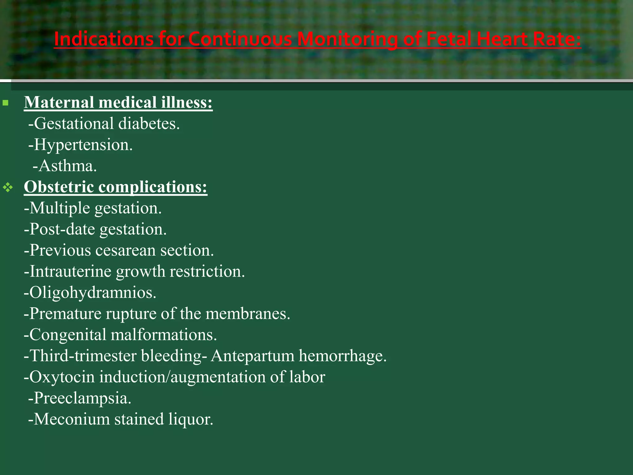
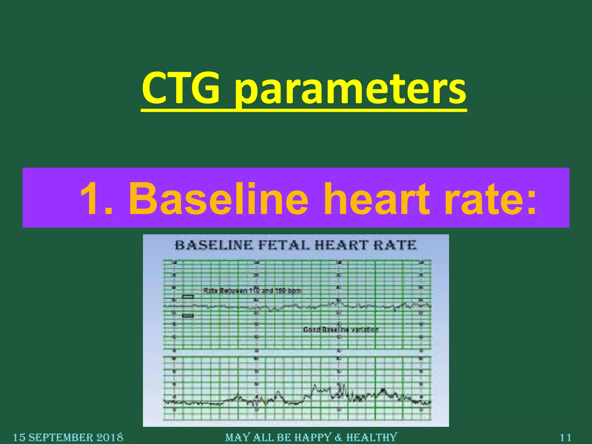
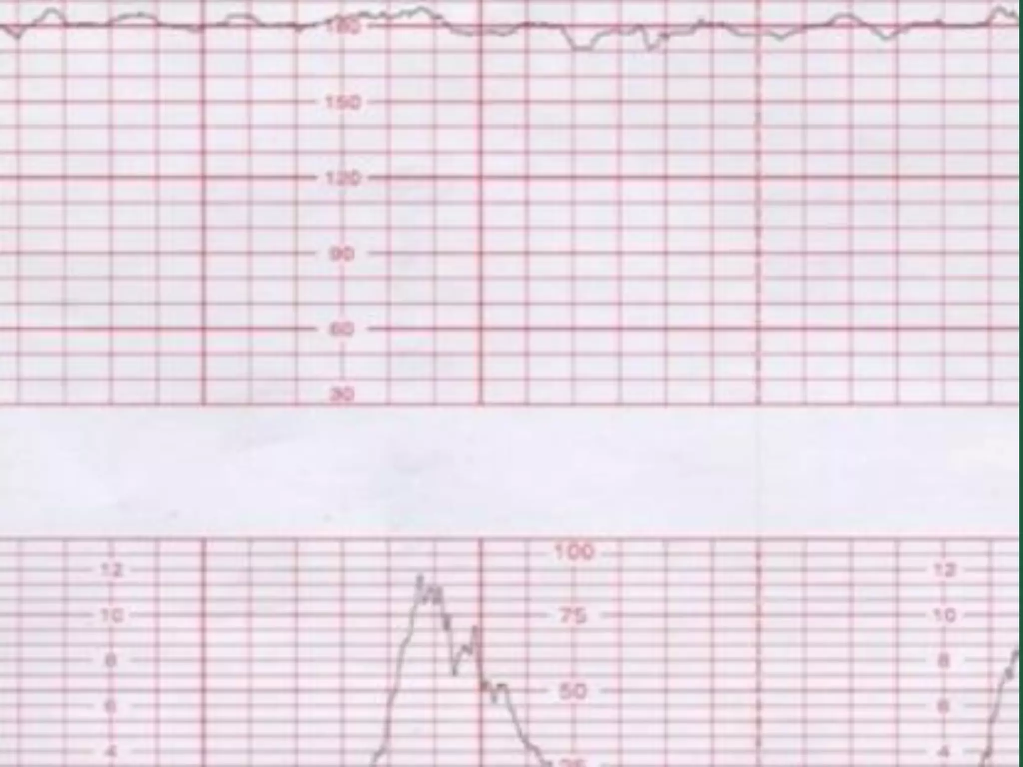
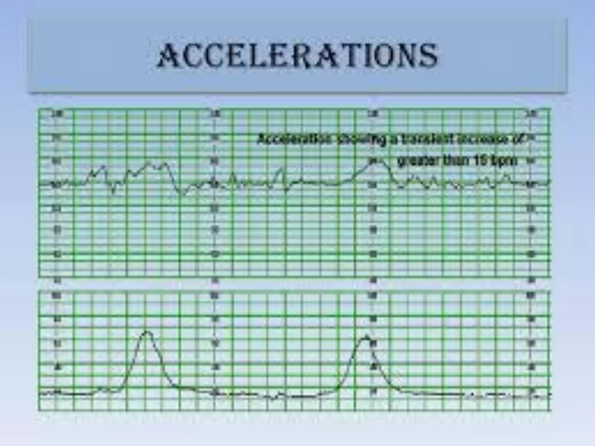
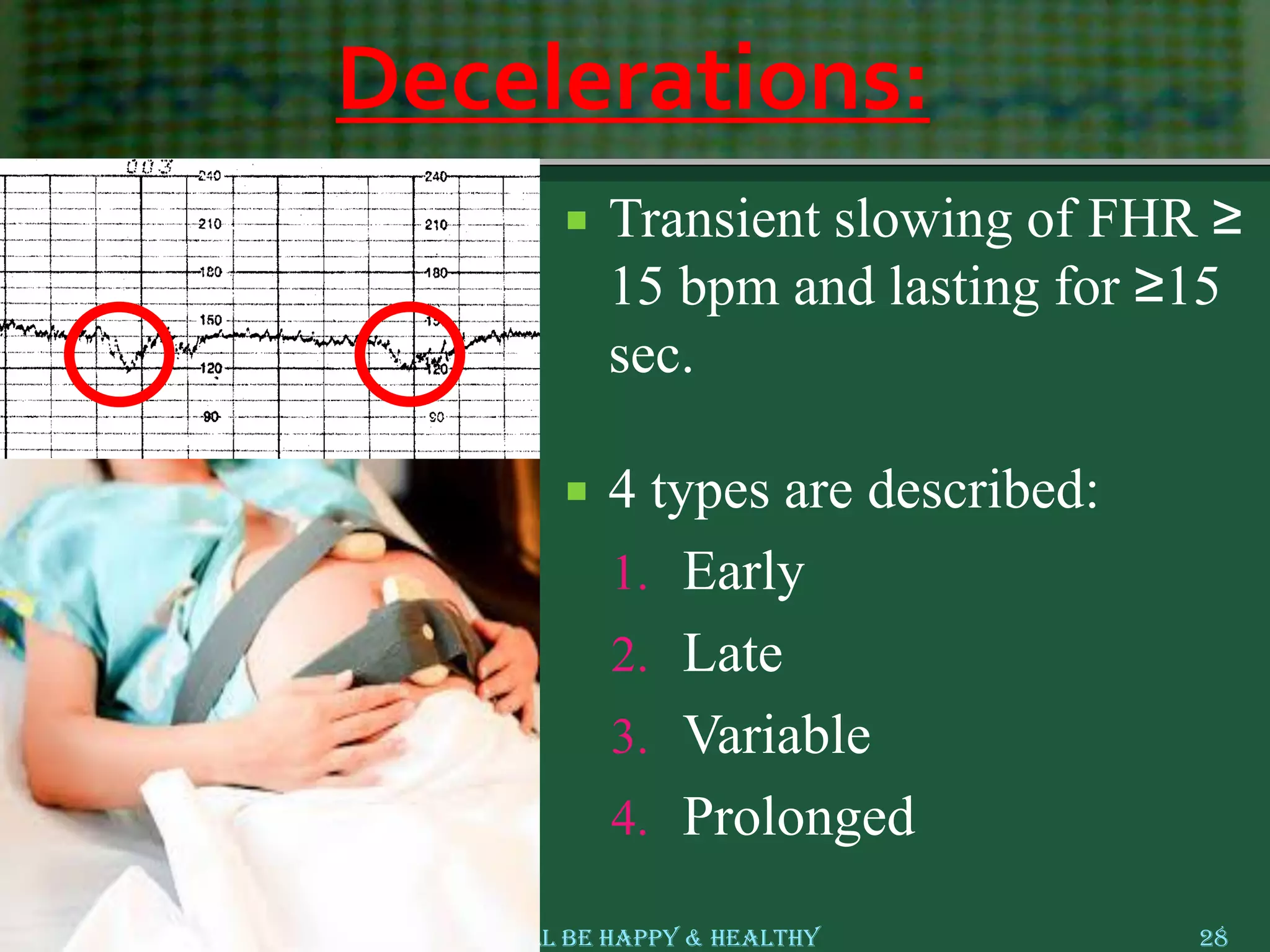
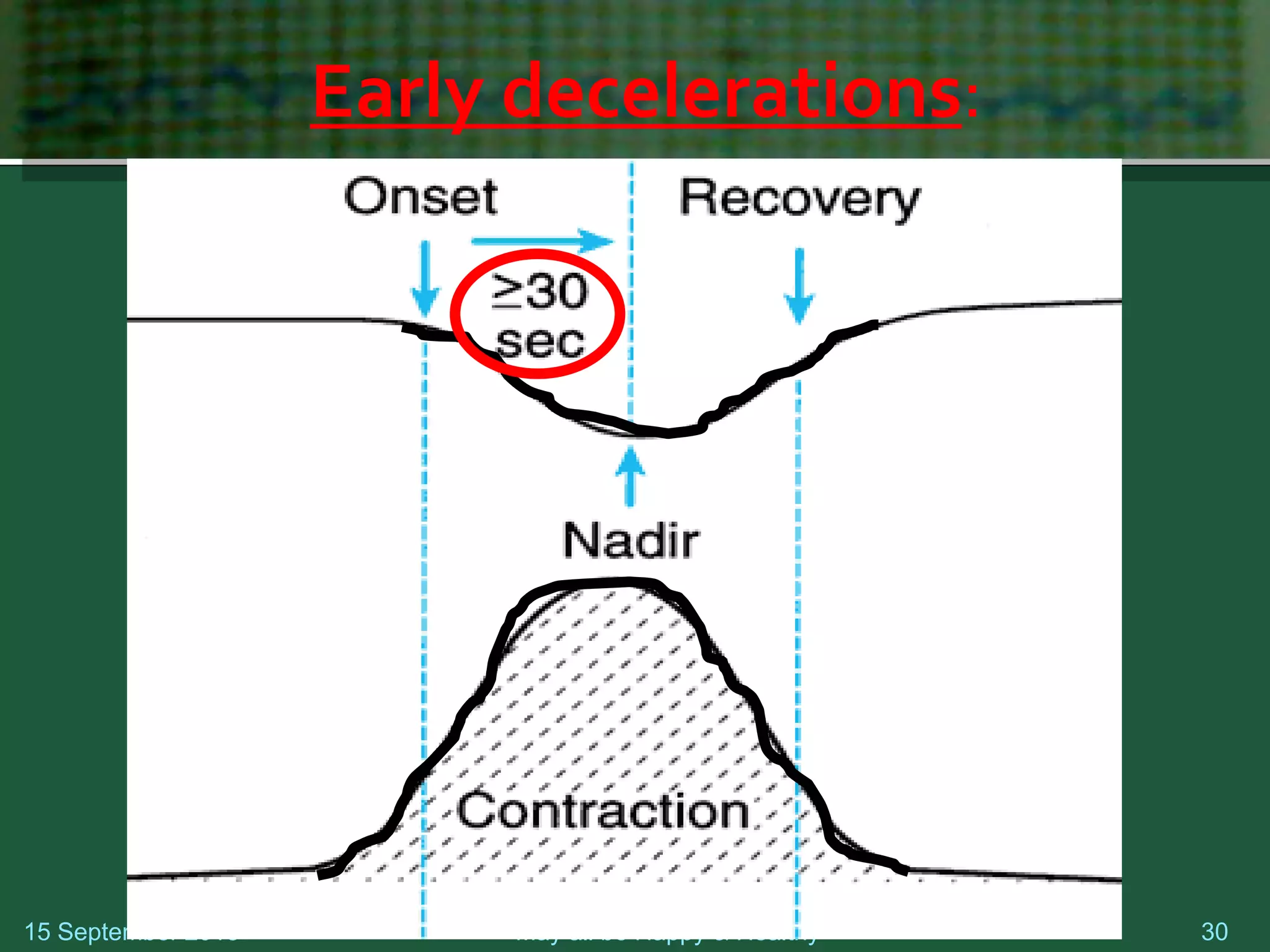
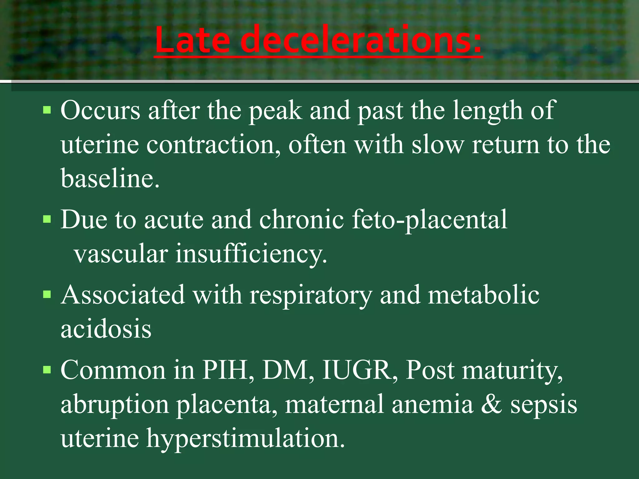
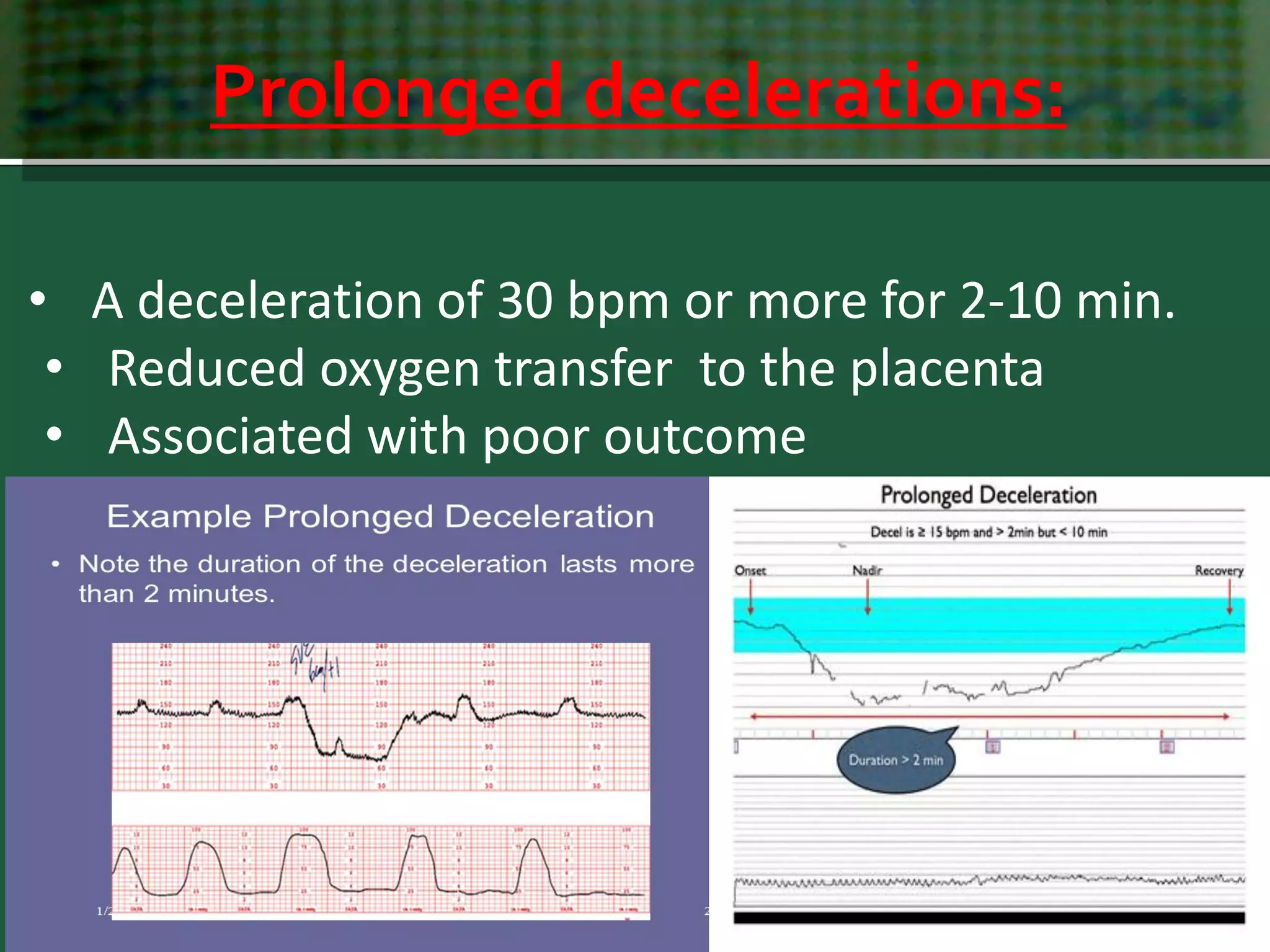
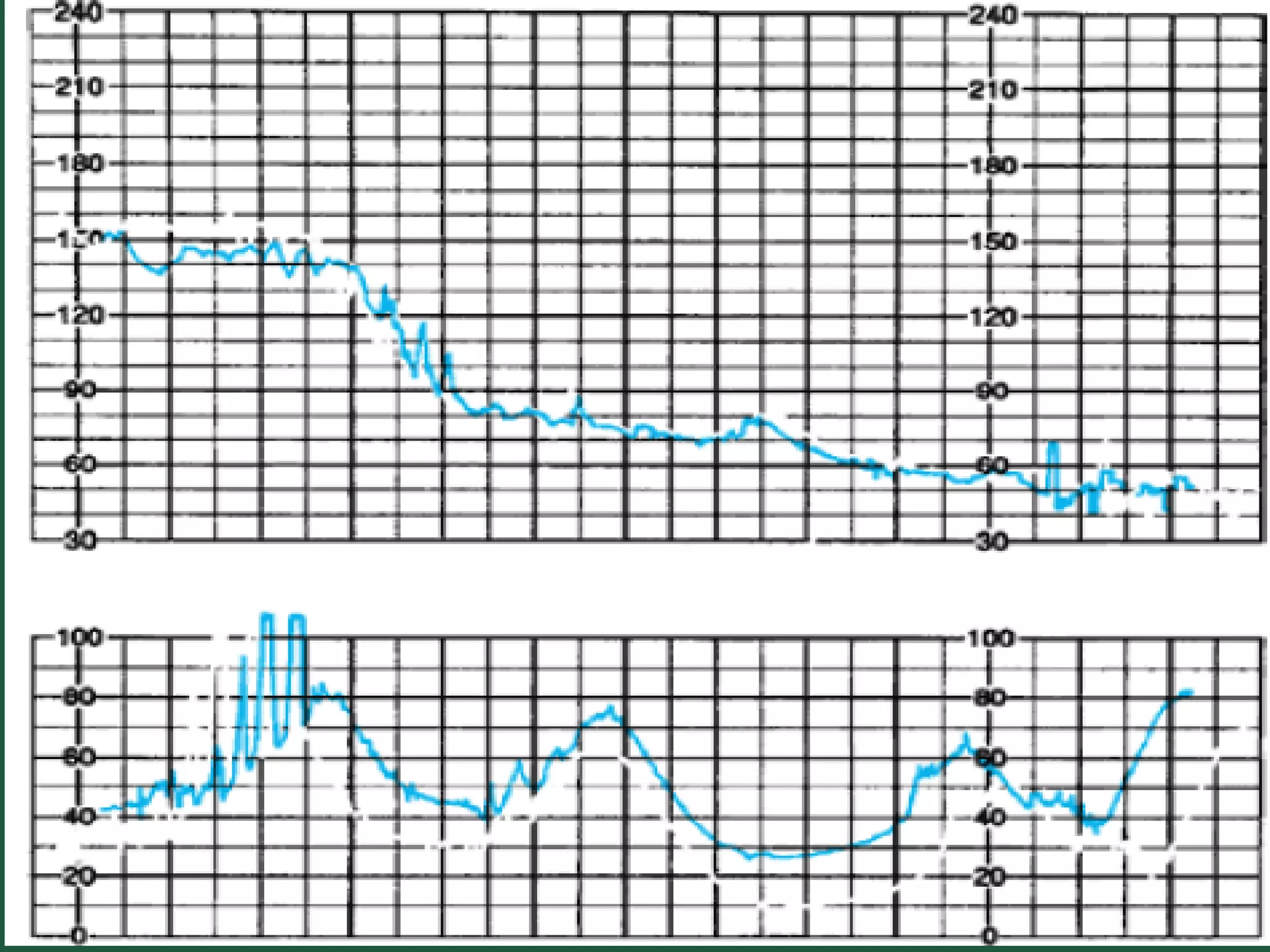
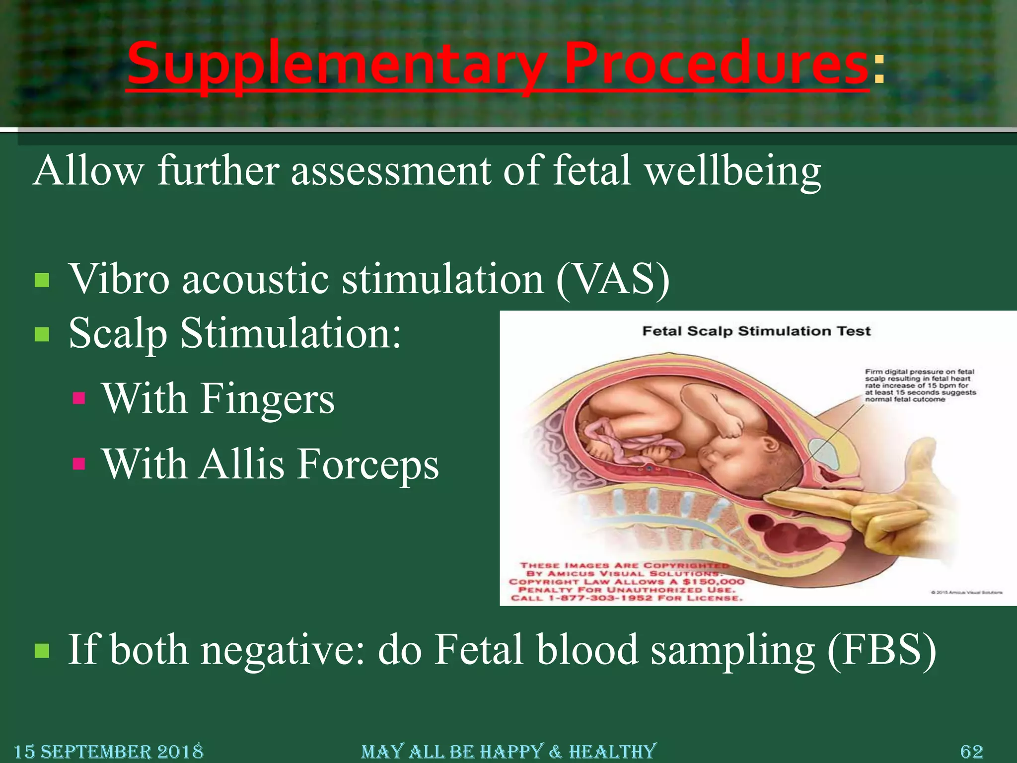
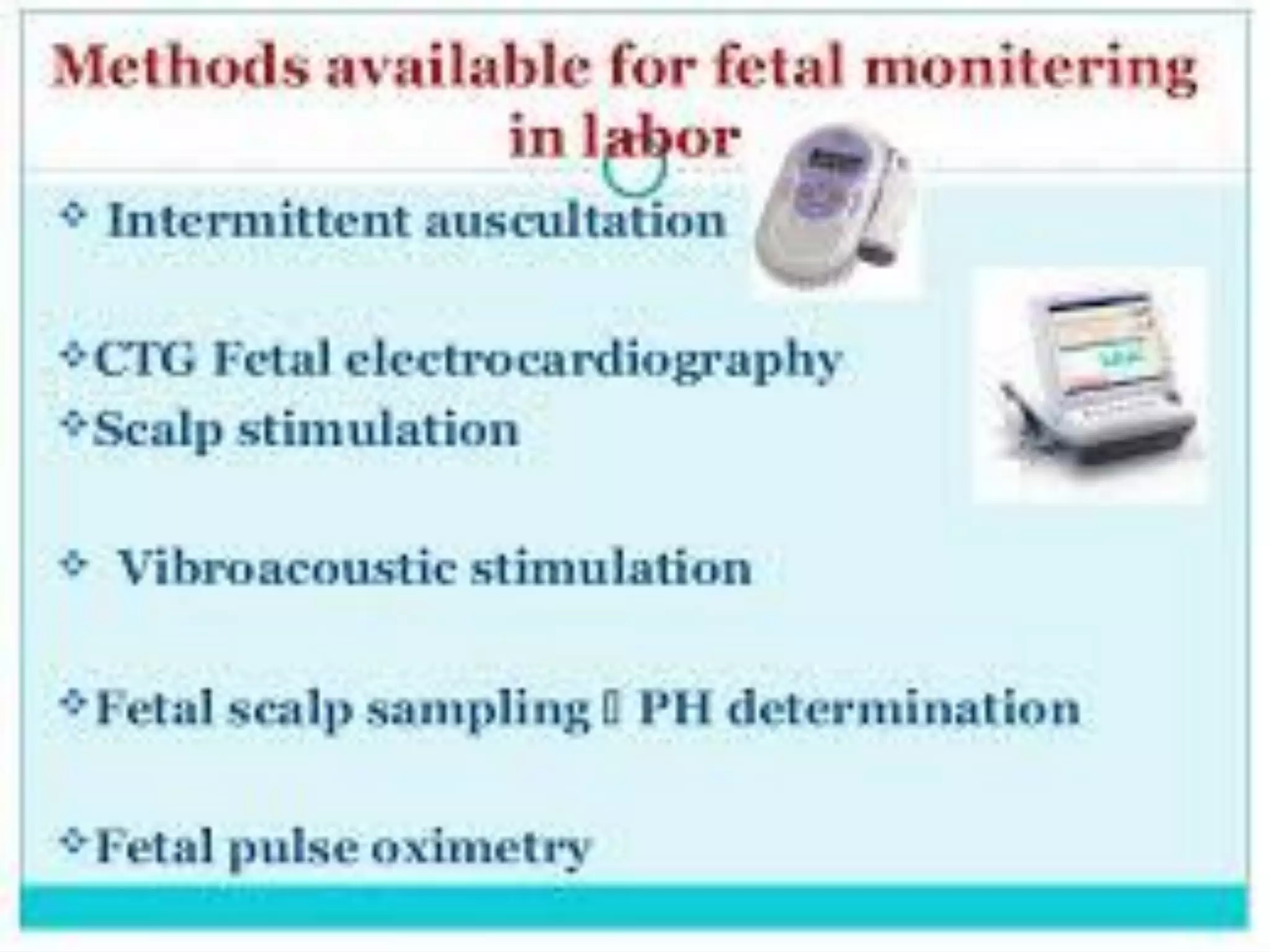
Cardiotocography (CTG)
For undergraduates
Associate Clinical Prof. Dr. Aisha M. El-Bareg, MD, PhD
Senior Consultant in (Obs & Gyn)/ Reproductive Medicine
Faculty of Medicine, Misurata .LIBYA
15 September 2018 May All Be Happy & Healthy 2
Eye doesn’t see
what mind doesn’t know
-- Osler
History
 1818-Francios Major in Geneva-DDx between FH and Maternal Pulse
 1827- John C Ferguson –described FHR sounds.
 1849-Killian indicated FHr parameters requiring interventions.
 1876-Pinard produced his design for a fetal stethoscope.
 1893-Winkel set normal FHR120-169 bpm.
 1958-Hon in USA and Hammacher in Europe introduced first EFM.
 1964- Doppler ultrasound scan replaced phonocord.
 1966- Saling in Berlin introduced FBS.
 1968-Hamacher and Hewitt-Packard developed first fetal monitor.
 1985- Dublin RCT changed terminology for the CTG interpretation.
 Pioneered in 1958 by Hon.in USA and Hammacher in Europe
 Commercially available 1968
Timeline of FHM: Summery
15 September 2018 May All Be Happy & Healthy 4
Mayor described
hearing Fetal heart
sounds & association
of slow rate with SB &
NND
1818 1841
Kennedy described
association of
Meconium with SB &
NND
1958
Honn reported
EFM
1966
1966- Saling introduced
FBS
Why fetal assessment ?
 The goal of fetal surveillance
is:
1. To detect fetal hypoxia at
early stage
2. To prevent CP & IUFD
15 September 2018 May All Be Happy & Healthy 5
Fetal heart monitoring
 External fetal heart monitoring
 Internal electronic FH monitoring- QRS wave
15 September 2018 6May All Be Happy & Healthy
External FHR monitoring
 Continuous FHR monitoring :
using CTG.
 Intermittent FHR Auscultation
using:
 Pinard stethoscope.
 Sonicaid
(CTG)???
External Fetal Monitoring
Internal Fetal Monitoring
Indications for Continuous Monitoring of Fetal Heart Rate:
 Maternal medical illness:
-Gestational diabetes.
-Hypertension.
-Asthma.
 Obstetric complications:
-Multiple gestation.
-Post-date gestation.
-Previous cesarean section.
-Intrauterine growth restriction.
-Oligohydramnios.
-Premature rupture of the membranes.
-Congenital malformations.
-Third-trimester bleeding- Antepartum hemorrhage.
-Oxytocin induction/augmentation of labor
-Preeclampsia.
-Meconium stained liquor.
15 September 2018 May All Be Happy & Healthy 11
1. Baseline heart rate:
CTG parameters
Baseline FHR (110 – 160):
 FHR occurs between contractions, regardless to
acceleration or deceleration.
 Decrease gradually from 16 weeks gestation to
term as the parasympathetic system develops.
15 September 2018 May All Be Happy & Healthy 12
Fetal Bradycardia:
 FHR between < 110 beats/min for a period of 10
min or more.
 In the absence of other changes, is not considered
significant.
 Causes
1. Hypoxia, Drugs (eg. Beta-blocker)
2. Autonomic effect (pressure on fetal head)
3. Fetal heart block
4. Severe Pyelonephritis
5. Hypothermia, Maternal hypotension
15 September 2018 May All Be Happy & Healthy 13
FetalTachycardia (>160):
 FHR > 160 bpm for a period of 10 min or more.
 Causes:
1. Prematurity
2. Hypoxia
3. Maternal Fever, Maternal Hypotension
4. Maternal thyrotoxicosis
5. Chorioamnionitis
6. Fetal Cardiac arrhythmias
7. Drugs: Atropine, Ventolin, Hydralazine,
Nifedipine.
15 September 2018 May All Be Happy & Healthy 15
2. Baseline variability
(BLV)
CTG parameters
BaselineVariability:
 Best indicators of intact integration between
fetal CNS & heart .
 Normal BLV 5-25 bpm
 Increased> 25 bpm.
 Absent < 3bpm
15 September 2018 May All Be Happy & Healthy 18
• The oscillatory changes that occur during the
course of 1 min and result in the waviness of the
baseline (3 – 5 /min).
• Result from the continuous interaction between
symp & parasymp nervous system.
15 September 2018 May All Be Happy & Healthy 20
15 September 2018 May All Be Happy & Healthy 21
Factors Affecting BaselineVariability:
 ↓ Variability:
1. Prematurity, fetal sleep
2. Fetal heart abn.,
arrhythmias
3. Fetal CNS abn.
4. Drugs: Sedatives,
Analgesics, & MgSO4
 ↑ Variability:
1. High gestation
2. Early, Mild Hypoxia
15 September 2018 May All Be Happy & Healthy 23
3. Periodic Changes
• Acceleration
• Deceleration
CTG parameters
Accelerations:
 Accelerations: Transient, abrupt, increase in FHR of
≥ 15 bpm lasting for ≥ 15 sec , return to baseline <
2 min.
 Occurs with fetal activity.
 Presence of FHR Accelerations have Good
outcome.
 Absence of accelerations on an otherwise normal
CTG remains unclear.
Causes of loss of Accelerations:
 Sleeping fetus
 CNS depressant drugs: Sedatives, Narcotic,
Analgesics
 Hypertensive Crisis, Diabetic Keto Acidosis
 Smoking
15 September 2018 May All Be Happy & Healthy 26
Accelerations:
15 September 2018 May All Be Happy & Healthy 27
Please
Consider my
Age for
interpretation
of
Accelerations
Decelerations:
 Transient slowing of FHR ≥
15 bpm and lasting for ≥15
sec.
 4 types are described:
1. Early
2. Late
3. Variable
4. Prolonged
15 September 2018 May All Be Happy & Healthy 28
Early Decelerations:
 Begins with the onset of contraction and returns to
baseline as the contraction ends.
 Mostly due to Head compression
 Considered physiologic, not associated with fetal
acidosis.
 Significant, if appear during early labor or
Antenatal.
15 September 2018 May All Be Happy & Healthy 29
Early decelerations:
15 September 2018 May all be Happy & Healthy 30
Early Decelerations:
15 September 2018 May All Be Happy & Healthy 31
Late decelerations:
 Occurs after the peak and past the length of
uterine contraction, often with slow return to the
baseline.
 Due to acute and chronic feto-placental
vascular insufficiency.
 Associated with respiratory and metabolic
acidosis
 Common in PIH, DM, IUGR, Post maturity,
abruption placenta, maternal anemia & sepsis
uterine hyperstimulation.
15 September 2018 May all be Happy & Healthy 33
Late decelerations:
Late decelerations:
15 September 2018 May All Be Happy & Healthy 34
 Deceleration occurs before, during, or after the
onset of uterine contraction.
 Caused by umbilical cord compression between
fetal parts and uterine wall.
 Common in oligohydramnios.
Variable Decelerations:
Variable Deceleration:
15 September 2018 May All Be Happy & Healthy 37
Significant if
recurrent
Variable Decelerations:
15 September 2018 May All Be Happy & Healthy 38
Recurrent Decelerations:
 Decelerations occur with > 50% of uterine contractions in any 20
minute segment.
 Recurrent variable decelerations (at least 3 in 20 minutes) may be
observed. However, close follow up is recommended because cord
accidents with subsequent fetal death may occur even in the
presence of normal amounts of amniotic fluid.
 Recurrent late decelerations should lead to consideration of
cesarean delivery unless the abnormal results are believed to be the
result of a reversible maternal condition such as diabetic
ketoacidosis or pneumonia with hypoxemia.
Prolonged decelerations:
15 September 2018 May All Be Happy & Healthy 42
• A deceleration of 30 bpm or more for 2-10 min.
• Reduced oxygen transfer to the placenta
• Associated with poor outcome
Causes of Prolonged Deceleration:
1. Impending birth (head compression).
2. Fetal hypoxia:
 Uterine hyperactivity- hypoperfusion/hypoxia
 Placental abruption
 Umbilical cord knots or prolapse.
 Maternal hypotension
 Maternal seizures including eclampsia and epilepsy.
 Sympathetic blockade (regional anesthesia)
 Maternal Valsalva maneuver.
15 September 2018 May All Be Happy & Healthy 44
The Contraction StressTest (CST):
 The Contraction stress test is used by some antepartum testing
centers to evaluate placental function under stress. The test is
performed by placing transducers (ultrasound and toco), on patient's
abdomen as with the non-stress test.
 The tracing is then observed for late decelerations.
 The test requires three contractions in 10 minutes to be
 present with the contractions lasting 40 to 60 seconds.
 If uterine activity is absent then oxytocin is infused or nipple
stimulation is used.
CST:
 The test is positive if late decelerations are consistent and present
with more than 50% of the contractions.
 A positive CST has been associated with an increased incidence of
intrauterine death, late decelerations in labor, low 5-minute Apgar
scores, and intrauterine growth restriction.
 The CST is equivocal or suspicious if there are intermittent late
decelerations
 A suspicious or equivocal CST should be repeated in 24 hours
15 September 2018 May All Be Happy & Healthy 48
CTG interpretation:
CTG Features:
 Baseline(beats per minute)
 Short term variability( beats per minute)
 Phase rectified signal averaging (beats per minute)
 Signal stability index
 Number of decelerations in 15 min
 Number of contractions in 15 min
 Contraction duration (seconds)
 Resting time between contractions (seconds)
CTG Interpretation:
 Consider :
1. Intranatal or Antenatal
2. Stage of labour
3. Gestational Age
4. Fetal presentation ?Malpresentation
5. Induction or augmentation of labour
6. Medicines (especially OTC)
7. Maternal Vitals & medical disorders
CTG Interpretation:
 It has to be taken into consideration the
following:
 There are different differences in the way clinicians
interpret CTG tracings, depending on the guidelines they
use.
 Differences in guideline structure as well as in clarity and
complexity of definitions, have a profound effect on inter-
observer agreement and reliability, as well as on the
sensitivity and specificity of CT classifications in
predicting acidemia.
Normal Antenatal CTG
Features of reassuring (Normal) CTG:
15 September 2018 May All Be Happy & Healthy 53
Normal Base line FHR (110 – 160)
Normal
BLV
(6 to 25)
≥ 2 Accelerations in 20 minutes
No
Significant
Deceleration
Non reassuring (Nonreactive) CTG:
 Absence of one or more features of normal CTG
15 September 2018 May All Be Happy & Healthy 56
Normal Base line FHR
Normal
Base line
Variability
Presence of Acceleration
No
Significant
Deceleration
Sinusoidal pattern
Sinusoidal pattern
 Regular Oscillation of the Baseline long-term
Variability resembling a Sine wave ,with no B-b
Variability.
 Has fixed cycle of 3-5 p min. with amplitude of 5-
15 bpm and above but not below the baseline.
 Should be viewed with suspicion as poor outcome
has been seen (eg Feto-maternal hemorrhage)
 Sinusoidal pattern - distinctive smooth undulating
 Sine-wave baseline with no B-b variability.
 0.3 % (Young 1980)
 cord compression
 Hypovolemia
 ascites
 idiopathic(fetal thumb sucking)
 Analgesics
 Anemia
 Abruption
DeadlyTrio:
 Accelerations absent
 Loss of BLV
 Recurrent late decelerations at least for 20 minutes
15 September 2018 May All Be Happy & Healthy 60
Management of non reassuring CTG
 Change maternal position.
 Provide oxygen by face mask
 Reversal of anesthetic effect.
 Regulation of uterine activity:
 Stop oxytocin
 Good hydration
 Correction of cord compression
 Change maternal position
 Amnioinfusion
Supplementary Procedures:
Allow further assessment of fetal wellbeing
 Vibro acoustic stimulation (VAS)
 Scalp Stimulation:
 With Fingers
 With Allis Forceps
 If both negative: do Fetal blood sampling (FBS)
15 September 2018 May All Be Happy & Healthy 62
Fetal Blood Sampling:
 Useful in the presence of a non reassuring CTG.
 A scalp blood sample for pH or lactate
determination.
 Specificity is high ( normal value rules out
asphyxia).
 The sensitivity and positive predictive value of a
low scalp pH in identifying a newborn with
Hypoxic-ischemic encephalopathy is low.
FBS Contraindications
 Premature –less than 34 weeks
 Active Herpes
 Known HIV, Hep B,C positive status.
 Thrombocytopenia.
 Maternal-
 Unfavorable Cx
 Mobile PP
 Malpresentation(face etc.) uncertain??
 Pl Praevia or APH
 Sepsis
FBS-Sampling errors:
 Between decelerations if possible
 Avoid Excess pressure on PP reduces perfusion
 Do not sample on the caput.
 Failure of scalp to bleed –due to peripheral shut
down.
FBS:
• Normal pH 7.25—7.35
• If <7.20 – significant
acidosis/ immediate
delivery
Fetal blood oximetry
False positive diagnosis is reduced to 10 %
Problems with CTG:
1-Has a high sensitivity but with limited specificity
in the prediction of fetal hypoxia/acidosis.
2. Increases operative vaginal delivery
3. No change in incidence of CP
4. Reduction in Neonatal seizures rates only 0.51%
5. No significant difference in APGAR scores
6. ? About the efficacy
15 September 2018 May All Be Happy & Healthy 69
CTG lecture for undergraduates by Associate Prof.Dr Aisha Elbareg

CTG lecture for undergraduates by Associate Prof.Dr Aisha Elbareg

  • 1.
    Cardiotocography (CTG) For undergraduates AssociateClinical Prof. Dr. Aisha M. El-Bareg, MD, PhD Senior Consultant in (Obs & Gyn)/ Reproductive Medicine Faculty of Medicine, Misurata .LIBYA
  • 2.
    15 September 2018May All Be Happy & Healthy 2 Eye doesn’t see what mind doesn’t know -- Osler
  • 3.
    History  1818-Francios Majorin Geneva-DDx between FH and Maternal Pulse  1827- John C Ferguson –described FHR sounds.  1849-Killian indicated FHr parameters requiring interventions.  1876-Pinard produced his design for a fetal stethoscope.  1893-Winkel set normal FHR120-169 bpm.  1958-Hon in USA and Hammacher in Europe introduced first EFM.  1964- Doppler ultrasound scan replaced phonocord.  1966- Saling in Berlin introduced FBS.  1968-Hamacher and Hewitt-Packard developed first fetal monitor.  1985- Dublin RCT changed terminology for the CTG interpretation.  Pioneered in 1958 by Hon.in USA and Hammacher in Europe  Commercially available 1968
  • 4.
    Timeline of FHM:Summery 15 September 2018 May All Be Happy & Healthy 4 Mayor described hearing Fetal heart sounds & association of slow rate with SB & NND 1818 1841 Kennedy described association of Meconium with SB & NND 1958 Honn reported EFM 1966 1966- Saling introduced FBS
  • 5.
    Why fetal assessment?  The goal of fetal surveillance is: 1. To detect fetal hypoxia at early stage 2. To prevent CP & IUFD 15 September 2018 May All Be Happy & Healthy 5
  • 6.
    Fetal heart monitoring External fetal heart monitoring  Internal electronic FH monitoring- QRS wave 15 September 2018 6May All Be Happy & Healthy
  • 7.
    External FHR monitoring Continuous FHR monitoring : using CTG.  Intermittent FHR Auscultation using:  Pinard stethoscope.  Sonicaid
  • 8.
  • 9.
  • 10.
    Indications for ContinuousMonitoring of Fetal Heart Rate:  Maternal medical illness: -Gestational diabetes. -Hypertension. -Asthma.  Obstetric complications: -Multiple gestation. -Post-date gestation. -Previous cesarean section. -Intrauterine growth restriction. -Oligohydramnios. -Premature rupture of the membranes. -Congenital malformations. -Third-trimester bleeding- Antepartum hemorrhage. -Oxytocin induction/augmentation of labor -Preeclampsia. -Meconium stained liquor.
  • 11.
    15 September 2018May All Be Happy & Healthy 11 1. Baseline heart rate: CTG parameters
  • 12.
    Baseline FHR (110– 160):  FHR occurs between contractions, regardless to acceleration or deceleration.  Decrease gradually from 16 weeks gestation to term as the parasympathetic system develops. 15 September 2018 May All Be Happy & Healthy 12
  • 13.
    Fetal Bradycardia:  FHRbetween < 110 beats/min for a period of 10 min or more.  In the absence of other changes, is not considered significant.  Causes 1. Hypoxia, Drugs (eg. Beta-blocker) 2. Autonomic effect (pressure on fetal head) 3. Fetal heart block 4. Severe Pyelonephritis 5. Hypothermia, Maternal hypotension 15 September 2018 May All Be Happy & Healthy 13
  • 15.
    FetalTachycardia (>160):  FHR> 160 bpm for a period of 10 min or more.  Causes: 1. Prematurity 2. Hypoxia 3. Maternal Fever, Maternal Hypotension 4. Maternal thyrotoxicosis 5. Chorioamnionitis 6. Fetal Cardiac arrhythmias 7. Drugs: Atropine, Ventolin, Hydralazine, Nifedipine. 15 September 2018 May All Be Happy & Healthy 15
  • 17.
  • 18.
    BaselineVariability:  Best indicatorsof intact integration between fetal CNS & heart .  Normal BLV 5-25 bpm  Increased> 25 bpm.  Absent < 3bpm 15 September 2018 May All Be Happy & Healthy 18 • The oscillatory changes that occur during the course of 1 min and result in the waviness of the baseline (3 – 5 /min). • Result from the continuous interaction between symp & parasymp nervous system.
  • 20.
    15 September 2018May All Be Happy & Healthy 20
  • 21.
    15 September 2018May All Be Happy & Healthy 21
  • 22.
    Factors Affecting BaselineVariability: ↓ Variability: 1. Prematurity, fetal sleep 2. Fetal heart abn., arrhythmias 3. Fetal CNS abn. 4. Drugs: Sedatives, Analgesics, & MgSO4  ↑ Variability: 1. High gestation 2. Early, Mild Hypoxia
  • 23.
    15 September 2018May All Be Happy & Healthy 23 3. Periodic Changes • Acceleration • Deceleration CTG parameters
  • 24.
    Accelerations:  Accelerations: Transient,abrupt, increase in FHR of ≥ 15 bpm lasting for ≥ 15 sec , return to baseline < 2 min.  Occurs with fetal activity.  Presence of FHR Accelerations have Good outcome.  Absence of accelerations on an otherwise normal CTG remains unclear.
  • 26.
    Causes of lossof Accelerations:  Sleeping fetus  CNS depressant drugs: Sedatives, Narcotic, Analgesics  Hypertensive Crisis, Diabetic Keto Acidosis  Smoking 15 September 2018 May All Be Happy & Healthy 26
  • 27.
    Accelerations: 15 September 2018May All Be Happy & Healthy 27 Please Consider my Age for interpretation of Accelerations
  • 28.
    Decelerations:  Transient slowingof FHR ≥ 15 bpm and lasting for ≥15 sec.  4 types are described: 1. Early 2. Late 3. Variable 4. Prolonged 15 September 2018 May All Be Happy & Healthy 28
  • 29.
    Early Decelerations:  Beginswith the onset of contraction and returns to baseline as the contraction ends.  Mostly due to Head compression  Considered physiologic, not associated with fetal acidosis.  Significant, if appear during early labor or Antenatal. 15 September 2018 May All Be Happy & Healthy 29
  • 30.
    Early decelerations: 15 September2018 May all be Happy & Healthy 30
  • 31.
    Early Decelerations: 15 September2018 May All Be Happy & Healthy 31
  • 32.
    Late decelerations:  Occursafter the peak and past the length of uterine contraction, often with slow return to the baseline.  Due to acute and chronic feto-placental vascular insufficiency.  Associated with respiratory and metabolic acidosis  Common in PIH, DM, IUGR, Post maturity, abruption placenta, maternal anemia & sepsis uterine hyperstimulation.
  • 33.
    15 September 2018May all be Happy & Healthy 33 Late decelerations:
  • 34.
    Late decelerations: 15 September2018 May All Be Happy & Healthy 34
  • 36.
     Deceleration occursbefore, during, or after the onset of uterine contraction.  Caused by umbilical cord compression between fetal parts and uterine wall.  Common in oligohydramnios. Variable Decelerations:
  • 37.
    Variable Deceleration: 15 September2018 May All Be Happy & Healthy 37 Significant if recurrent
  • 38.
    Variable Decelerations: 15 September2018 May All Be Happy & Healthy 38
  • 40.
    Recurrent Decelerations:  Decelerationsoccur with > 50% of uterine contractions in any 20 minute segment.  Recurrent variable decelerations (at least 3 in 20 minutes) may be observed. However, close follow up is recommended because cord accidents with subsequent fetal death may occur even in the presence of normal amounts of amniotic fluid.  Recurrent late decelerations should lead to consideration of cesarean delivery unless the abnormal results are believed to be the result of a reversible maternal condition such as diabetic ketoacidosis or pneumonia with hypoxemia.
  • 42.
    Prolonged decelerations: 15 September2018 May All Be Happy & Healthy 42 • A deceleration of 30 bpm or more for 2-10 min. • Reduced oxygen transfer to the placenta • Associated with poor outcome
  • 44.
    Causes of ProlongedDeceleration: 1. Impending birth (head compression). 2. Fetal hypoxia:  Uterine hyperactivity- hypoperfusion/hypoxia  Placental abruption  Umbilical cord knots or prolapse.  Maternal hypotension  Maternal seizures including eclampsia and epilepsy.  Sympathetic blockade (regional anesthesia)  Maternal Valsalva maneuver. 15 September 2018 May All Be Happy & Healthy 44
  • 45.
    The Contraction StressTest(CST):  The Contraction stress test is used by some antepartum testing centers to evaluate placental function under stress. The test is performed by placing transducers (ultrasound and toco), on patient's abdomen as with the non-stress test.  The tracing is then observed for late decelerations.  The test requires three contractions in 10 minutes to be  present with the contractions lasting 40 to 60 seconds.  If uterine activity is absent then oxytocin is infused or nipple stimulation is used.
  • 46.
    CST:  The testis positive if late decelerations are consistent and present with more than 50% of the contractions.  A positive CST has been associated with an increased incidence of intrauterine death, late decelerations in labor, low 5-minute Apgar scores, and intrauterine growth restriction.  The CST is equivocal or suspicious if there are intermittent late decelerations  A suspicious or equivocal CST should be repeated in 24 hours
  • 48.
    15 September 2018May All Be Happy & Healthy 48 CTG interpretation:
  • 49.
    CTG Features:  Baseline(beatsper minute)  Short term variability( beats per minute)  Phase rectified signal averaging (beats per minute)  Signal stability index  Number of decelerations in 15 min  Number of contractions in 15 min  Contraction duration (seconds)  Resting time between contractions (seconds)
  • 50.
    CTG Interpretation:  Consider: 1. Intranatal or Antenatal 2. Stage of labour 3. Gestational Age 4. Fetal presentation ?Malpresentation 5. Induction or augmentation of labour 6. Medicines (especially OTC) 7. Maternal Vitals & medical disorders
  • 51.
    CTG Interpretation:  Ithas to be taken into consideration the following:  There are different differences in the way clinicians interpret CTG tracings, depending on the guidelines they use.  Differences in guideline structure as well as in clarity and complexity of definitions, have a profound effect on inter- observer agreement and reliability, as well as on the sensitivity and specificity of CT classifications in predicting acidemia.
  • 52.
  • 53.
    Features of reassuring(Normal) CTG: 15 September 2018 May All Be Happy & Healthy 53 Normal Base line FHR (110 – 160) Normal BLV (6 to 25) ≥ 2 Accelerations in 20 minutes No Significant Deceleration
  • 56.
    Non reassuring (Nonreactive)CTG:  Absence of one or more features of normal CTG 15 September 2018 May All Be Happy & Healthy 56 Normal Base line FHR Normal Base line Variability Presence of Acceleration No Significant Deceleration
  • 57.
  • 58.
    Sinusoidal pattern  RegularOscillation of the Baseline long-term Variability resembling a Sine wave ,with no B-b Variability.  Has fixed cycle of 3-5 p min. with amplitude of 5- 15 bpm and above but not below the baseline.  Should be viewed with suspicion as poor outcome has been seen (eg Feto-maternal hemorrhage)
  • 59.
     Sinusoidal pattern- distinctive smooth undulating  Sine-wave baseline with no B-b variability.  0.3 % (Young 1980)  cord compression  Hypovolemia  ascites  idiopathic(fetal thumb sucking)  Analgesics  Anemia  Abruption
  • 60.
    DeadlyTrio:  Accelerations absent Loss of BLV  Recurrent late decelerations at least for 20 minutes 15 September 2018 May All Be Happy & Healthy 60
  • 61.
    Management of nonreassuring CTG  Change maternal position.  Provide oxygen by face mask  Reversal of anesthetic effect.  Regulation of uterine activity:  Stop oxytocin  Good hydration  Correction of cord compression  Change maternal position  Amnioinfusion
  • 62.
    Supplementary Procedures: Allow furtherassessment of fetal wellbeing  Vibro acoustic stimulation (VAS)  Scalp Stimulation:  With Fingers  With Allis Forceps  If both negative: do Fetal blood sampling (FBS) 15 September 2018 May All Be Happy & Healthy 62
  • 65.
    Fetal Blood Sampling: Useful in the presence of a non reassuring CTG.  A scalp blood sample for pH or lactate determination.  Specificity is high ( normal value rules out asphyxia).  The sensitivity and positive predictive value of a low scalp pH in identifying a newborn with Hypoxic-ischemic encephalopathy is low.
  • 66.
    FBS Contraindications  Premature–less than 34 weeks  Active Herpes  Known HIV, Hep B,C positive status.  Thrombocytopenia.  Maternal-  Unfavorable Cx  Mobile PP  Malpresentation(face etc.) uncertain??  Pl Praevia or APH  Sepsis
  • 67.
    FBS-Sampling errors:  Betweendecelerations if possible  Avoid Excess pressure on PP reduces perfusion  Do not sample on the caput.  Failure of scalp to bleed –due to peripheral shut down.
  • 68.
    FBS: • Normal pH7.25—7.35 • If <7.20 – significant acidosis/ immediate delivery Fetal blood oximetry False positive diagnosis is reduced to 10 %
  • 69.
    Problems with CTG: 1-Hasa high sensitivity but with limited specificity in the prediction of fetal hypoxia/acidosis. 2. Increases operative vaginal delivery 3. No change in incidence of CP 4. Reduction in Neonatal seizures rates only 0.51% 5. No significant difference in APGAR scores 6. ? About the efficacy 15 September 2018 May All Be Happy & Healthy 69