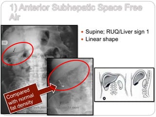
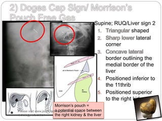
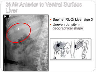
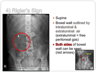
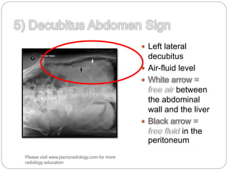
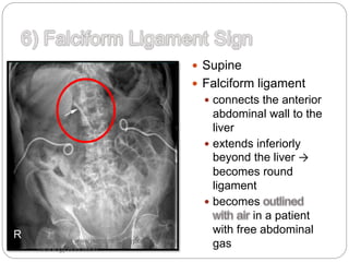
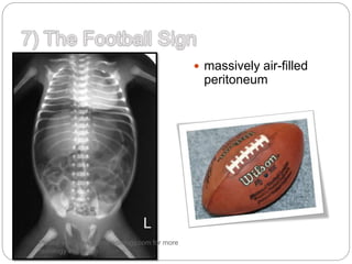
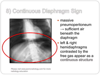
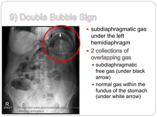
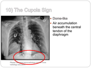
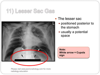
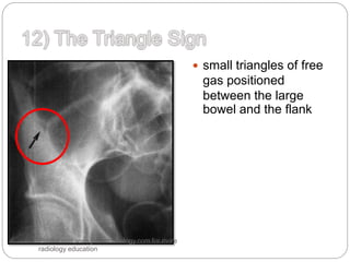
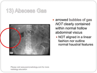
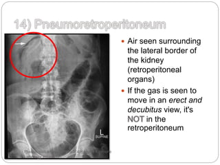
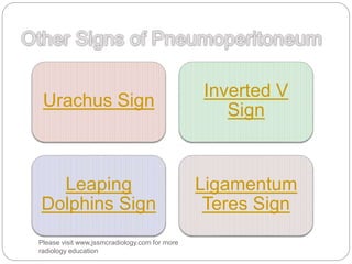
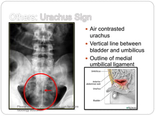
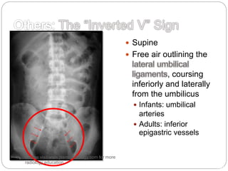
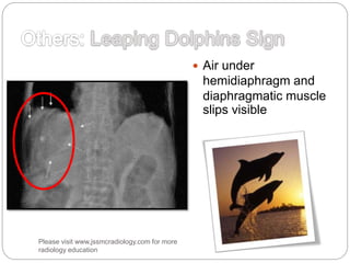
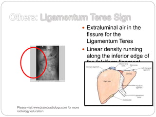
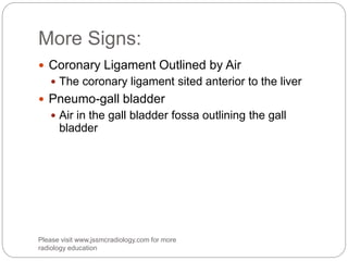
Please visit www.jssmcradiology.com
for more radiology education
 Supine; RUQ/Liver sign 1
 Linear shape
Please visit www.jssmcradiology.com for more
radiology education
 Supine; RUQ/Liver sign 2
shaped
lateral
corner
border outlining the
medial border of the
liver
4. Positioned inferior to
the 11thrib
5. Positioned superior
to the right kidney
Morrison’s pouch =
a potential space between
the right kidney & the liver
Please visit www.jssmcradiology.com for more
radiology education
 Supine; RUQ/ Liver sign 3
 Uneven density in
Please visit www.jssmcradiology.com for more
radiology education
 Supine
 Bowel wall
(extraluminal = free
peritoneal gas)
of bowel
wall can be seen
(red arrows)
Please visit www.jssmcradiology.com for more
radiology education
 Left lateral
decubitus
 Air-fluid level
between
the abdominal
wall and the liver
in the
peritoneum
Please visit www.jssmcradiology.com for more
radiology education
 Supine
 Falciform ligament
 connects the anterior
abdominal wall to the
liver
 extends inferiorly
beyond the liver →
becomes round
ligament
 becomes
in a patient
with free abdominal
gasPlease visit www.jssmcradiology.com for more
radiology education
 massively air-filled
peritoneum
Please visit www.jssmcradiology.com for more
radiology education
 massive
pneumoperitoneum
→ sufficient air
beneath the
diaphragm
 left & right
hemidiaphragms
contrasted by the
free gas appear as a
Please visit www.jssmcradiology.com for more
radiology education
 subdiaphragmatic gas
under the left
hemidiaphragm
 subdiaphragmatic
free gas (under black
arrow)
 normal gas within the
fundus of the stomach
(under white arrow)
Please visit www.jssmcradiology.com for more
radiology education
 Air accumulation
beneath the central
tendon of the
diaphragm
Please visit www.jssmcradiology.com for more
radiology education
 The lesser sac
 positioned posterior to
the stomach
 usually a potential
space
Note:
White arrow = Cupola
sign
Please visit www.jssmcradiology.com for more
radiology education
 small triangles of free
gas positioned
between the large
bowel and the flank
Please visit www.jssmcradiology.com for more
radiology education
 arrowed
NOT clearly contained
within normal hollow
abdominal viscus
 NOT aligned in a linear
fashion nor outline
normal haustral features
Please visit www.jssmcradiology.com for more
radiology education
 Air seen surrounding
the lateral border of
the kidney
(retroperitoneal
organs)
 If the gas is seen to
move in an erect and
decubitus view, it's
in the
retroperitoneum
Please visit www.jssmcradiology.com for more
radiology education
Urachus Sign
Inverted V
Sign
Leaping
Dolphins Sign
Ligamentum
Teres Sign
Please visit www.jssmcradiology.com for more
radiology education
 Air contrasted
urachus
 Vertical line between
bladder and umbilicus
 Outline of medial
umbilical ligament
Please visit www.jssmcradiology.com for more
radiology education
 Supine
 Free air outlining the
, coursing
inferiorly and laterally
from the umbilicus
 Infants: umbilical
arteries
 Adults: inferior
epigastric vessels
http://dx.doi.org/10.1148/radiology.151.1.62306
89
Please visit www.jssmcradiology.com for more
radiology education
 Air under
hemidiaphragm and
diaphragmatic muscle
slips visible
Please visit www.jssmcradiology.com for more
radiology education
 Extraluminal air in the
fissure for the
Ligamentum Teres
 Linear density running
along the inferior edge of
the falciform ligament
Emerg Med J 2011;28:728
doi:10.1136/emj.2010.098699
Please visit www.jssmcradiology.com for more
radiology education
More Signs:
 Coronary Ligament Outlined by Air
 The coronary ligament sited anterior to the liver
 Pneumo-gall bladder
 Air in the gall bladder fossa outlining the gall
bladder
Please visit www.jssmcradiology.com for more
radiology education
Please visit www.jssmcradiology.com
for more radiology education.
You can contribute for the collection
by email to jssmcrad@gmail.com

Signs in pneumoperitoneum

  • 1.
  • 2.
     Supine; RUQ/Liversign 1  Linear shape Please visit www.jssmcradiology.com for more radiology education
  • 3.
     Supine; RUQ/Liversign 2 shaped lateral corner border outlining the medial border of the liver 4. Positioned inferior to the 11thrib 5. Positioned superior to the right kidney Morrison’s pouch = a potential space between the right kidney & the liver Please visit www.jssmcradiology.com for more radiology education
  • 4.
     Supine; RUQ/Liver sign 3  Uneven density in Please visit www.jssmcradiology.com for more radiology education
  • 5.
     Supine  Bowelwall (extraluminal = free peritoneal gas) of bowel wall can be seen (red arrows) Please visit www.jssmcradiology.com for more radiology education
  • 6.
     Left lateral decubitus Air-fluid level between the abdominal wall and the liver in the peritoneum Please visit www.jssmcradiology.com for more radiology education
  • 7.
     Supine  Falciformligament  connects the anterior abdominal wall to the liver  extends inferiorly beyond the liver → becomes round ligament  becomes in a patient with free abdominal gasPlease visit www.jssmcradiology.com for more radiology education
  • 8.
     massively air-filled peritoneum Pleasevisit www.jssmcradiology.com for more radiology education
  • 9.
     massive pneumoperitoneum → sufficientair beneath the diaphragm  left & right hemidiaphragms contrasted by the free gas appear as a Please visit www.jssmcradiology.com for more radiology education
  • 10.
     subdiaphragmatic gas underthe left hemidiaphragm  subdiaphragmatic free gas (under black arrow)  normal gas within the fundus of the stomach (under white arrow) Please visit www.jssmcradiology.com for more radiology education
  • 11.
     Air accumulation beneaththe central tendon of the diaphragm Please visit www.jssmcradiology.com for more radiology education
  • 12.
     The lessersac  positioned posterior to the stomach  usually a potential space Note: White arrow = Cupola sign Please visit www.jssmcradiology.com for more radiology education
  • 13.
     small trianglesof free gas positioned between the large bowel and the flank Please visit www.jssmcradiology.com for more radiology education
  • 14.
     arrowed NOT clearlycontained within normal hollow abdominal viscus  NOT aligned in a linear fashion nor outline normal haustral features Please visit www.jssmcradiology.com for more radiology education
  • 15.
     Air seensurrounding the lateral border of the kidney (retroperitoneal organs)  If the gas is seen to move in an erect and decubitus view, it's in the retroperitoneum Please visit www.jssmcradiology.com for more radiology education
  • 16.
    Urachus Sign Inverted V Sign Leaping DolphinsSign Ligamentum Teres Sign Please visit www.jssmcradiology.com for more radiology education
  • 17.
     Air contrasted urachus Vertical line between bladder and umbilicus  Outline of medial umbilical ligament Please visit www.jssmcradiology.com for more radiology education
  • 18.
     Supine  Freeair outlining the , coursing inferiorly and laterally from the umbilicus  Infants: umbilical arteries  Adults: inferior epigastric vessels http://dx.doi.org/10.1148/radiology.151.1.62306 89 Please visit www.jssmcradiology.com for more radiology education
  • 19.
     Air under hemidiaphragmand diaphragmatic muscle slips visible Please visit www.jssmcradiology.com for more radiology education
  • 20.
     Extraluminal airin the fissure for the Ligamentum Teres  Linear density running along the inferior edge of the falciform ligament Emerg Med J 2011;28:728 doi:10.1136/emj.2010.098699 Please visit www.jssmcradiology.com for more radiology education
  • 21.
    More Signs:  CoronaryLigament Outlined by Air  The coronary ligament sited anterior to the liver  Pneumo-gall bladder  Air in the gall bladder fossa outlining the gall bladder Please visit www.jssmcradiology.com for more radiology education
  • 22.
    Please visit www.jssmcradiology.com formore radiology education. You can contribute for the collection by email to jssmcrad@gmail.com