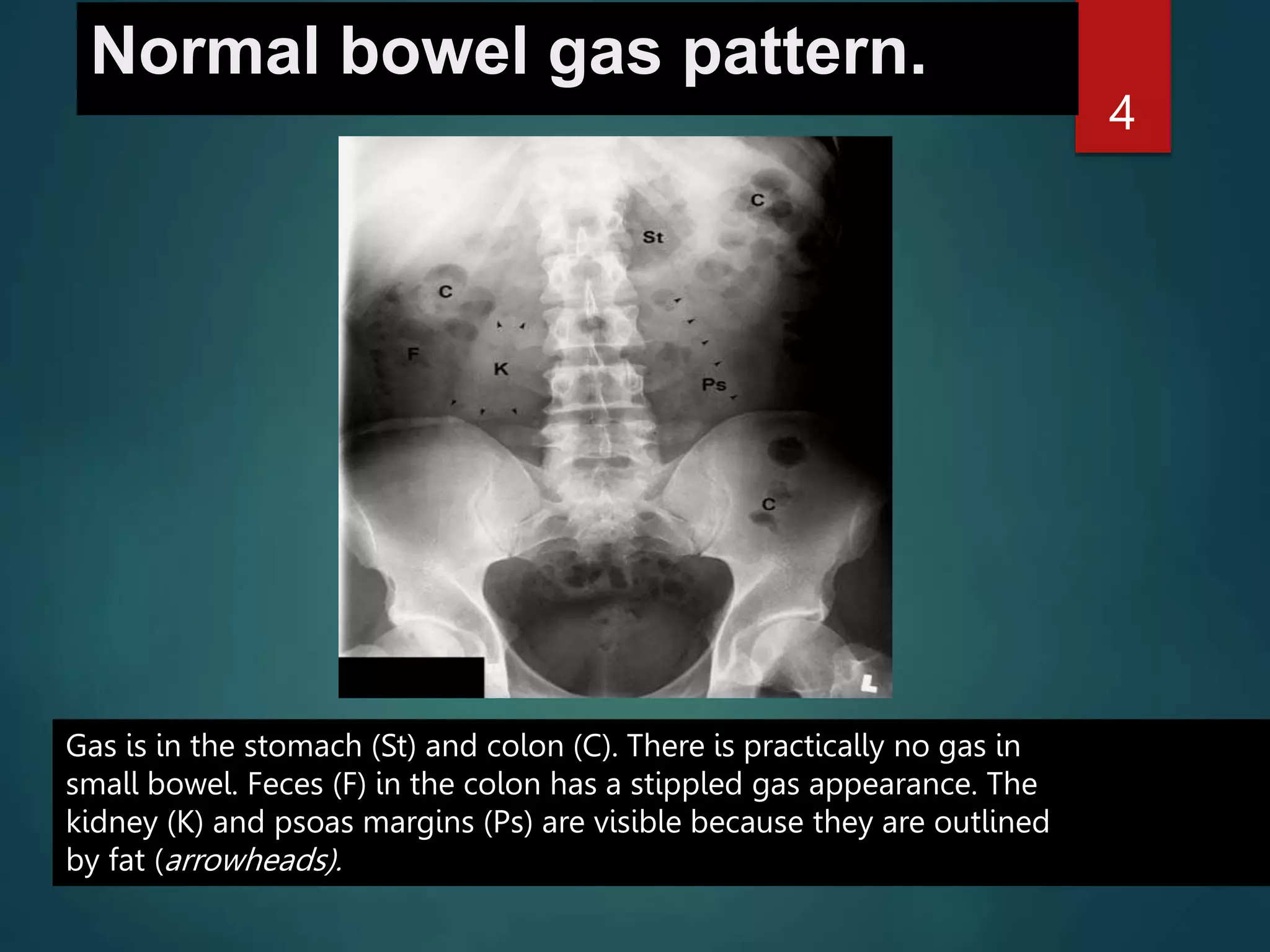
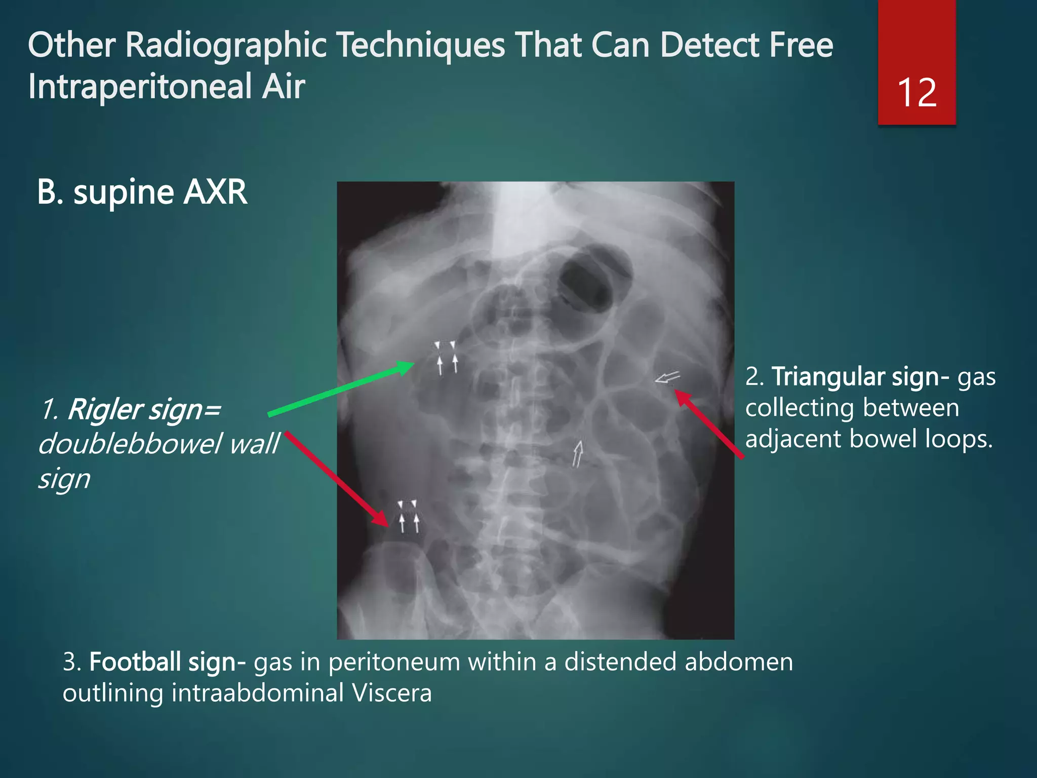
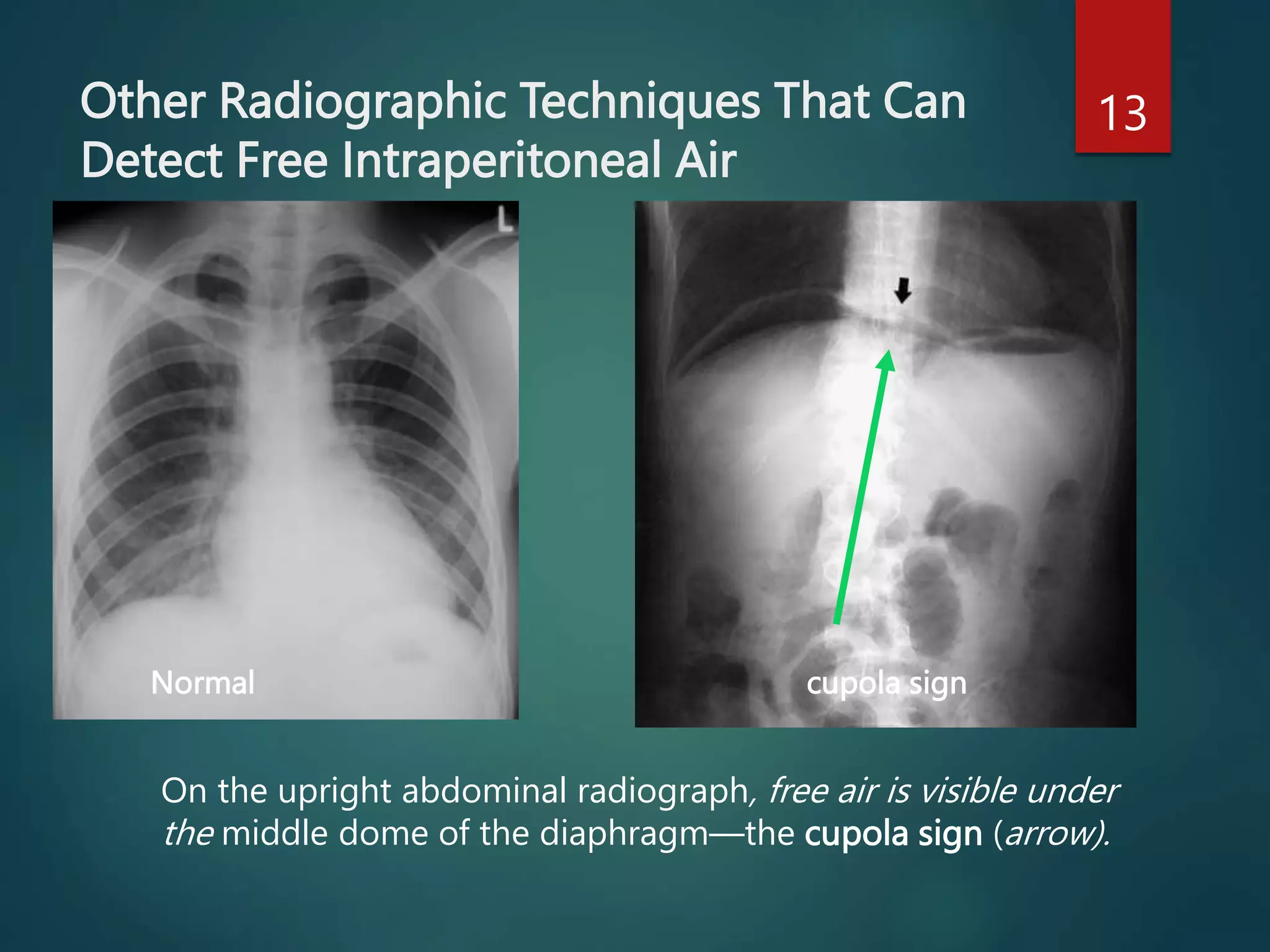
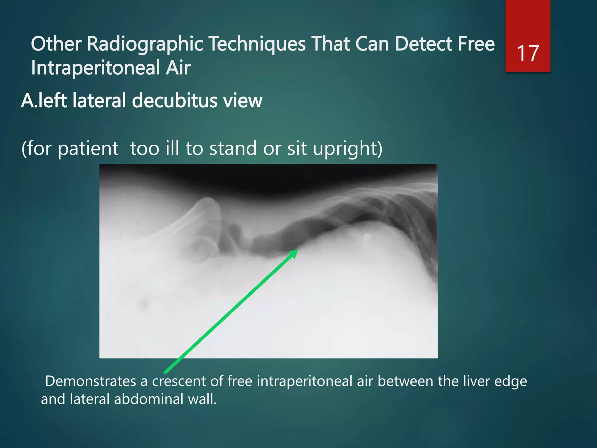
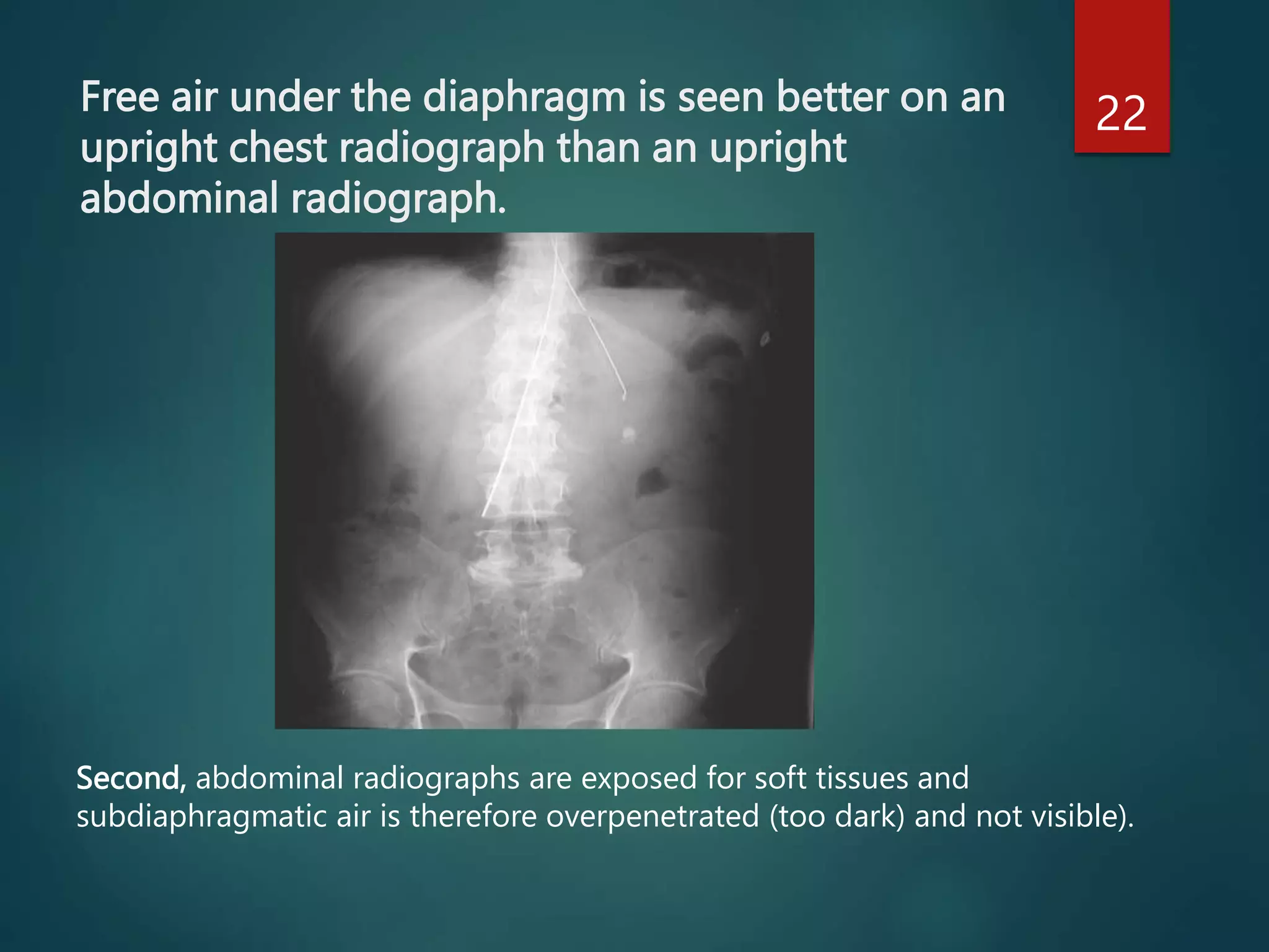
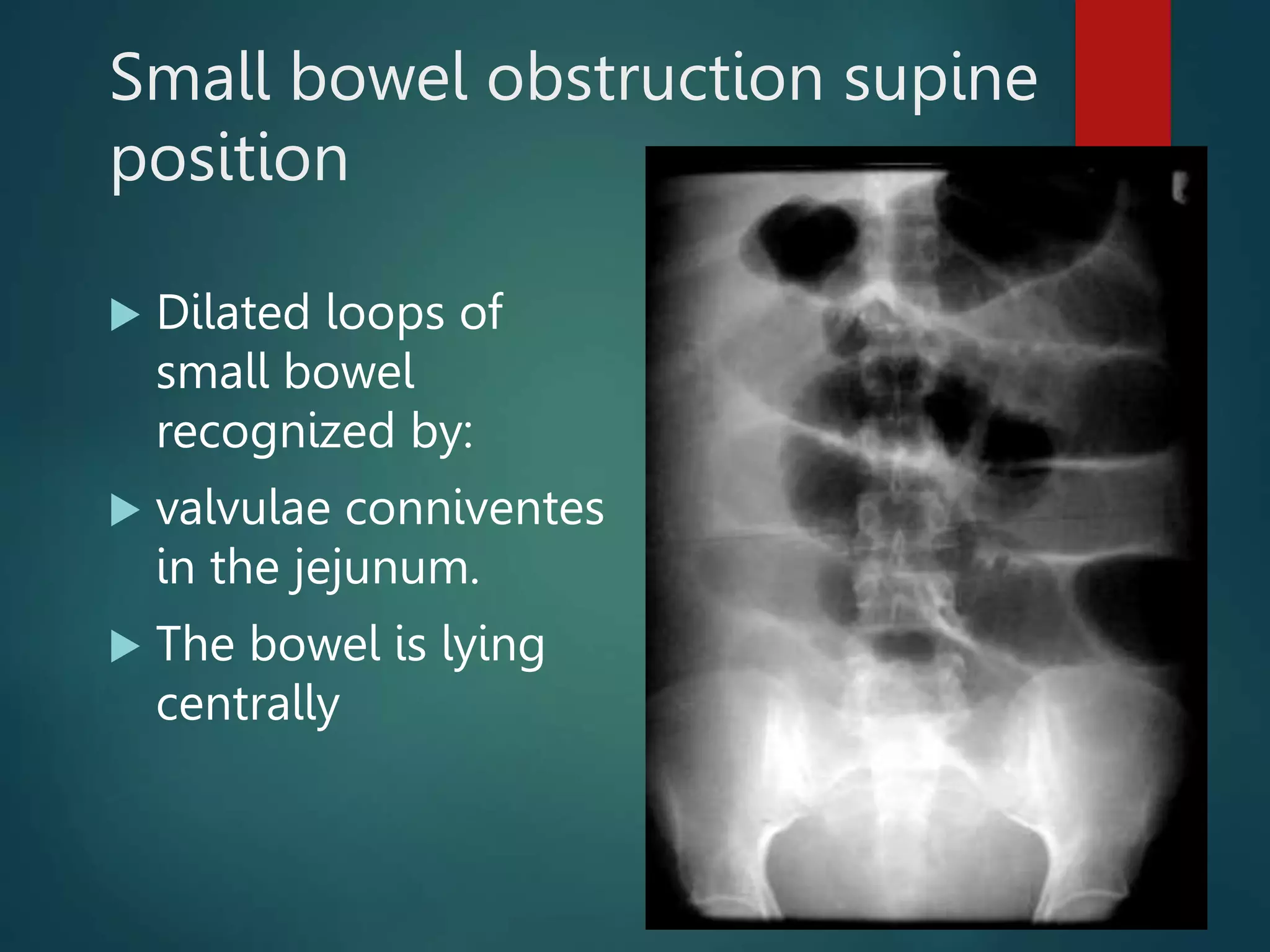
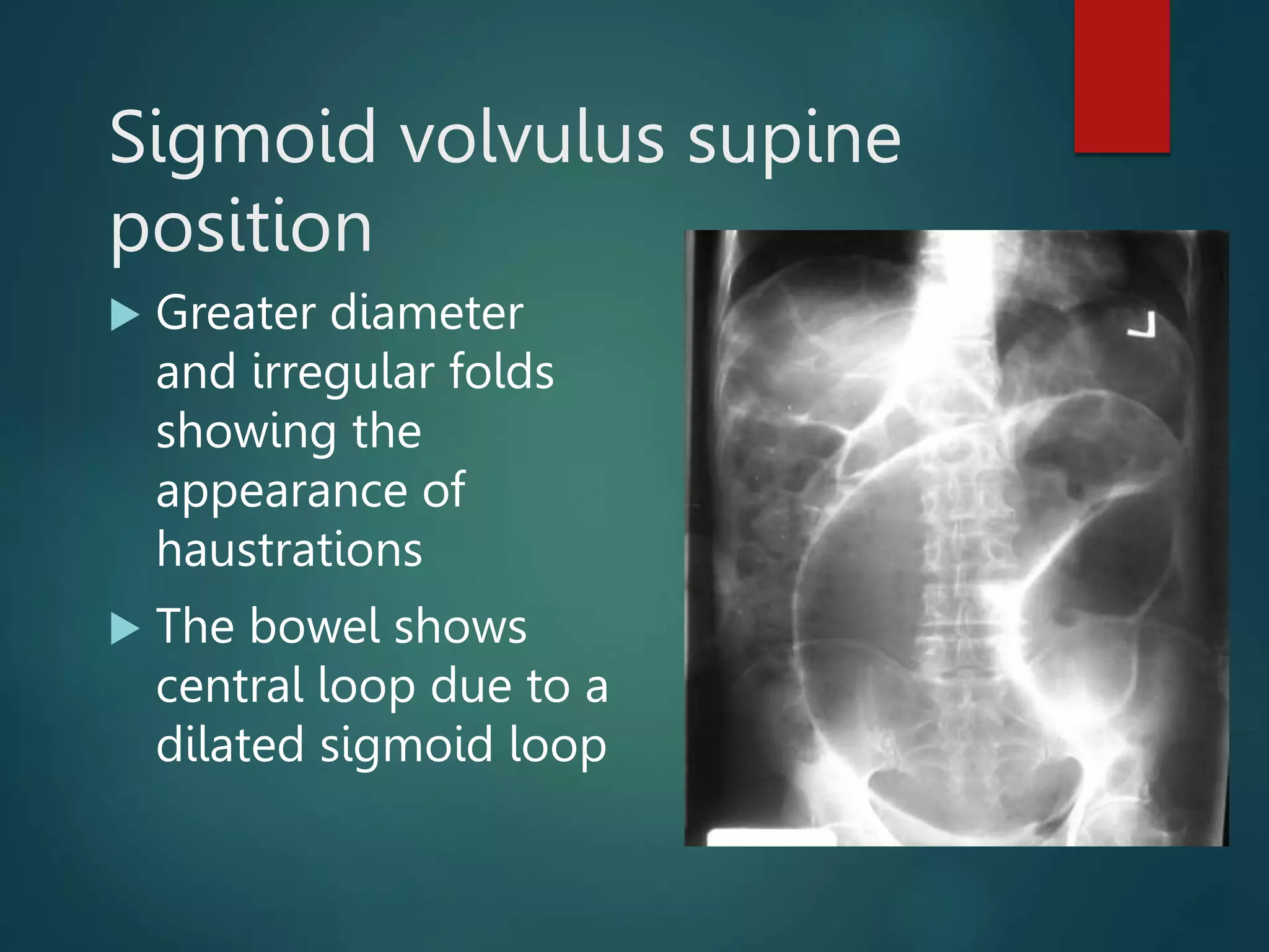
ABDOMINAL
RADIOGRAPHY
INTERPRETATIO
N
DR MALEYA
NORMAL
ABDOMINAL
RADIOGRAPHY
Normal bowel gas pattern.
4
Gas is in the stomach (St) and colon (C). There is practically no gas in
small bowel. Feces (F) in the colon has a stippled gas appearance. The
kidney (K) and psoas margins (Ps) are visible because they are outlined
by fat (arrowheads).
ABNORMAL
ABDOMINAL
RADIOGRAPHY
: Pneumoperitoneum
10
Free gas under diaphragm’ – indicates free peritoneal
gas between the dome of the hemidiaphragm and
superior surface of liver.
Pneumoperitoneum
Other Radiographic Techniques That Can Detect Free
Intraperitoneal Air 12
1. Rigler sign=
doublebbowel wall
sign
2. Triangular sign- gas
collecting between
adjacent bowel loops.
B. supine AXR
3. Football sign- gas in peritoneum within a distended abdomen
outlining intraabdominal Viscera
Other Radiographic Techniques That Can
Detect Free Intraperitoneal Air
13
On the upright abdominal radiograph, free air is visible under
the middle dome of the diaphragm—the cupola sign (arrow).
Normal cupola sign
Large pneumoperitoneum (CXR)
 A very large amount
of free air within the
peritoneal cavity lying
beneath both
diaphragms
 The liver and spleen
are pushed
downwards and the
diaphragms upwards.
Pneumoperitoneum (supine plain
abd film)
 Large collection of
air in the L flank
 There is a ‘double
wall sign’ the outer
wall of the bowel
can be well seen.
 The small bowel is
dilated due to ileus
If clinical suspicion remains high despite a negative CXR
16
1. A CT scan is better at demonstrating the extent of intraperitoneal
free gas and detecting small amounts of gas collects anterior to the
liver
2. A watersoluble contrast examination of the bowel may help to
confirm/rule out the diagnosis (by demonstrating contrast leakage from
the site of perforation
Other Radiographic Techniques That Can Detect Free
Intraperitoneal Air
17
Demonstrates a crescent of free intraperitoneal air between the liver edge
and lateral abdominal wall.
A.left lateral decubitus view
(for patient too ill to stand or sit upright)
Chest Radiographic Findings That Can Mimic Free Intraperitoneal Air
19
The hepatic flexure of the colon has migrated superior to the liver.
This can mimic free air under the right hemidiaphragm on a chest radiograph.
A. Hepatodiaphragmantic interposition of the colon
Chest Radiographic Findings That Can Mimic Free Intraperitoneal Air
21
Subdiaphragmatic air mimicked by
the inferior margin of a rib (arrow).
By following the inferior margin of
the rib to the lateral chest wall
(arrowhead), it can be determined
that this is not free air but a rib.
B . Rib margin that parallels diaphragm
Free air under the diaphragm is seen better on an
upright chest radiograph than an upright
abdominal radiograph.
22
Second, abdominal radiographs are exposed for soft tissues and
subdiaphragmatic air is therefore overpenetrated (too dark) and not visible).
Free air under the diaphragm is seen better on an upright
chest radiograph than an upright abdominal radiograph.
24
The chest radiograph clearly
shows free air on the right
(arrowhead
Volumes as small as 1–2 ml of free
air may be detected (Miller et al.
1971 and 1980).
25
First, on an upright chest radiograph,
the x-ray beam is centered near the
diaphragm rather than the
midabdomen, and is therefore parallel
to the intra-abdominal air/fluid level
x-ray beam is obliquely
oriented at this level.
Are we together?
Yes, we are together!
26
Gastric ulcer 27
Gastric ulcer on the lesser curve. Note how the gastric folds are pulled
towards it instead of passing longitudinally down the body of the stomach
Normal mucosal pattern
Small bowel obstruction supine
position
 Dilated loops of
small bowel
recognized by:
 valvulae conniventes
in the jejunum.
 The bowel is lying
centrally
Small bowel obstruction upright position
 Dilated loops of
small bowel showing
multiple short air-
fluid levels
Ileus supine position
 Dilated loops of
small bowel
Ileus upright position
 Few and long air-
fluid levels
Sigmoid volvulus supine
position
 Greater diameter
and irregular folds
showing the
appearance of
haustrations
 The bowel shows
central loop due to a
dilated sigmoid loop
Carcinoma of esophagus
 A long stricture and
irregularity of the
lumen due to
mucosal destruction.
NORMAL ABDOMINAL X-RAY
 NORMAL BOWEL GAS PARTEN
 NORMAL SOFT TISUES
 THERE IS GAS IN THE RECTUM
 NO AIR FLUID LEVELS
 CONCLUSION:
1. NORMAL ABDOMINAL X-RAY
35
QUESTIONS

ABDOMINAL RADIOGRAPHY.pptx.pptx

  • 1.
  • 2.
  • 4.
    Normal bowel gaspattern. 4 Gas is in the stomach (St) and colon (C). There is practically no gas in small bowel. Feces (F) in the colon has a stippled gas appearance. The kidney (K) and psoas margins (Ps) are visible because they are outlined by fat (arrowheads).
  • 9.
  • 10.
    : Pneumoperitoneum 10 Free gasunder diaphragm’ – indicates free peritoneal gas between the dome of the hemidiaphragm and superior surface of liver. Pneumoperitoneum
  • 12.
    Other Radiographic TechniquesThat Can Detect Free Intraperitoneal Air 12 1. Rigler sign= doublebbowel wall sign 2. Triangular sign- gas collecting between adjacent bowel loops. B. supine AXR 3. Football sign- gas in peritoneum within a distended abdomen outlining intraabdominal Viscera
  • 13.
    Other Radiographic TechniquesThat Can Detect Free Intraperitoneal Air 13 On the upright abdominal radiograph, free air is visible under the middle dome of the diaphragm—the cupola sign (arrow). Normal cupola sign
  • 14.
    Large pneumoperitoneum (CXR) A very large amount of free air within the peritoneal cavity lying beneath both diaphragms  The liver and spleen are pushed downwards and the diaphragms upwards.
  • 15.
    Pneumoperitoneum (supine plain abdfilm)  Large collection of air in the L flank  There is a ‘double wall sign’ the outer wall of the bowel can be well seen.  The small bowel is dilated due to ileus
  • 16.
    If clinical suspicionremains high despite a negative CXR 16 1. A CT scan is better at demonstrating the extent of intraperitoneal free gas and detecting small amounts of gas collects anterior to the liver 2. A watersoluble contrast examination of the bowel may help to confirm/rule out the diagnosis (by demonstrating contrast leakage from the site of perforation
  • 17.
    Other Radiographic TechniquesThat Can Detect Free Intraperitoneal Air 17 Demonstrates a crescent of free intraperitoneal air between the liver edge and lateral abdominal wall. A.left lateral decubitus view (for patient too ill to stand or sit upright)
  • 18.
    Chest Radiographic FindingsThat Can Mimic Free Intraperitoneal Air
  • 19.
    Chest Radiographic FindingsThat Can Mimic Free Intraperitoneal Air 19 The hepatic flexure of the colon has migrated superior to the liver. This can mimic free air under the right hemidiaphragm on a chest radiograph. A. Hepatodiaphragmantic interposition of the colon
  • 20.
    Chest Radiographic FindingsThat Can Mimic Free Intraperitoneal Air
  • 21.
    Chest Radiographic FindingsThat Can Mimic Free Intraperitoneal Air 21 Subdiaphragmatic air mimicked by the inferior margin of a rib (arrow). By following the inferior margin of the rib to the lateral chest wall (arrowhead), it can be determined that this is not free air but a rib. B . Rib margin that parallels diaphragm
  • 22.
    Free air underthe diaphragm is seen better on an upright chest radiograph than an upright abdominal radiograph. 22 Second, abdominal radiographs are exposed for soft tissues and subdiaphragmatic air is therefore overpenetrated (too dark) and not visible).
  • 24.
    Free air underthe diaphragm is seen better on an upright chest radiograph than an upright abdominal radiograph. 24 The chest radiograph clearly shows free air on the right (arrowhead Volumes as small as 1–2 ml of free air may be detected (Miller et al. 1971 and 1980).
  • 25.
    25 First, on anupright chest radiograph, the x-ray beam is centered near the diaphragm rather than the midabdomen, and is therefore parallel to the intra-abdominal air/fluid level x-ray beam is obliquely oriented at this level.
  • 26.
    Are we together? Yes,we are together! 26
  • 27.
    Gastric ulcer 27 Gastriculcer on the lesser curve. Note how the gastric folds are pulled towards it instead of passing longitudinally down the body of the stomach Normal mucosal pattern
  • 28.
    Small bowel obstructionsupine position  Dilated loops of small bowel recognized by:  valvulae conniventes in the jejunum.  The bowel is lying centrally
  • 29.
    Small bowel obstructionupright position  Dilated loops of small bowel showing multiple short air- fluid levels
  • 30.
    Ileus supine position Dilated loops of small bowel
  • 31.
    Ileus upright position Few and long air- fluid levels
  • 32.
    Sigmoid volvulus supine position Greater diameter and irregular folds showing the appearance of haustrations  The bowel shows central loop due to a dilated sigmoid loop
  • 33.
    Carcinoma of esophagus A long stricture and irregularity of the lumen due to mucosal destruction.
  • 34.
    NORMAL ABDOMINAL X-RAY NORMAL BOWEL GAS PARTEN  NORMAL SOFT TISUES  THERE IS GAS IN THE RECTUM  NO AIR FLUID LEVELS  CONCLUSION: 1. NORMAL ABDOMINAL X-RAY
  • 35.