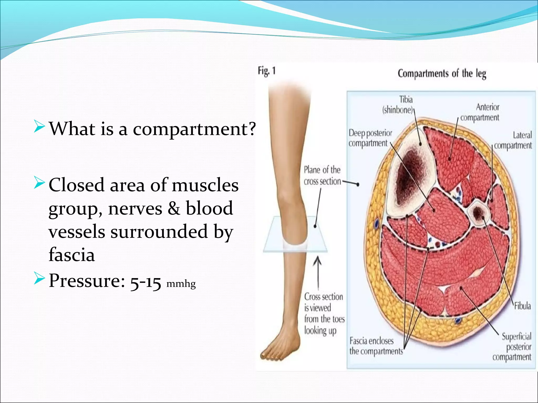
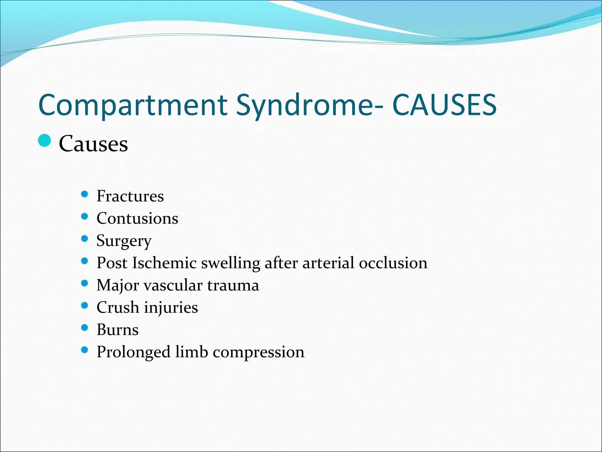
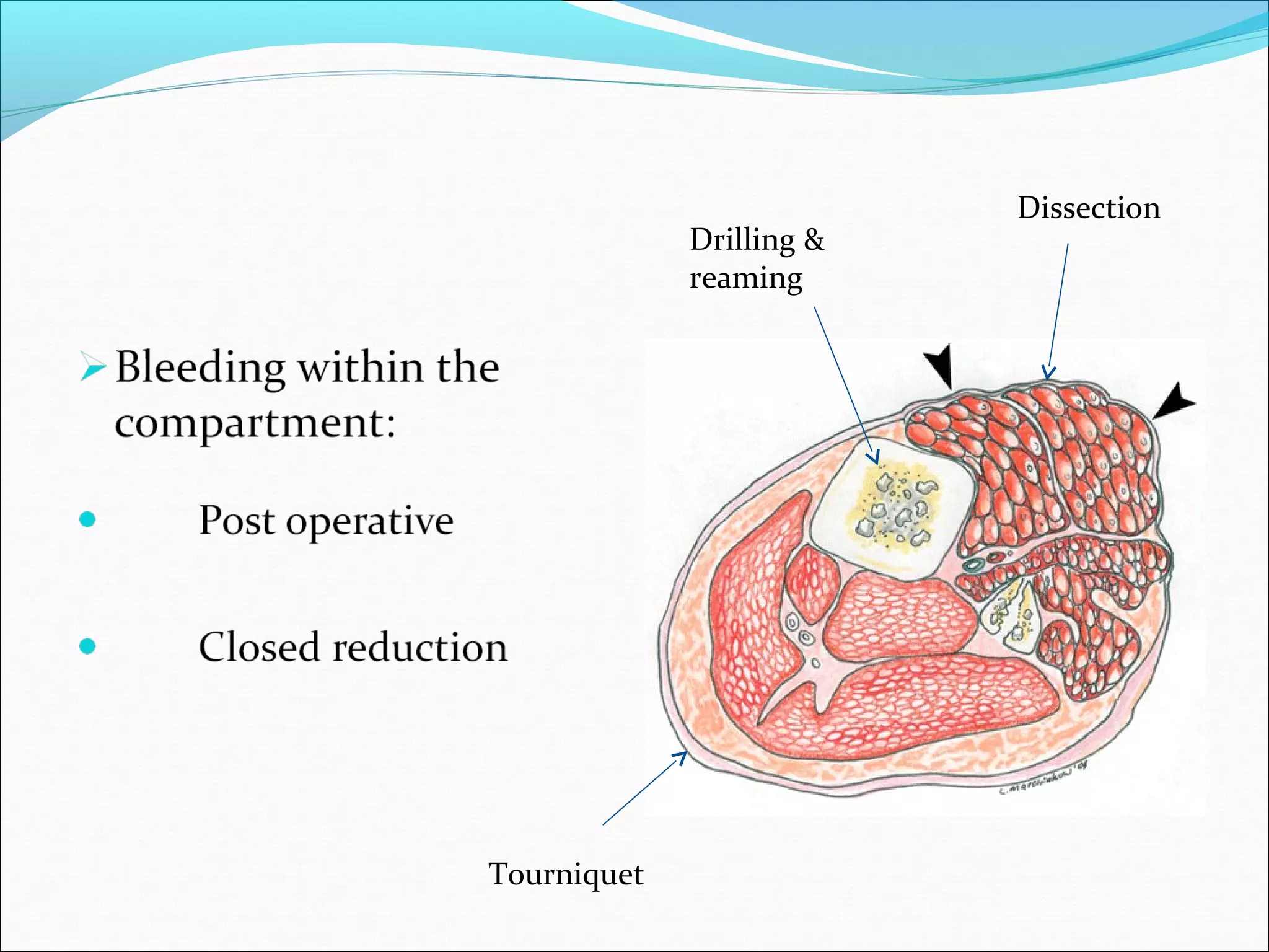
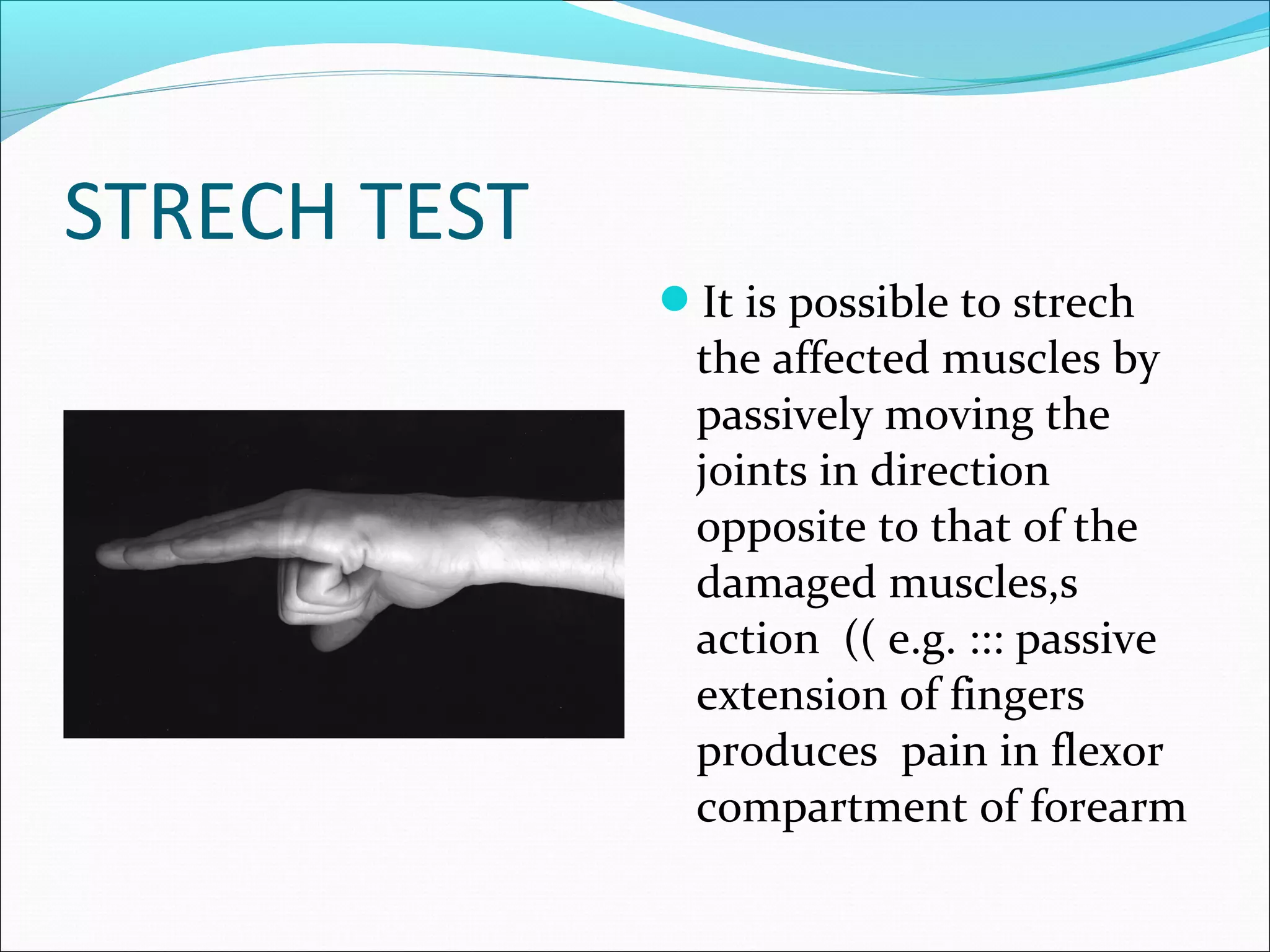
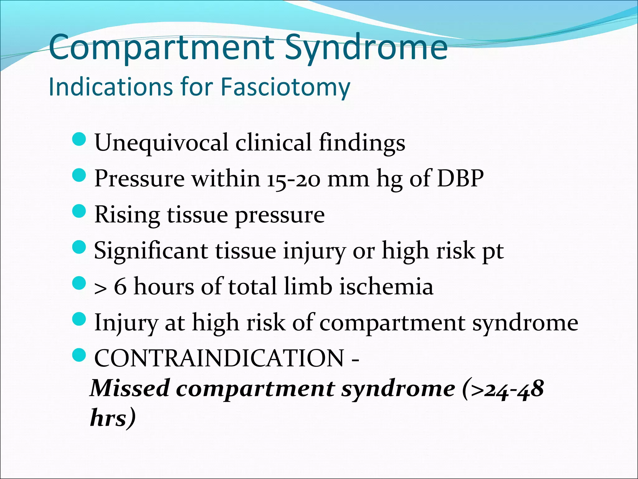
DR ASHWANI PANCHAL
P.G IN ORTHOPAEDICS
JSS HOSPITAL
MYSORE
 What is a compartment?
 Closed area of muscles

group, nerves & blood
vessels surrounded by
fascia
 Pressure: 5-15 mmhg
Definition:
An increased pressure within enclosed

osteofascial space that reduces capillary
perfusion below level necessary for
tissue viability;
the underlying mechanism is:
- increased volume within space
- decreased space for contents
- combination of both
 What is a compartment

syndrome?
 intra comp. pressure
(35-40 mmhg)
 capillaries collapse

 Blood flow to muscles

and nerves

 Bl.Vs collapse
Pathophysiology:
Increased compartment pressure
leads to increased venous pressure
which decreases A-V gradient
resulting in muscle and nerve
ischemia.
Consequences –vicious cycle
Why is it dangerous?
 Nerves:
neuropraxia: will

regenerate
Ischemia: cell death

 Muscles: contracture

(Volkmann's ischemic
contracture)

 Gangrene
Compartment Syndrome- CAUSES
Causes
Fractures
 Contusions
 Surgery
 Post Ischemic swelling after arterial occlusion
 Major vascular trauma
 Crush injuries
 Burns
 Prolonged limb compression

Causes
 Fracture of a long bone
(Supracondylar humerus,
forearm, hand,tibia and
foot)
CAUSES
Drilling &
reaming

Tourniquet

Dissection
CAUSES
swelling

Tight cast

numbness

Bluish
discoloration
CAUSES
 Severe bruised muscle
(even if there is no fracture)
 Don’t take contusion lightly
COMPARTMENT SYNDROME
Signs and Symptoms
Increased Pressure and Tightness
Progressive pain out of proportion to initial

injury
Markedly swollen area
Progressive neurologic deficit
Seven P’s
 Pain
 Pressure
 Pain with passive stretch
 Parethesia
 Paresis/ Paralysis
 Pulses
 Pallor
SYMPTOMS
 Severe pain

inappropriate to the
injury(not relieved even
with morphia)
SYMPTOMS
 Burning of the affected limb
 Tight muscle(rigid)tightness feeling
 Numbness: bad sign
SIGNS & DIAGNOSIS
 Passive stretching of fingers or toes (muscle

stretch)will lead to severe pain (diagnostic sign)

 Never wait for signs of ischemia (5 Ps):irreversible

damage
STRECH TEST
It is possible to strech

the affected muscles by
passively moving the
joints in direction
opposite to that of the
damaged muscles,s
action (( e.g. ::: passive
extension of fingers
produces pain in flexor
compartment of forearm
Technique
STRYKER TECHNIQUE
MERCURY

MANOMETER
Wick hand held
instrument
Whiteside maneuver

Wick hand held instrument
Direct
reading

syringe

mmhg
mano.

3 way stopcock
electrode
Stryker Stic System
Easy to use
Can check multiple compartments
Different areas in one compartment
Complications related to CS
Late Sequelae
Volckmann’s contracture
 Weak dorsiflexors
 Claw toes
 Sensory loss
 Chronic pain
 Amputation

COMPARTMENT SYNDROME
Management
 Non surgical management:
 Remove any tight bandage, tubigrip or soaked

dressing

 Cast should be removed completely
 Elevation
•management
 Surgical management:

(FASCIOTOMY)
Open skin and fascia
down to a compartment
It is a surgical procedure
where the fascia is cut to
relieve tension or
pressure commonly to
treat the resulting loss of
circulation to the tissue
Fasciotomy Principles
Make early diagnosis
Long extensile incisions
Release all fascial compartments
Preserve neurovascular structures
Debride necrotic tissues
Coverage within 7-10 days
Compartment Syndrome
Indications for Fasciotomy

Unequivocal clinical findings
Pressure within 15-20 mm hg of DBP
Rising tissue pressure
Significant tissue injury or high risk pt
> 6 hours of total limb ischemia
Injury at high risk of compartment syndrome
CONTRAINDICATION -

Missed compartment syndrome (>24-48
hrs)
Forearm Fasciotomy
Volar-Henry

approach

Include a carpal

tunnel release

Release lacertus

fibrosus and fascia
Protect median
nerve, brachial artery
and tendons after
release
Gastroc-soleus

Flexor digitorum
longus
Intermuscular septum

Superficial peroneal ner
Fasciotomy of Hand
10 separate osteofascial

compartments

dorsal interossei (4)
palmar interossei (3)
thenar and hypothenar

(2)
adductor pollicis (1)
Close skin by 2ry sutures

after oedema subsides
It may need skin graft
Wound Management
Wound is not closed at initial surgery
Second look debridement with consideration for

coverage after 48-72 hrs

Limb should not be at risk for further swelling
Pt should be adequately stabilized
Usually requires skin graft
DPC possible if residual swelling is minimal
Flap coverage needed if nerves, vessels, or bone

exposed

Goal is to obtain definitive coverage within 7-10

days
Wound Management

After the fasciotomy, a bulky compression dressing and

a splint are applied.
“VAC” (Vacuum Assisted Closure) can be used
 Foot should be placed in neutral to prevent equinus
contracture.
Incision for the fasciotomy usually can be closed after
three to five days
Complications Related to
Fasciotomies
Altered sensation within the margins of the wound (77%)
Dry, scaly skin (40%)
Pruritus (33%)
Discolored wounds (30%)
Swollen limbs (25%)
Tethered scars (26%)
Recurrent ulceration (13%)
Muscle herniation (13%)
Pain related to the wound (10%)
Tethered tendons (7%)
Fitzgerald, McQueen Br J Plast Surg 2000
Compartment syndrome is a serious syndrome, Which needs

to be diagnosed early.
Palpable pulse doesn’t exclude compartment syndrome
If diagnosis and fasciotomy were done within 24 hrs, the
prognosis is good.
If delayed, complications will develop.

The earlier you diagnose, the safer you are

compartment syndrome

Editor's Notes

  • #19 Seven P’s: Pain- pain in excess of the presenting injury Pressure- affected compartment may be tense to palpation Pain with passive stretch- pain increases with passive ROM Paresthesia- Numbness over the cutaneus distribution of the nerve that runs through the affected compartment Paresis/Paralysis- Ischemia or necrosis of the nerves or muscles develops within the affected compartment Pulses- could be absent but frequently palpable Pallor- skin discoloration may be visible due to impaired venous drainage Co-Morbidities include: Mild: ~ mm weakness ~fatigue ~ Myositis ossificans Severe ~ Severe life threatening vascular compromise ~ decreased arteriovenous gradient **To Remember** ~ Do not elevate limb above hear because it decreases the arteriovenous gradient therefore decreasing blood flow ~ Instead limb should be elevated at heart level to maintain arteriovenous gradient and assist with venous drainage ~ Remove any compression casts or dressings
  • #32 These are indications for surgical decompression. A missed CS > 24-48 hours should not be opened. (see Rockwood and Green 5th edition and Campbell’s 10 th edition) The damage cannot be reversed and there is a significant infection rate when the dead tissue is exposed to the hospital environment. The surgeon must deal with the residual contractures of the ischemic muscle and not risk the chance of infection. Some have suggested that the scarred ant tib muscle can serve as a check rein and limit foot drop sequelae.
  • #33 Volar approach use Henry Approach to release superficial and deep flexors flexors followed by the pronators and supinator. The dorsal approach is centered over the proximal forearm and can be used to release the Mobile Wad (BR, ECRL,ECRB). It is important to remember when treating and electrical injury with a compartment syndrome, the most damage is deep along the bone (i.e. the Pro and Sup muscles) and may require debridement at the initial surgery…Do not miss this!