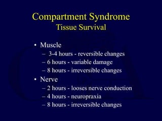
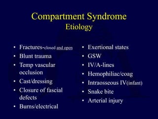
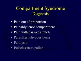
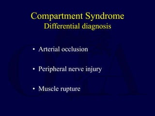
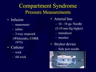
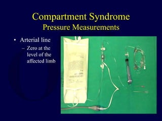
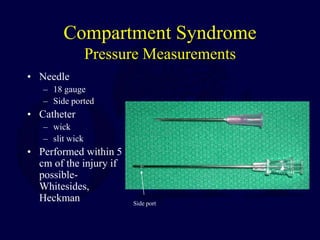
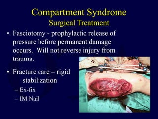
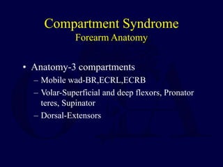
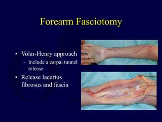
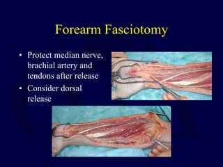
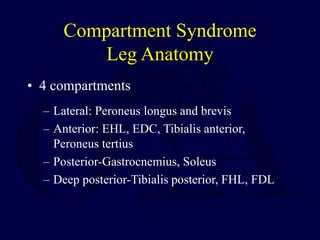
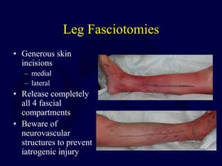
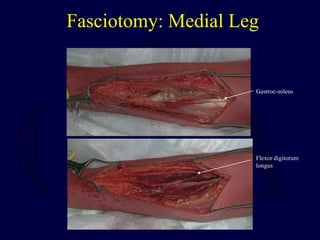
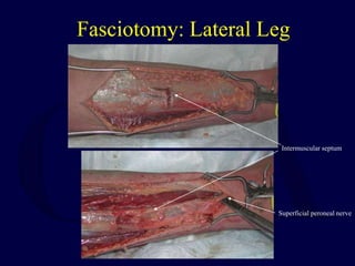
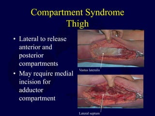
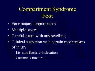
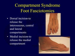
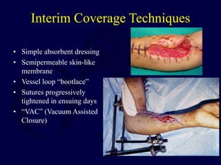
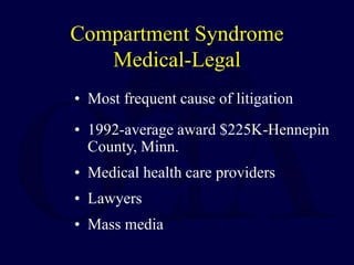
Compartment
Syndromes
Robert M. Harris, MD
Compartment Syndrome
Definition
• Elevated tissue pressure within a closed fascial
space
• Reduces tissue perfusion
• Results in cell death
• Pathogenesis
– Too much inflow (edema, hemorrhage)
– Decreased outflow (venous obstruction, tight
dressing/cast)
Compartment Syndrome
Historical Review
• Late complications of ischemic contracture
– Volkmann, 1881
• Ischemia of forearm
venous stasis leading
to irreversible contracture
– Ellis, 1958; Seddon, 1966
• Lower extremity
• Retrospective reviews
– Advised the early recognition of the syndrome and
fasciotomies of the affected limbs
Compartment Syndrome
Pathophysiology
• Normal tissue pressure
– 0-4 mm Hg
– 8-10 with exertion
• Absolute pressure theory
– 30 mm Hg - Mubarak
– 45 mm Hg - Matsen
• Pressure gradient theory
– < 20 mm Hg of diastolic pressure – Whitesides
– McQueen, et al
Compartment Syndrome
Tissue Survival
• Muscle
– 3-4 hours - reversible changes
– 6 hours - variable damage
– 8 hours - irreversible changes
• Nerve
– 2 hours - looses nerve conduction
– 4 hours - neuropraxia
– 8 hours - irreversible changes
Compartment Syndrome
Etiology
• Fractures-closed and open
• Blunt trauma
• Temp vascular
occlusion
• Cast/dressing
• Closure of fascial
defects
• Burns/electrical
• Exertional states
• GSW
• IV/A-lines
• Hemophiliac/coag
• Intraosseous IV(infant)
• Snake bite
• Arterial injury
Compartment Syndrome
Diagnosis
• Pain out of proportion
• Palpably tense compartment
• Pain with passive stretch
• Paresthesia/hypoesthesia
• Paralysis
• Pulselessness/pallor
Compartment Syndrome
Differential diagnosis
• Arterial occlusion
• Peripheral nerve injury
• Muscle rupture
Compartment Syndrome
Pressure Measurements
• Suspected compartment syndrome
• Equivocal or unreliable exam
• Clinical adjunct
• Contraindication
– Clinically evident compartment
syndrome
Compartment Syndrome
Pressure Measurements
• Infusion
– manometer
– saline
– 3-way stopcock
(Whitesides, CORR
1975)
• Catheter
– wick
– slit wick
• Arterial line
– 16 - 18 ga. Needle
(5-19 mm Hg higher)
– transducer
– monitor
• Stryker device
– Side port needle
Compartment Syndrome
Pressure Measurements
• Arterial line
– Zero at the
level of the
affected limb
Compartment Syndrome
Pressure Measurements
• Needle
– 18 gauge
– Side ported
• Catheter
– wick
– slit wick
• Performed within 5
cm of the injury if
possible-
Whitesides,
Heckman Side port
Compartment Syndrome
Emergent Treatment
• Remove cast or dressing
• Place at level of heart
(DO NOT ELEVATE to optimize
perfusion)
• Alert OR and Anesthesia
• Bedside procedure
• Medical treatment
Compartment Syndrome
Surgical Treatment
• Fasciotomy - prophylactic release of
pressure before permanent damage
occurs. Will not reverse injury from
trauma.
• Fracture care – rigid
stabilization
– Ex-fix
– IM Nail
Compartment Syndrome
Indications for Fasciotomy
• Unequivocal clinical findings
• Pressure within 15-20 mm hg of DBP
• Rising tissue pressure
• Significant tissue injury or high risk pt
• > 6 hours of total limb ischemia
• Injury at high risk of compartment
syndrome
• CONTRAINDICATION - Missed CS (>24-
48 hrs)
Fasciotomy Principles
• Make early diagnosis
• Long extensile incisions
• Release all fascial compartments
• Preserve neurovascular structures
• Debride necrotic tissues
• Coverage within 7-10 days
Compartment Syndrome
Forearm Anatomy
• Anatomy-3 compartments
– Mobile wad-BR,ECRL,ECRB
– Volar-Superficial and deep flexors, Pronator
teres, Supinator
– Dorsal-Extensors
Forearm Fasciotomy
• Volar-Henry approach
– Include a carpal tunnel
release
• Release lacertus
fibrosus and fascia
Forearm Fasciotomy
• Protect median nerve,
brachial artery and
tendons after release
• Consider dorsal
release
Compartment Syndrome
Leg Anatomy
• 4 compartments
– Lateral: Peroneus longus and brevis
– Anterior: EHL, EDC, Tibialis anterior,
Peroneus tertius
– Posterior-Gastrocnemius, Soleus
– Deep posterior-Tibialis posterior, FHL, FDL
Leg Fasciotomies
• Generous skin
incisions
– medial
– lateral
• Release completely
all 4 fascial
compartments
• Beware of
neurovascular
structures to prevent
iatrogenic injury
Fasciotomy: Medial Leg
Flexor digitorum
longus
Gastroc-soleus
Fasciotomy: Lateral Leg
Superficial peroneal nerve
Intermuscular septum
Compartment Syndrome
Thigh
• Lateral to release
anterior and
posterior
compartments
• May require medial
incision for
adductor
compartment
Lateral septum
Vastus lateralis
Compartment Syndrome
Foot
• Four major compartments
• Multiple layers
• Careful exam with any swelling
• Clinical suspicion with certain mechanisms
of injury
– Lisfranc fracture dislocation
– Calcaneus fracture
Compartment Syndrome
Foot Fasciotomies
• Dorsal incision-to
release the
interosseous, central
and lateral
compartments
• Medial incision-to
release the medial
compartment
Compartment Syndrome
Other Areas
• Can occur anywhere in the body
• Hand-dorsal incisions, thenar, hypothenar
• Arm-lateral incision
• Buttock-posterior (Kocher) approach
• Abdominal- with the Trauma surgeons
Interim Coverage Techniques
• Simple absorbent dressing
• Semipermeable skin-like
membrane
• Vessel loop “bootlace”
• Sutures progressively
tightened in ensuing days
• “VAC” (Vacuum Assisted
Closure)
Aftercare
• Wound is not closed at initial surgery
• Second look debridement with consideration for
coverage after 48-72 hrs
– Limb should not be at risk for further swelling
– Pt should be adequately stabilized
– Usually requires skin graft
– DPC possible if residual swelling is minimal
• Goal is to obtain definitive coverage within 7-10
days
– DPC/STSG/flap if nerves, vessels, bone exposed
Compartment Syndrome
Medical-Legal
• Most frequent cause of litigation
• 1992-average award $225K-Hennepin
County, Minn.
• Medical health care providers
• Lawyers
• Mass media
Questions
Return to
General Index

Compartment Syndrome.ppt

  • 1.
  • 2.
    Compartment Syndrome Definition • Elevatedtissue pressure within a closed fascial space • Reduces tissue perfusion • Results in cell death • Pathogenesis – Too much inflow (edema, hemorrhage) – Decreased outflow (venous obstruction, tight dressing/cast)
  • 3.
    Compartment Syndrome Historical Review •Late complications of ischemic contracture – Volkmann, 1881 • Ischemia of forearm venous stasis leading to irreversible contracture – Ellis, 1958; Seddon, 1966 • Lower extremity • Retrospective reviews – Advised the early recognition of the syndrome and fasciotomies of the affected limbs
  • 4.
    Compartment Syndrome Pathophysiology • Normaltissue pressure – 0-4 mm Hg – 8-10 with exertion • Absolute pressure theory – 30 mm Hg - Mubarak – 45 mm Hg - Matsen • Pressure gradient theory – < 20 mm Hg of diastolic pressure – Whitesides – McQueen, et al
  • 5.
    Compartment Syndrome Tissue Survival •Muscle – 3-4 hours - reversible changes – 6 hours - variable damage – 8 hours - irreversible changes • Nerve – 2 hours - looses nerve conduction – 4 hours - neuropraxia – 8 hours - irreversible changes
  • 6.
    Compartment Syndrome Etiology • Fractures-closedand open • Blunt trauma • Temp vascular occlusion • Cast/dressing • Closure of fascial defects • Burns/electrical • Exertional states • GSW • IV/A-lines • Hemophiliac/coag • Intraosseous IV(infant) • Snake bite • Arterial injury
  • 7.
    Compartment Syndrome Diagnosis • Painout of proportion • Palpably tense compartment • Pain with passive stretch • Paresthesia/hypoesthesia • Paralysis • Pulselessness/pallor
  • 8.
    Compartment Syndrome Differential diagnosis •Arterial occlusion • Peripheral nerve injury • Muscle rupture
  • 9.
    Compartment Syndrome Pressure Measurements •Suspected compartment syndrome • Equivocal or unreliable exam • Clinical adjunct • Contraindication – Clinically evident compartment syndrome
  • 10.
    Compartment Syndrome Pressure Measurements •Infusion – manometer – saline – 3-way stopcock (Whitesides, CORR 1975) • Catheter – wick – slit wick • Arterial line – 16 - 18 ga. Needle (5-19 mm Hg higher) – transducer – monitor • Stryker device – Side port needle
  • 11.
    Compartment Syndrome Pressure Measurements •Arterial line – Zero at the level of the affected limb
  • 12.
    Compartment Syndrome Pressure Measurements •Needle – 18 gauge – Side ported • Catheter – wick – slit wick • Performed within 5 cm of the injury if possible- Whitesides, Heckman Side port
  • 13.
    Compartment Syndrome Emergent Treatment •Remove cast or dressing • Place at level of heart (DO NOT ELEVATE to optimize perfusion) • Alert OR and Anesthesia • Bedside procedure • Medical treatment
  • 14.
    Compartment Syndrome Surgical Treatment •Fasciotomy - prophylactic release of pressure before permanent damage occurs. Will not reverse injury from trauma. • Fracture care – rigid stabilization – Ex-fix – IM Nail
  • 15.
    Compartment Syndrome Indications forFasciotomy • Unequivocal clinical findings • Pressure within 15-20 mm hg of DBP • Rising tissue pressure • Significant tissue injury or high risk pt • > 6 hours of total limb ischemia • Injury at high risk of compartment syndrome • CONTRAINDICATION - Missed CS (>24- 48 hrs)
  • 16.
    Fasciotomy Principles • Makeearly diagnosis • Long extensile incisions • Release all fascial compartments • Preserve neurovascular structures • Debride necrotic tissues • Coverage within 7-10 days
  • 17.
    Compartment Syndrome Forearm Anatomy •Anatomy-3 compartments – Mobile wad-BR,ECRL,ECRB – Volar-Superficial and deep flexors, Pronator teres, Supinator – Dorsal-Extensors
  • 18.
    Forearm Fasciotomy • Volar-Henryapproach – Include a carpal tunnel release • Release lacertus fibrosus and fascia
  • 19.
    Forearm Fasciotomy • Protectmedian nerve, brachial artery and tendons after release • Consider dorsal release
  • 20.
    Compartment Syndrome Leg Anatomy •4 compartments – Lateral: Peroneus longus and brevis – Anterior: EHL, EDC, Tibialis anterior, Peroneus tertius – Posterior-Gastrocnemius, Soleus – Deep posterior-Tibialis posterior, FHL, FDL
  • 21.
    Leg Fasciotomies • Generousskin incisions – medial – lateral • Release completely all 4 fascial compartments • Beware of neurovascular structures to prevent iatrogenic injury
  • 22.
    Fasciotomy: Medial Leg Flexordigitorum longus Gastroc-soleus
  • 23.
    Fasciotomy: Lateral Leg Superficialperoneal nerve Intermuscular septum
  • 24.
    Compartment Syndrome Thigh • Lateralto release anterior and posterior compartments • May require medial incision for adductor compartment Lateral septum Vastus lateralis
  • 25.
    Compartment Syndrome Foot • Fourmajor compartments • Multiple layers • Careful exam with any swelling • Clinical suspicion with certain mechanisms of injury – Lisfranc fracture dislocation – Calcaneus fracture
  • 26.
    Compartment Syndrome Foot Fasciotomies •Dorsal incision-to release the interosseous, central and lateral compartments • Medial incision-to release the medial compartment
  • 27.
    Compartment Syndrome Other Areas •Can occur anywhere in the body • Hand-dorsal incisions, thenar, hypothenar • Arm-lateral incision • Buttock-posterior (Kocher) approach • Abdominal- with the Trauma surgeons
  • 28.
    Interim Coverage Techniques •Simple absorbent dressing • Semipermeable skin-like membrane • Vessel loop “bootlace” • Sutures progressively tightened in ensuing days • “VAC” (Vacuum Assisted Closure)
  • 29.
    Aftercare • Wound isnot closed at initial surgery • Second look debridement with consideration for coverage after 48-72 hrs – Limb should not be at risk for further swelling – Pt should be adequately stabilized – Usually requires skin graft – DPC possible if residual swelling is minimal • Goal is to obtain definitive coverage within 7-10 days – DPC/STSG/flap if nerves, vessels, bone exposed
  • 30.
    Compartment Syndrome Medical-Legal • Mostfrequent cause of litigation • 1992-average award $225K-Hennepin County, Minn. • Medical health care providers • Lawyers • Mass media
  • 31.