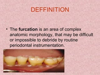
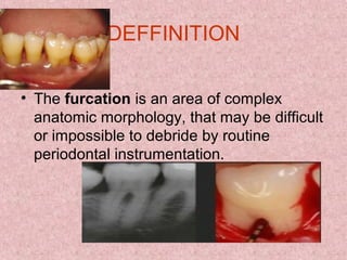
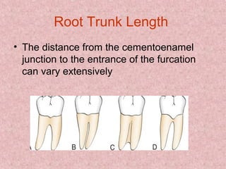
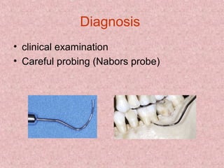
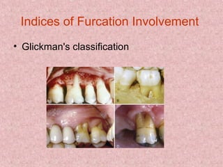
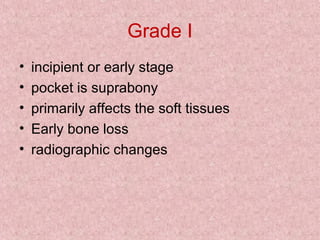
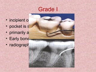
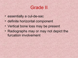
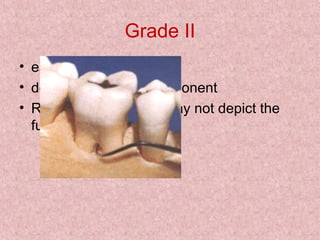
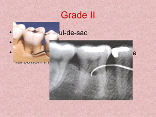
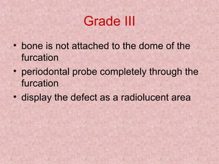
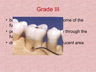
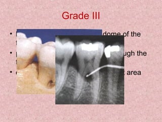
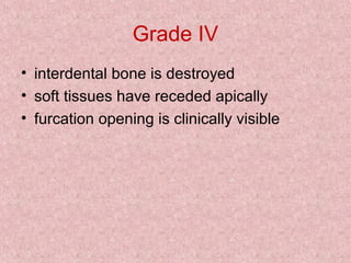
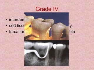
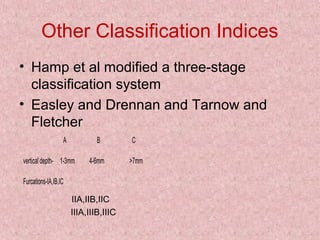
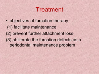
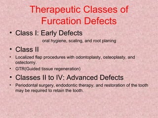
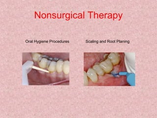
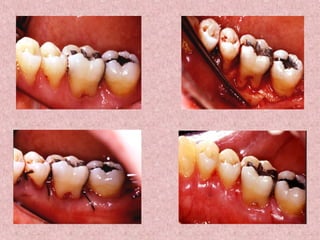
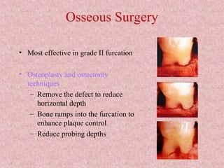
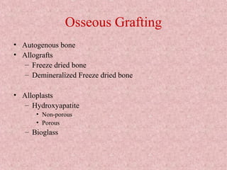
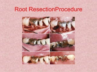
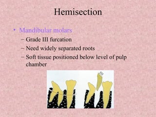
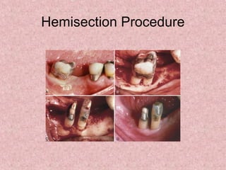
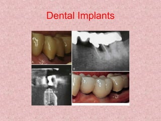
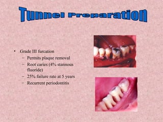
FURCATION
By Azkah Qazi
DEFFINITION
• The furcation is an area of complex
anatomic morphology, that may be difficult
or impossible to debride by routine
periodontal instrumentation.
DEFFINITION
• The furcation is an area of complex
anatomic morphology, that may be difficult
or impossible to debride by routine
periodontal instrumentation.
DEFFINITION
• The furcation is an area of complex
anatomic morphology, that may be difficult
or impossible to debride by routine
periodontal instrumentation.
Etiologic Factors
• Bacterial plaque
• Local anatomic factors (e.g., root trunk
length, root morphology)
• Local developmental anomalies (e.g.,
cervical enamel projections)
Local Anatomic Factors
• Root trunk length
• Root length
• Root form
• Interradicular dimension
• Anatomy of furcation
• Cervical enamel projections
Root Trunk Length
• The distance from the cementoenamel
junction to the entrance of the furcation
can vary extensively
Root length
• This is directly related to the quantity of
attachment supporting the tooth
• Teeth with long roots and short to medium
root trunks are more readily treated
because sufficient attachments remains to
meet functional demands.
Anatomy of furcation
• Presence of bifurcational ridges, a
concavity in the dome and possibly
accesory canals complicates scaling, root
planning and surgical therapy
Interradicular Dimension
• Degree of seperation is an imp factor in
Rx planning
• Closely approximated or fused roots can
preclude adequate instrumentation during
scaling,root planning & surgery
• Teeth with widely seperated roots present
more treatment options.
Cervical Enamel Projections
• 13% of molars have
CEPs
• These projections may
favor the onset of
periodontal lesions in
the affected furcations
Diagnosis
• clinical examination
• Careful probing (Nabors probe)
Indices of Furcation Involvement
• Glickman's classification
Grade I
• incipient or early stage
• pocket is suprabony
• primarily affects the soft tissues
• Early bone loss
• radiographic changes
Grade I
• incipient or early stage
• pocket is suprabony
• primarily affects the soft tissues
• Early bone loss
• radiographic changes
Grade II
• essentially a cul-de-sac
• definite horizontal component
• Vertical bone loss may be present
• Radiographs may or may not depict the
furcation involvement
Grade II
• essentially a cul-de-sac
• definite horizontal component
• Radiographs may or may not depict the
furcation involvement
Grade II
• essentially a cul-de-sac
• definite horizontal component
• Radiographs may or may not depict the
furcation involvement
Grade III
• bone is not attached to the dome of the
furcation
• periodontal probe completely through the
furcation
• display the defect as a radiolucent area
Grade III
• bone is not attached to the dome of the
furcation
• periodontal probe completely through the
furcation
• display the defect as a radiolucent area
Grade III
• bone is not attached to the dome of the
furcation
• periodontal probe completely through the
furcation
• display the defect as a radiolucent area
Grade IV
• interdental bone is destroyed
• soft tissues have receded apically
• furcation opening is clinically visible
Grade IV
• interdental bone is destroyed
• soft tissues have receded apically
• furcation opening is clinically visible
Other Classification Indices
• Hamp et al modified a three-stage
classification system
• Easley and Drennan and Tarnow and
Fletcher
A B C
vertical depth- 1-3mm 4-6mm >7mm
Furcations-IA,IB,IC
IIA,IIB,IIC
IIIA,IIIB,IIIC
Treatment
• objectives of furcation therapy
(1) facilitate maintenance
(2) prevent further attachment loss
(3) obliterate the furcation defects as a
periodontal maintenance problem
Therapeutic Classes of
Furcation Defects
• Class I: Early Defects
oral hygiene, scaling, and root planing
• Class II
• Localized flap procedures with odontoplasty, osteoplasty, and
ostectomy.
• GTR(Guided tissue regeneration)
• Classes II to IV: Advanced Defects
• Periodontal surgery, endodontic therapy, and restoration of the tooth
may be required to retain the tooth.
Nonsurgical Therapy
Oral Hygiene Procedures Scaling and Root Planing
Antimicrobials
• Adjunct to scaling and root planning
– Chlorhexidine
– Tetracycline fibers
Open Debridement
• Greater calculus removal than closed
• Ultrasonic
– Narrow furcations
– Dome of furcation
• Surgical access and increased operator
experience significantly enhance calculus
removal in molar furcation.
Surgical Therapy
• Osseous Resection
• Regeneration(GTR)
• Root Resection
• Hemesection
• Extraction
• Dental Implants
Osseous Surgery
• Most effective in grade II furcation
• Osteoplasty and ostectomy
techniques
– Remove the defect to reduce
horizontal depth
– Bone ramps into the furcation to
enhance plaque control
– Reduce probing depths
Osseous Grafting
• Autogenous bone
• Allografts
– Freeze dried bone
– Demineralized Freeze dried bone
• Alloplasts
– Hydroxyapatite
• Non-porous
• Porous
– Bioglass
Root ResectionProcedure
Hemisection
• Mandibular molars
– Grade III furcation
– Need widely separated roots
– Soft tissue positioned below level of pulp
chamber
Hemisection Procedure
Dental Implants
• Grade III furcation
– Permits plaque removal
– Root caries (4% stannous
fluoride)
– 25% failure rate at 5 years
– Recurrent periodontitis
Prognosis
• The keys to long time success appear to
be
1. thorough diagnosis
2.Selection of patients with good oral
hygiene
3.Excellence in nonsurgical therapy
4.Careful surgical and restorative
management
Furcation ppt

Furcation ppt

  • 1.
  • 2.
    DEFFINITION • The furcationis an area of complex anatomic morphology, that may be difficult or impossible to debride by routine periodontal instrumentation.
  • 3.
    DEFFINITION • The furcationis an area of complex anatomic morphology, that may be difficult or impossible to debride by routine periodontal instrumentation.
  • 4.
    DEFFINITION • The furcationis an area of complex anatomic morphology, that may be difficult or impossible to debride by routine periodontal instrumentation.
  • 5.
    Etiologic Factors • Bacterialplaque • Local anatomic factors (e.g., root trunk length, root morphology) • Local developmental anomalies (e.g., cervical enamel projections)
  • 6.
    Local Anatomic Factors •Root trunk length • Root length • Root form • Interradicular dimension • Anatomy of furcation • Cervical enamel projections
  • 7.
    Root Trunk Length •The distance from the cementoenamel junction to the entrance of the furcation can vary extensively
  • 8.
    Root length • Thisis directly related to the quantity of attachment supporting the tooth • Teeth with long roots and short to medium root trunks are more readily treated because sufficient attachments remains to meet functional demands.
  • 9.
    Anatomy of furcation •Presence of bifurcational ridges, a concavity in the dome and possibly accesory canals complicates scaling, root planning and surgical therapy
  • 10.
    Interradicular Dimension • Degreeof seperation is an imp factor in Rx planning • Closely approximated or fused roots can preclude adequate instrumentation during scaling,root planning & surgery • Teeth with widely seperated roots present more treatment options.
  • 11.
    Cervical Enamel Projections •13% of molars have CEPs • These projections may favor the onset of periodontal lesions in the affected furcations
  • 12.
    Diagnosis • clinical examination •Careful probing (Nabors probe)
  • 13.
    Indices of FurcationInvolvement • Glickman's classification
  • 14.
    Grade I • incipientor early stage • pocket is suprabony • primarily affects the soft tissues • Early bone loss • radiographic changes
  • 15.
    Grade I • incipientor early stage • pocket is suprabony • primarily affects the soft tissues • Early bone loss • radiographic changes
  • 16.
    Grade II • essentiallya cul-de-sac • definite horizontal component • Vertical bone loss may be present • Radiographs may or may not depict the furcation involvement
  • 17.
    Grade II • essentiallya cul-de-sac • definite horizontal component • Radiographs may or may not depict the furcation involvement
  • 18.
    Grade II • essentiallya cul-de-sac • definite horizontal component • Radiographs may or may not depict the furcation involvement
  • 19.
    Grade III • boneis not attached to the dome of the furcation • periodontal probe completely through the furcation • display the defect as a radiolucent area
  • 20.
    Grade III • boneis not attached to the dome of the furcation • periodontal probe completely through the furcation • display the defect as a radiolucent area
  • 21.
    Grade III • boneis not attached to the dome of the furcation • periodontal probe completely through the furcation • display the defect as a radiolucent area
  • 22.
    Grade IV • interdentalbone is destroyed • soft tissues have receded apically • furcation opening is clinically visible
  • 23.
    Grade IV • interdentalbone is destroyed • soft tissues have receded apically • furcation opening is clinically visible
  • 24.
    Other Classification Indices •Hamp et al modified a three-stage classification system • Easley and Drennan and Tarnow and Fletcher A B C vertical depth- 1-3mm 4-6mm >7mm Furcations-IA,IB,IC IIA,IIB,IIC IIIA,IIIB,IIIC
  • 25.
    Treatment • objectives offurcation therapy (1) facilitate maintenance (2) prevent further attachment loss (3) obliterate the furcation defects as a periodontal maintenance problem
  • 26.
    Therapeutic Classes of FurcationDefects • Class I: Early Defects oral hygiene, scaling, and root planing • Class II • Localized flap procedures with odontoplasty, osteoplasty, and ostectomy. • GTR(Guided tissue regeneration) • Classes II to IV: Advanced Defects • Periodontal surgery, endodontic therapy, and restoration of the tooth may be required to retain the tooth.
  • 27.
    Nonsurgical Therapy Oral HygieneProcedures Scaling and Root Planing
  • 28.
    Antimicrobials • Adjunct toscaling and root planning – Chlorhexidine – Tetracycline fibers
  • 29.
    Open Debridement • Greatercalculus removal than closed • Ultrasonic – Narrow furcations – Dome of furcation • Surgical access and increased operator experience significantly enhance calculus removal in molar furcation.
  • 31.
    Surgical Therapy • OsseousResection • Regeneration(GTR) • Root Resection • Hemesection • Extraction • Dental Implants
  • 32.
    Osseous Surgery • Mosteffective in grade II furcation • Osteoplasty and ostectomy techniques – Remove the defect to reduce horizontal depth – Bone ramps into the furcation to enhance plaque control – Reduce probing depths
  • 33.
    Osseous Grafting • Autogenousbone • Allografts – Freeze dried bone – Demineralized Freeze dried bone • Alloplasts – Hydroxyapatite • Non-porous • Porous – Bioglass
  • 34.
  • 35.
    Hemisection • Mandibular molars –Grade III furcation – Need widely separated roots – Soft tissue positioned below level of pulp chamber
  • 36.
  • 37.
  • 38.
    • Grade IIIfurcation – Permits plaque removal – Root caries (4% stannous fluoride) – 25% failure rate at 5 years – Recurrent periodontitis
  • 39.
    Prognosis • The keysto long time success appear to be 1. thorough diagnosis 2.Selection of patients with good oral hygiene 3.Excellence in nonsurgical therapy 4.Careful surgical and restorative management