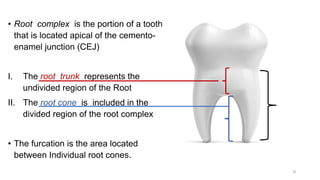
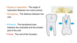
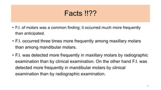
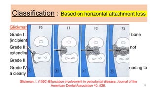
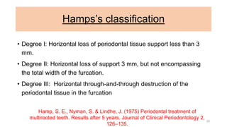
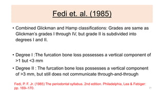
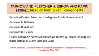
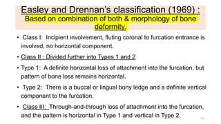
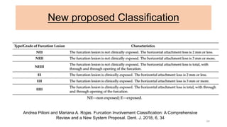
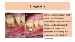
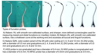
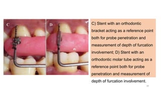
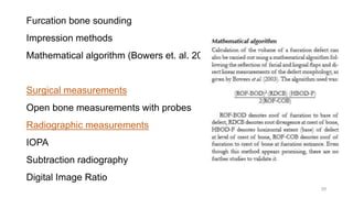
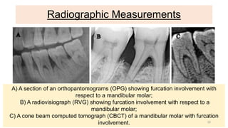
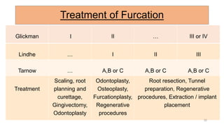
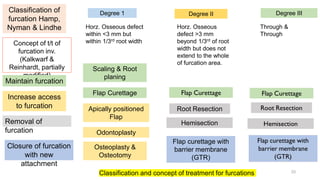
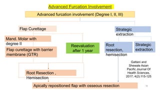
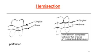
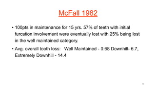
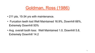
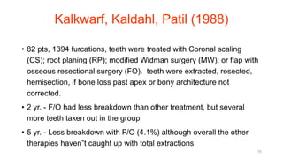
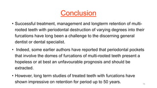
SlideShare a Scribd company logo
1
GOOD MORNING
Most of the important things in the world have been accomplished by people who have
kept on trying when there seemed to be no hope at all.
Furcation
Dr. Suman Mukherjee
MDS 2ND Year
VSDCH
2
Contents :
1. Introduction
2. Definition
3. Terminologies
4. Classification
5. Diagnosis
6. Management
7. Conclusion
8. Reference
3
Introduction
• A furcation is defined as ‘‘the anatomic area of a multirooted tooth
where the roots diverge’’, and furcation invasion refers to the
‘‘pathologic resorption of bone within a furcation’’
~ AAP 1992
• Glickman (1950): Commonly occurring condition in which the
bifurcation and trifurcation of multi-rooted teeth are denuded by
periodontal disease.
4
• Prichard (1965) Bifurcation and trifurcation involvements are
common periodontal lesions which occur as a result of gingival
inflammation and bone resorption adjacent to and within the
furcation of multi-rooted teeth.
• Goldman & Cohen (1968) Extension of pocket into the inter-radicular
area of bone in multi-rooted teeth.
5
• Characterized by bone resorption & attachment loss in the inter
radicular space. (Newmann, Takei & Caranzza 2003).
• The mandibular first molars are the most common sites, and
maxillary premolars are the least common; the number of furcation
involvements increases with age.
6
Terminologies
7
• Root complex is the portion of a tooth
that is located apical of the cemento-
enamel junction (CEJ)
I. The root trunk represents the
undivided region of the Root
II. The root cone is included in the
divided region of the root complex
• The furcation is the area located
between Individual root cones.
8
• Degree of separation: The angle of
separation Between two roots (cones)
• Divergence: The distance between two
root
• Entrance: The transitional area
between the undivided and the divided
part of the root
• Fornix: The roof of the furcation
9
Facts !!??
• F.I. of molars was a common finding; it occurred much more frequently
than anticipated.
• F.I. occurred three times more frequently among maxillary molars
than among mandibular molars.
• F.I. was detected more frequently in maxillary molars by radiographic
examination than by clinical examination. On the other hand F.I. was
detected more frequently in mandibular molars by clinical
examination than by radiographic examination.
10
• Highest frequency of furcation involvement is the distal of Maxillary
1st molar (53%). Lowest frequency of furcation involvement is the
mesial of the Maxillary 2nd molar (20%).
~ Svardstrom (1996)
11
Etiology
1. Primary factor
2. Predisposing factors
3. Contributing factor
4. Local factors
12
Primary Factors
Bacterial plaque:
• Waerhaug (1980): Plaque is the main contributor, subgingival
plaque even in areas where no supragingival plaque was
evident, GI and PI do not reflect actual level of destruction of
furcation, loss of attachment did not increase with increasing
mobility.
13
Kalkwarf& Reinhardt (1988)
Diagnosis not complete until surgical access.
1. Anatomic factors: carious lesions, restorations, furcation
morphology-width, shape, root trunk length
2. Enamel projections: role as contributing factor uncertain.
3. Occlusal trauma: still may be controversial. Waerhaug (1980):
Plaque is the main contributing factors
14
Predisposing factors
• Location relative to CEJ,
• Root trunk length, Root length, Root form.
• Inter-radicular dimension.
• Furcation shape, Location of entrance, Furcation entrance
diameter,
• Facial and lingual radicular bone,
• Enamel projections, Enamel pearls,
• Bifurcation ridges,
• Root concavities and Carious lesions
15
Contributing factors
• Plaque-associated Inflammation,
• Trauma From Occlusion,
• Pulpal Pathology,
• Vertical Root Fractures And
• Iatrogenic Factors
17
Classification : Based on horizontal attachment loss
Glickman’s classification
Grade I : Pocket formation into the flute, but intact interradicular bone
(incipient).
Grade II: Loss of interradicular bone and pocket formation, but not
extending through to the opposite side.
Grade III: Through-and-through lesion.
Grade IV: Through-and-through lesion with gingival recession, leading to
a clearly visible furcation area
Glickman, I. (1950) Bifurcation involvement in periodontal disease. Journal of the
American Dental Association 40, 528. 18
Goldman’s classification
• Grade I: Incipient.
• Grade II: Cul-de-sac.
• Grade III: Through-and-through
Goldman, H. M. (1958) Therapy of the incipient bifurcation involvement. Journal of
Periodontology 29, 112
19
Hamps’s classification
• Degree I: Horizontal loss of periodontal tissue support less than 3
mm.
• Degree II: Horizontal loss of support 3 mm, but not encompassing
the total width of the furcation.
• Degree III: Horizontal through-and-through destruction of the
periodontal tissue in the furcation
Hamp, S. E., Nyman, S. & Lindhe, J. (1975) Periodontal treatment of
multirooted teeth. Results after 5 years. Journal of Clinical Periodontology 2,
126–135.
20
Fedi et. al. (1985)
• Combined Glickman and Hamp classifications: Grades are same as
Glickman’s grades I through IV, but grade II is subdivided into
degrees I and II.
• Degree I :The furcation bone loss possesses a vertical component of
>1 but <3 mm
• Degree II : The furcation bone loss possesses a vertical component
of >3 mm, but still does not communicate through-and-through
Fedi, P. F. Jr. (1985) The periodontal syllabus. 2nd edition. Philadelphia, Lea & Febiger:
pp. 169–170. 21
TARNOW AND FLETCHER & ESKOW AND KAPIN
(1984) : Based on horz. & vert. components
• Sub-classification based on the degree of vertical involvement
• Subclass A. 0–3 mm
• Subclass B. 4–6 mm
• Subclass C. >7 mm
• Eskow and Kapin same subclasses as Tarnow & Fletcher (1984), but
thirds instead of 3-mm units are used.
Tarnow, Fletcher. Classification of the Vertical Component of Furcation Involvement. J.
Periodontol. May, 198
22
Easley and Drennan’s classification (1969) :
Based on combination of both & morphology of bone
deformity.
• Class I: Incipient involvement, fluting coronal to furcation entrance is
involved, no horizontal component.
• Class II : Divided further into Types 1 and 2
• Type 1: A definite horizontal loss of attachment into the furcation, but
pattern of bone loss remains horizontal.
• Type 2: There is a buccal or lingual bony ledge and a definite vertical
component to the furcation.
• Class III: Through-and-through loss of attachment into the furcation,
and the pattern is horizontal in Type 1 and vertical in Type 2. 23
New proposed Classification
24
Andrea Pilloni and Mariana A. Rojas. Furcation Involvement Classification: A Comprehensive
Review and a New System Proposal. Dent. J. 2018, 6, 34
However, there is no one classification system that is
accepted and followed universally.
25
Diagnosis
Probes
I. Straight periodontal probe (a variant of which is the TPS probe)
II. automated probes, such as the Florida probe with disc attachment
III. furcation probes, such as the Nabers, ZA2, ZA3, HO2, NS2, NP2C and
ACE probes
IV. To measure the depth of furcation involvement, a straight probe, like the
UNC-15 probe with 1 mm markings, is inserted into the periodontal
pocket along the root surface to locate the initial fluting of the furcation
Clinical furcation measurement
techniques: A) Furcation
measurement using intersection
of two periodontal probes; B)
Rubber stop placed on a
periodontal probe acting as a
reference point for depth of
penetration;
26
Types of furcation probes:
A) Nabers 1N, with smooth non-calibrated surface, and sharper, more defined curves/angles used for
measuring mesial and distal furcations on maxillary molars; B) Nabers 2N, with smooth non-calibrated
surface, has a shallower curve at the working end and accesses all buccal and lingual furcations;
C) Nabers Q2N, color-coded variant of the 2N with color coding at 3, 6, 9 and 12 mm; D) ZA2 probe,
with a diameter of 0.5 mm and graduations at 2, 4, 6 and 8 mm; E) ZA3 probe, with a diameter of 0.5
mm and graduations at 3, 6, 9 and 12 mm;
F) HO2 probe is non-graduated and has a diameter of 0.4 mm; G) NS2 probe is nongraduated and
has a diameter of 0.5 mm; H) NP2C probe has a diameter of 0.5mm and graduations at 3-5 mm.27
C) Stent with an orthodontic
bracket acting as a reference point
both for probe penetration and
measurement of depth of furcation
involvement; D) Stent with an
orthodontic molar tube acting as a
reference point both for probe
penetration and measurement of
depth of furcation involvement.
28
Furcation bone sounding
Impression methods
Mathematical algorithm (Bowers et. al. 2003)
Surgical measurements
Open bone measurements with probes
Radiographic measurements
IOPA
Subtraction radiography
Digital Image Ratio
29
Radiographic Measurements
A) A section of an orthopantomograms (OPG) showing furcation involvement with
respect to a mandibular molar;
B) A radiovisiograph (RVG) showing furcation involvement with respect to a
mandibular molar;
C) A cone beam computed tomograph (CBCT) of a mandibular molar with furcation
involvement. 30
New Frontiers
• Natural frequency analysis
• Optical coherence tomography (OCT)
• Fiberscopes
• Ultrasonography
31
Karthikeyan et al.: Furcation Measurements Journal of the International Academy of Periodontology
(2015) 17/4
Treatment of Furcation
Glickman I II … III or IV
Lindhe … I II III
Tarnow … A,B or C A,B or C A,B or C
Treatment
Scaling, root
planning and
curettage,
Gingivectomy,
Odontoplasty
Odontoplasty,
Osteoplasty,
Furcationplasty,
Regenerative
procedures
Root resection, Tunnel
preparation, Regenerative
procedures, Extraction / implant
placement
32
Classification of
furcation Hamp,
Nyman & Lindhe
Degree 1 Degree II Degree III
Concept of t/t of
furcation inv.
(Kalkwarf &
Reinhardt, partially
modified)
Maintain furcation
Increase access
to furcation
Removal of
furcation
Closure of furcation
with new
attachment
Scaling & Root
planing
Flap Curettage
Apically positioned
Flap
Odontoplasty
Osteoplasty &
Osteotomy
Root Resection
Hemisection
Flap curettage with
barrier membrane
(GTR)
Classification and concept of treatment for furcations
Horz. Osseous defect
within <3 mm but
within 1/3rd root width
Through &
Through
Horz. Osseous
defect >3 mm
beyond 1/3rd of root
width but does not
extend to the whole
of furcation area.
33
Advanced Furcation Involvement
Advanced furcation involvement (Degree I, II, III)
Flap Curettage Strategic
extraction
Root
resection,
hemisection
Strategic
extraction
Mand. Molar with
degree II
Flap curettage with barrier
membrane (GTR)
Root Resection ,
Hemisection
Apically repositioned flap with osseous resection
Reevaluation
after 1 year
Gattani and
Shewale Asian
Pacific Journal Of
Health Sciences,
2017; 4(2):115-125
34
Therapy
Treatment of a bony defect in the furcation region is intended to meet
two objectives:
1. Elimination of the microbial plaque from the exposed surfaces of
the root complex.
2. Establishment of an anatomy of the affected surfaces that facilitates
proper self-performed plaque control.
35
Factors to
be
considered
for
successful
treatment of
furcation
involvement:
1. Degree of Involvement
2. Crown: Root ratio
3. Length of roots
4. Root
anatomy/morphology
5. Degree of root
separation
6. Strategic value of the
tooth
7. Residual tooth mobility
8. Need for endodontic
treatment
9. Prosthetic
requirements
10. Periodontal condition
of adjacent teeth
11. Ability to maintain oral
hygiene
12. Quality of bone/
ability to place implants
13. Financial
considerations
14. Long term prognosis
36
Three broad strategies of furcation therapy
(Kalkwarf & Reinhardt R.A 1988)
I. Maintenance of the existing Furcation.
• Scaling and root planning
• Obstruction of Furcation
II. Increasing access to the Furcation
• Gingivectomy/Apical positioned flap
• Odontoplasty
• Osteoplasty /ostectomy
III. Elimination of the Furcation
• Root amputation/ Tooth resection Bicuspidization
furcationplasty
37
Furcation Involvement Degree I
• Non-surgical Treatment
• Oral Hygiene measurements and Scaling and Root planning
• Obliteration of furcation by restorative materials
• Furcationplasty
38
Root Curettage : Non Surgical therapy
• This practice works well when the interradicular fluting is broad and
access is not a problem.
• Loos et al. (1989): In sites of > 7 mm regressed after initial treatment,
Overall 25% of molar furcation sites demonstrated loss of attachment
compared to 7% for non-molar sites and 10% of molar flat-surface
sites.
• Badersten: Non-surgical therapy works, but in non molar teeth only.
• Nordland (1987): Furcations with initial pocket depth > 4mm had
poorer response to non-surg therapy verses flat molar and non-molar
sites. 0.5mm loss in 24 months 39
• Leon and Vogel (1987) Compared hand and ultrasonic scaling in
furcations Class I No difference between modalities Class II and III
ultrasonic scaler better.
• Parashis (1993) Calculus removal in furcations best with open
scaling and rotary diamonds.
• Bower (1979) Width of furcation entrance is too narrow for most
scalers.
40
Odontoplasty
• This term means, “The reshaping of the tooth.” With respect to
furcation invasions, it means the widening of the furcal area in a
buccolingual or mesiodistal as well as apicocoronal direction with a
high-speed diamond.
• The net effect is to widen the inter-radicular area and to remove or
reshape the horizontal component of the furcation invasion. The
furcation is thus made more accessible for oral hygiene efforts.
42
• The initial reshaping is done with round diamonds and is refined with
curetts. This procedure is really limited to Class I and shallow Class
II furcation invasions.
• The deeper the invasion, the more reshaping that is required, and
thus the more tooth structure that must be removed.
• Such removal increases the likelihood of dentinal sensitivity, which
can be so severe that root canal therapy is required.
43
Osteoplasty
• If the fluting is narrow or there is restricted access to the furcation,
osteoplasty/ odontoplasty procedures may be necessary. The tooth
and alveolar bone in the furcation area are reshaped.
• The elimination of bony ledges and the placement of “vertical
grooves" in the bone just coronal to the furcation make the contours
more gradual in an apical-coronal direction, which improves access
for home care devices and curets.
• This technique is generally employed as part of a segment of
periodontal surgery, and is not often used alone.
44
Root Resection
• Root resections (amputations) are utilized when the furcation
invasion is too advanced to be corrected by the previous techniques.
Access to the furcation can be gained by removing one or more of
the affected roots.
• A flap is reflected to expose the underlying bone. The bony plate
covering the involved root is removed to about 2/3rds of its length. A
bur is used to cut through the root to be removed. This cut is apical
to the
45
• Opening of the furcation. Once resected, the root is extracted. The
remaining stump is contoured to smooth out any sharp angles and to
remove any undercuts.
• This is critical so the restorative dentist can prepare the tooth for the
eventual crown. If endodontic therapy has not been previously
performed, ZOE is placed into the opening to the canal.
46
Indications & Contraindications (Basaraba
1969)
1. Uncorrected bone loss involving one root of a mandibular molar,
one or two buccal roots or a palatal root of a maxillary molar.
2. Furcation invasion such that odontoplasty is not indicated (greater
than Class I).
3. When root proximity prevents proper maintenance.
4. When osseous recontouring (ostectomy) would cause the exposure
of a furcation.
47
5. To improve the prognosis of teeth within a fixed bridge.
6. Fracture of a tooth or root
7. Failure of endodontic therapy in one canal and correction or
retreatment of this canal is not possible
8. When recession exposing the entire length of a root cannot be
corrected with mucogingival procedures.
48
Contraindications:
1. RCT cannot be done on the remaining roots, e.g. partly calcified
canal or fused roots
2. Bone loss around the remaining roots is too severe to be corrected
via periodontal procedures.
3. Class I furcation invasion.
49
Hemisection
• In a hemisection the tooth is cut in half. The technique is used
virtually exclusively on mandibular molars to treat Class II or III
furcation invasions.
• The tooth is sectioned from buccal to lingual, parallel to a line joining
the buccal and lingual furcas. In contrast to root amputations,
extraction of one of the sections does not necessarily need to be
performed.
50
• A hemisection often will be followed by the extraction of one of the
sectioned halves. This is done primarily when the severe attachment
loss is restricted to one root, the other root can be treated, and there
is no other stable distal abutment.
• A bur (highspeed) is used to cut through the coronal portion of the
tooth separating it into two halves. It is advisable to make the coronal
cut prior to flap reflection to minimize the amount of tooth structure
and restorative material that gets into the surgical site.
51
• Langer et al, made a 10-year evaluation of root resections. Of 100
teeth so treated, 38 had failed by the end of 10 years.
• Interestingly although the primary reason for performing the root
resections was to treat periodontal lesions, most of the failures (28 of
38) were due to endodontic or restorative problems such as root
fractures, cemental washouts, caries, and recurrent periapical
pathoses.
• Mandibular molars failed twice as often as did maxillary molars. On a
more positive note 62% of the cases did last a decade.
52
Root Amputation
• Consider implants, may have better long term success.
Langer et al (1981): 10yr, 100pts, results are as follows for resected
teeth: 38% of resected teeth failed by 10 yr. mark (62% success rate),
15.8 % in 5yr. Of the failures:
1. 47% (greatest number of teeth) failed due to root fractures
2. 26.3 failed to progression of periodontal disease, most were
maxillary molars
3. 18.4% failed to endodontic procedures
4. 7.9% failed due to cement washout
53
• Carnevale et al (1991): 500 teeth with either root amps or
hemisections. Overall 5.7% failures, highest being caries and root
fractures, 97.6% of these teeth were treated for periodontal reasons,
only 0.6% had recurrence of periodontal breakdown.
• Buhler (1994): 337 cases, 7 yr. period of hemisection, reported
failure rate of 13.1%.
54
Tunneling
• The “tunneling” procedure has been used in Class III furcation
inversions.
• A flap is reflected, ostectomy and osteoplasty usually are required,
and the flap is sutured in an apical position exposing the furcation to
the oral cavity so that it is accessible for oral hygiene measures.
• This is generally limited to molar teeth with well separated roots.
Roots in close proximity are not good candidates because of the
difficulty in obtaining access for plaque control.
55
• The tunneling procedure is not done frequently. Caries may develop
because of the difficulty in removing plaque from the furcal “roof”
which often is concave. Interproximal brushes dipped in fluoride and
irrigation using a Mono-Jet syringe and chlorhexidine may help to
slow caries activity.
• Little (1995): 18 pts with 5 maxillary and 13 mandibular furcations
treated by tunneling. Adjacent teeth were used to evaluate bone
loss. After 5 yr., 3/18 had developed root caries. No difference seen
in CAL or bone loss when compared to adjacent teeth.
56
• Hellden, Steffensen et al (1989): 149 teeth with Grade III furcations
at 3 yrs, 75% caries free.
• Hamp, Nyman, Lindhe (1975): Treatment of teeth with furcations
revealed the following 5 yr. results:
44% of the teeth were extracted during initial treatment.
50% of the remaining teeth received root resections, one root
preserved 64% of the time ,none of the teeth were lost in 5 yrs.
Tunneling procedure had root caries 4/7.
57
Regeneration
• Regeneration procedures designed to recreate lost periodontal
attachment have not been particularly rewarding especially in
furcation invasions.
• Recent interest has focused on “guided tissue regeneration” in the
treatment of Class II & III defects. In this technique, full-thickness
flaps are reflected, the areas are thoroughly debrided, and a
synthetic membrane or other material is placed over the bony defect.
58
GTR in Furcation's
59
• Gantes (1991): Class 3 furcations, citric acid and coronally positioned
flaps with moderate results.
• Lu (1992): Complete circumferential adaptation of the membrane to
the root is not possible, gaps will remain. Occlusal border should be
placed 1-2mm below CEJ. GTR success may be more from clot
stabilization than from epithelial exclusion.
• Pontoriero et al (1988): GTR in class II furcations, 14/21 complete
closure, 5/21 had residual of < 1 mm. 90% closure of Class II‟s with
membrane, OFD 2/21 completely closed, No reentries. GTR better than
OFD in Class II furcas.
60
Pontoriero (1989): Class III, 8/21 closure with GTR, control 0/21, no re-
entry, clinical probing depth only.
Pontoriero (1995) Buccal furcations more predictable than interproximal
furcas, GTR doesn‟t work in class III furcas.
Lindhe et al (1995): Flap management and bioresorbable membranes in
class III molar furcations in dogs:
-Large furcation defects can be treated provided soft tissue flaps
covering membranes prevented from recession
-Resolute equally as effective as e-PTFE.
61
• Anderegg et al (1991): 15 pts, molar furcas, GTR alone vs. GTR and
DFDBA (BETTER), 6 more-entry, combined more fill, more PD
reduction, greater attach gain both horizontal and vertical.
• Mellonig (1991): IJPDR: Class 2s, membrane better than OFD,
improved HOPA/VOPA, GTR will improve clinical results, rarely
complete closure.
• Mellonig (1994): 13 pts with grade II furcas. Comparison of ePTFE
vs. debridement-6 month reentry. ePTFE sites showed more PDR,
ALG, as well as recession in man II defects. There was no difference
between the 2 txs in max grade II furcas 62
• Anderegg (1995): Gingival thickness in GTR. 37 pts with grade I or II
max or man furcas were txd with GTR. Pts with <1mm of gingival
thickness had 2.1mm of recession at 6 mos. postop. Pts with >1mm
gingival thickness only had 0.6mm recession. Less recession with
thicker tissues.
• Lekovic and Kenney (1989): class II furcations, ePTFE vs. OFD, test
site showed PD reduction, gain in attachment levels of 2.86mm, vs.
controls which didn’t change from preoperative levels.
• Nygaard Ostby (1996) GTR vs. OFD. GTR has no significant
advantage over OFD. No grafts were used.
63
ePTFE + GRAFT in furcations
• Lekovic et al (1990): grade II furcations PTFE+HA vs. PTFE alone, 30
defects, PTFE+HA had 2.9 mm attach gain vs. PTFE alone of 2.4 mm.
PTFE+HA had greater vertical/horizontal bone gain and less recession.
• McClain and Schallhorn(1993): GTR + GRAFT = Long term stability
DFDBA + autog + GTR + C.A. = 4.0 mm mean clinical AGAIN, including
furcation fill.5 yr. follow-up of GTR with and without CA root conditioning
and composite grafts. Long term results enhanced with CA + graft, 5yr
stability of CPAL. 93% stable with graft, 30% stable with membrane
only. 64
• Garrett (1994): Grade III man furcations treated with DFDBA alone or
DFDBA + ePTFE. Both covered by CPF.. No benefit was seen with the
use of ePTFE.
• Wallace (1994): Grade II mandibular furcations treated with either
ePTFE alone or ePTFE + DFDBA. 6 month re-entry showed similar
results as far as recession, and reduction of horizontal defect depth
were concerned. The ePTFE + DFDBA group showed greater vertical
defect fill and greater PALG when compared to the ePTFE only group.
• Mellado(1995):ePTFE with and without DFDBA more bone formed
without DFDBA Anti-DFDBA study.
65
Tooth Extraction
• This therapy is indicated when the destruction of the
periodontium has progressed to such a level that no tooth can
be preserved.
66
Restorative Management
• Crowns used to restore root-resected teeth should follow the form
created during the amputation procedure described previously.
Proximal walls should taper evenly into the remaining root surface.
• No spurs of overhangs should remain to complicate maintenance.
Interproximal areas should be open to facilitate cleaning. Root
concavities in the furcation areas should be reproduced in the
restoration.
• Contours should be flat for access for effective plaque removal.
Hemisected teeth should not be cantilevered unless supported by 67
• Endodontic therapy should be conservative with minimal
enlargement of the root canal for root strength. Condensation should
not be excessive.
• Gutta-percha permits the placement of posts without disturbing the
apical seal. Badly broken-down teeth may be built up with a post and
core before final restoration is attempted.
68
Furcation long term
maintainan
69
Hirschfeld, Wasserman (1978):
22 yr. maintenance study of 600 pts.
1. 31% of teeth with original furcation invasion were lost
2. Breakdown of teeth lost according to groups WM 19.3%, D 69.9%, ED
84.4%
3. Average overall tooth loss by patient groups WM 0.68, D 5.7, ED 13%
4. Order of tooth loss: Max 2nd, Max 1st, Mn 2nd, Mn 1st
5. 300 lost no teeth, 199 lost 1-3 teeth, 76 lost 4-9 teeth, 25 lost 10-23
teet
6. Mortality of teeth correlated more closely to case type rather than type
of surgery
7. Periodontal disease is symmetrical PATIENT PERCENTAGES: Well
70
• Pearlman (1993): 172 pts classified similar to Hirschfeld and
Wasserman with similar breakdown of results.
• Finding was that even in the well maintained group, there were
more molars lost with furcation involvement over those without
involvement.
71
Ross and Thompson (1978):
• 100 pts treated with 387 furcations.
• Conservative treatment only OFD, no resection or osseous
treatments, 5 yr.
• Minimum follow up, 88% of teeth were functioning after 5-24 yrs, and
radiographs were the only diagnostic tool of success.
• Maxillary Furcation involvement three times that of Mandibular.
• Maxillary furcations were detected most frequently by radiographs,
• Mandibular furcations detected most frequently by clinical exam.
72
McFall 1982
• 100pts in maintenance for 15 yrs. 57% of teeth with initial
furcation involvement were eventually lost with 25% being lost
in the well maintained category.
• Avg. overall tooth loss: Well Maintained - 0.68 Downhill- 6.7,
Extremely Downhill - 14.4
73
Goldman, Ross (1986)
• 211 pts, 15-34 yrs with maintenance.
• Furcation teeth lost Well Maintained 16.9%, Downhill 66%,
Extremely Downhill 93%
• Avg. overall tooth loss: Well Maintained 1.0, Downhill 5.8,
Extremely Downhill 14.2
74
Kalkwarf, Kaldahl, Patil (1988)
• 82 pts, 1394 furcations, teeth were treated with Coronal scaling
(CS); root planing (RP); modified Widman surgery (MW); or flap with
osseous resectional surgery (FO). teeth were extracted, resected,
hemisection, if bone loss past apex or bony architecture not
corrected.
• 2 yr. - F/O had less breakdown than other treatment, but several
more teeth taken out in the group
• 5 yr. - Less breakdown with F/O (4.1%) although overall the other
therapies haven‟t caught up with total extractions
75
Conclusion
• Successful treatment, management and longterm retention of multi-
rooted teeth with periodontal destruction of varying degrees into their
furcations have long been a challenge to the discerning general
dentist or dental specialist.
• Indeed, some earlier authors have reported that periodontal pockets
that involve the domes of furcations of multi-rooted teeth present a
hopeless or at best an unfavourable prognosis and should be
extracted.
• However, long term studies of treated teeth with furcations have
shown impressive on retention for period up to 50 years. 76
References
1. Novak MJ. Classification of Diseases and Conditions Affecting the Periodontium. In:
Newman MG, Takei HH, Klokkevold PR, Carranza FA, editors. Carranza‟s Clinical
Periodontology. 10th Ed. WB Saunders Co; 2009: p. 103.
2. Fermin A. Carranza. Bone Loss and Patterns of Bone Destruction. In: Newman MG,
Takei HH, Klokkevold PR, Carranza FA, editors. Carranza‟s Clinical Periodontology. 10th
Ed. Missouri: WB Saunders Co;2009: p. 462.
3. Svardstrom G, Wennstrom JL. Furcation Topography of the Maxillarv and Mandibular
First Molars. J Clin Periodontol 1988;15:271-5.
4. Bower RC. Furcation Morphology Relative to Periodontal Treatment: Furcation Entrance
Architecture. J Periodontol 1979;50:23-7.
5. Al-Shammari KF, Kazor CE. Molar Root Anatomy and Management of Furcation Defects.
J Clin Periodontol 2001;28:730-40.
6. Hamp SE, Nyman S, Lindhe J. Periodontal Treatment of Multirooted Teeth. J Clin
Periodontol 1975;2:126-35.
7. Muller HP, Eger T. Furcation Diagnosis. J Clin Periodontol 1999;26:485-98.
77
8. Recchetti, P. (1982) A furcation classification based upon pulp chamberfurcation
relationships and vertical radiographic bone loss. International Journal of Periodontics and
Restorative Dentistry 2, 51.
9. Al-Shammari KF, Kazor CE, Wang HL: Molar root anatomy and management of furcation
defects. J Clin Periodontol 2001; 28: 730–740.
10. Grant, D. A., Stern, I. B. & Listgarten, M. A., eds. (1988) Periodontics, 6th edition. St.
Louis: CV Mosby, 921–932.
11. Nevins, M., Cappetta, E. G. (1998) Treatment of maxillary furcations. In: Nevins, M.,
Mellonig, J. T. (eds.): Periodontal therapy: clinical approaches and evidence of success, vol.
1. Quintessence.
12. Gattani and Shewale. Furcation invasion- a literature review on its treatment modalities.
Asian Pac. J. Health Sci., 2017; 4(2):115-125 .
13. Karthikeyan et al.: Furcation Measurements. Journal of the International Academy of
Periodontology (2015) 17/4.
14. Parihar AS et al. Furcation Involvement & Its Treatment. Journal of Advanced Medical
and Dental Sciences Research |Vol. 3|Issue 1| January-March 2015 78
Questions
79
Drawbacks of the study
• Small sample size
• No control in the study to compare with the tests group in the
study.
• Results not compared to other studies for vertical augmentation.
80
Factors responsible for crestal bone loss
• Factors thought to influence the number of changes in crestal bone height
after implant placement include delayed vs immediate implant placement,
staging, timing of implant loading, requirement of bone graft at the implant
site, presence of infection, medical conditions that compromise wound
healing, smoking, status of oral hygiene, location of implant placement, and
size of the implants.
• Other mechanical factors such as periosteum elevation during surgery,
overheating of the instrument resulting in osteonecrosis, occlusal trauma,
cantilever effect, and physiologic bone remodeling from inflammatory
process and plaque accumulation have been also suggested.
81
Sang Y. Kim, Thomas B. Dodson, Duy T. Do, Gary Wadhwa, Sung-Kiang Chuang, Factors Associated With
Crestal Bone Loss Following Dental Implant Placement in a Longitudinal Follow-up Study, Journal of Oral
Implantology. 2015;41(5):579-585.
Sandwich technique better than others
• The inlay bone grafting technique (sandwich technique) carries many
advantages over other reconstructive techniques like the ability to
gain higher better bone quality in shorter time as compared to other
techniques.
- Mansour et. al. 2018
• SBA technique was used for its advantages such as short treatment
time, ideal restoration, less morbidity, more comfortable, and lower
costs.
- Anton Lee 2017
82
• Reduced treatment time and patient need not get another additional
surgical procedure done.
- Fu J
Fu, Jia-Hui & Wang, Hom-Lay. (2012). The Sandwich Bone Augmentation
Technique. Clinical Advances in Periodontics. 2. 172-177.
83
Commercially avl. Xenografts
• OsteoBiol – porcine derived
• BioCoral – calcium carbonate
• Bio-Oss
• Bio-Oss Collagen
• Pepgen-P15
84
Inter Implant distance 3mm why?
• The bone crest was more apically located at sites with <3 mm inter-
implant distance than at sites where the implants were standing
>3 mm apart, Tarnow et al. (2000) suggested that not only vertical
bone loss but also lateral bone loss at implants could have an effect
on the level of the bone crest between two implants.
• Violation of biological width happens.
Distance between implants has a potential impact of crestal bone resorption. Danza M et. al. Saudi
Dent J. 2011 Jul; 23(3): 129–133.
85
Furcation
Dr. Suman Mukherjee
MDS 2ND Year
VSDCH
8
6

More Related Content

What's hot

Wound healing [including healing after periodontal therapy]
Wound healing [including healing after periodontal therapy]Wound healing [including healing after periodontal therapy]
Wound healing [including healing after periodontal therapy]Jignesh Patel
 
Critical apprisal of 2018 classification of periodontal disease
Critical apprisal of 2018 classification of periodontal disease Critical apprisal of 2018 classification of periodontal disease
Critical apprisal of 2018 classification of periodontal disease yasmin parvin ss
 
Post operative complications of periodontal surgery
Post operative complications of periodontal surgeryPost operative complications of periodontal surgery
Post operative complications of periodontal surgeryRitam Kundu
 
RESECTIVE OSSEOUS SURGERY
RESECTIVE OSSEOUS SURGERYRESECTIVE OSSEOUS SURGERY
RESECTIVE OSSEOUS SURGERYAnkita Dadwal
 
Peri implantitis treatment protocol
Peri implantitis treatment protocolPeri implantitis treatment protocol
Peri implantitis treatment protocolFadi Al-Zaitoun
 
Furcation involvements and its treatments
Furcation  involvements and its treatmentsFurcation  involvements and its treatments
Furcation involvements and its treatmentsDiana Abo el Ola
 
Wound healing in Perio - Dr. Malvika Thakur
Wound healing in Perio - Dr. Malvika ThakurWound healing in Perio - Dr. Malvika Thakur
Wound healing in Perio - Dr. Malvika ThakurDr.Malvika Thakur
 
Periodontal plastic surgery
Periodontal plastic surgeryPeriodontal plastic surgery
Periodontal plastic surgeryRobert Cain
 
PLATELET RICH FIBRIN - ROLE IN PERIODONTICS
PLATELET RICH FIBRIN - ROLE IN PERIODONTICSPLATELET RICH FIBRIN - ROLE IN PERIODONTICS
PLATELET RICH FIBRIN - ROLE IN PERIODONTICSDr.Shraddha Kode
 
Modified Papilla Preservation Technique
Modified Papilla Preservation TechniqueModified Papilla Preservation Technique
Modified Papilla Preservation TechniqueWendy Jeng
 
Periodontal flap surgeries by Dr. Jerry
Periodontal flap surgeries by Dr. JerryPeriodontal flap surgeries by Dr. Jerry
Periodontal flap surgeries by Dr. JerryDeepesh Mehta
 
Stress on periodontium
Stress on periodontiumStress on periodontium
Stress on periodontiumVijay Apparaju
 
Diagnostic test and investigation in periodontology
Diagnostic test and investigation in periodontology Diagnostic test and investigation in periodontology
Diagnostic test and investigation in periodontology Dr Abhilasha
 
Root conditioning in periodontics
Root conditioning in periodonticsRoot conditioning in periodontics
Root conditioning in periodonticsR Viswa Chandra
 
Gingival pigmentation
Gingival pigmentationGingival pigmentation
Gingival pigmentationNida Sumra
 
Periodontal pocket
Periodontal pocketPeriodontal pocket
Periodontal pocketshyasaman
 
FURCATION INVOLVEMENT AND ITS MANAGEMENT
FURCATION INVOLVEMENT AND ITS MANAGEMENTFURCATION INVOLVEMENT AND ITS MANAGEMENT
FURCATION INVOLVEMENT AND ITS MANAGEMENTFarzana Nafi
 
periodontal regeneration
periodontal regenerationperiodontal regeneration
periodontal regenerationSupriyoGhosh15
 

What's hot (20)

Wound healing [including healing after periodontal therapy]
Wound healing [including healing after periodontal therapy]Wound healing [including healing after periodontal therapy]
Wound healing [including healing after periodontal therapy]
 
Critical apprisal of 2018 classification of periodontal disease
Critical apprisal of 2018 classification of periodontal disease Critical apprisal of 2018 classification of periodontal disease
Critical apprisal of 2018 classification of periodontal disease
 
Post operative complications of periodontal surgery
Post operative complications of periodontal surgeryPost operative complications of periodontal surgery
Post operative complications of periodontal surgery
 
RESECTIVE OSSEOUS SURGERY
RESECTIVE OSSEOUS SURGERYRESECTIVE OSSEOUS SURGERY
RESECTIVE OSSEOUS SURGERY
 
Peri implantitis treatment protocol
Peri implantitis treatment protocolPeri implantitis treatment protocol
Peri implantitis treatment protocol
 
Furcation involvements and its treatments
Furcation  involvements and its treatmentsFurcation  involvements and its treatments
Furcation involvements and its treatments
 
Wound healing in Perio - Dr. Malvika Thakur
Wound healing in Perio - Dr. Malvika ThakurWound healing in Perio - Dr. Malvika Thakur
Wound healing in Perio - Dr. Malvika Thakur
 
Periodontal plastic surgery
Periodontal plastic surgeryPeriodontal plastic surgery
Periodontal plastic surgery
 
Ridge augmentation
Ridge augmentationRidge augmentation
Ridge augmentation
 
PLATELET RICH FIBRIN - ROLE IN PERIODONTICS
PLATELET RICH FIBRIN - ROLE IN PERIODONTICSPLATELET RICH FIBRIN - ROLE IN PERIODONTICS
PLATELET RICH FIBRIN - ROLE IN PERIODONTICS
 
Modified Papilla Preservation Technique
Modified Papilla Preservation TechniqueModified Papilla Preservation Technique
Modified Papilla Preservation Technique
 
Periodontal flap surgeries by Dr. Jerry
Periodontal flap surgeries by Dr. JerryPeriodontal flap surgeries by Dr. Jerry
Periodontal flap surgeries by Dr. Jerry
 
Stress on periodontium
Stress on periodontiumStress on periodontium
Stress on periodontium
 
Diagnostic test and investigation in periodontology
Diagnostic test and investigation in periodontology Diagnostic test and investigation in periodontology
Diagnostic test and investigation in periodontology
 
Root conditioning in periodontics
Root conditioning in periodonticsRoot conditioning in periodontics
Root conditioning in periodontics
 
Gingival pigmentation
Gingival pigmentationGingival pigmentation
Gingival pigmentation
 
Periodontal pocket
Periodontal pocketPeriodontal pocket
Periodontal pocket
 
FURCATION INVOLVEMENT AND ITS MANAGEMENT
FURCATION INVOLVEMENT AND ITS MANAGEMENTFURCATION INVOLVEMENT AND ITS MANAGEMENT
FURCATION INVOLVEMENT AND ITS MANAGEMENT
 
periodontal regeneration
periodontal regenerationperiodontal regeneration
periodontal regeneration
 
Gingival curettage
Gingival curettageGingival curettage
Gingival curettage
 

Similar to Furcation - Session I

Furcation involvement in general dental practice
Furcation involvement in general dental practiceFurcation involvement in general dental practice
Furcation involvement in general dental practiceBelal Nabil Elmarhoumy
 
Periodontal probing
Periodontal probingPeriodontal probing
Periodontal probingSHIVANI IYER
 
Management of Impacted Third Molars
Management of Impacted Third MolarsManagement of Impacted Third Molars
Management of Impacted Third MolarsDibya Falgoon Sarkar
 
4.furcation involvement and its treatment
4.furcation involvement and its treatment4.furcation involvement and its treatment
4.furcation involvement and its treatmentpunitnaidu07
 
Attached Gingiva.pptx
Attached Gingiva.pptxAttached Gingiva.pptx
Attached Gingiva.pptxRinisha Sinha
 
2017 classification of periodontal and periimplant diseases
2017 classification of periodontal and periimplant diseases2017 classification of periodontal and periimplant diseases
2017 classification of periodontal and periimplant diseasesDr. Bibina George
 
Journal club on Mandibular fracture after third molar
Journal club on Mandibular fracture after third molarJournal club on Mandibular fracture after third molar
Journal club on Mandibular fracture after third molarDr Bhavik Miyani
 
Diagnosis of canine impaction by TABZ.pptx
Diagnosis of canine impaction by TABZ.pptxDiagnosis of canine impaction by TABZ.pptx
Diagnosis of canine impaction by TABZ.pptxthabassumparakkal
 
Furcation involvement
Furcation involvementFurcation involvement
Furcation involvementneeti shinde
 
Epidimiology periodontic
Epidimiology periodontic Epidimiology periodontic
Epidimiology periodontic dentalcare3
 
The Perio-Endo Relationship
The Perio-Endo RelationshipThe Perio-Endo Relationship
The Perio-Endo RelationshipEric Jewell
 
FURCATION INVOLVEMENT.pptx
FURCATION INVOLVEMENT.pptxFURCATION INVOLVEMENT.pptx
FURCATION INVOLVEMENT.pptxNandini K
 

Similar to Furcation - Session I (20)

Furcation involvement in general dental practice
Furcation involvement in general dental practiceFurcation involvement in general dental practice
Furcation involvement in general dental practice
 
Furcation case
Furcation caseFurcation case
Furcation case
 
Periodontal probing
Periodontal probingPeriodontal probing
Periodontal probing
 
Management of Impacted Third Molars
Management of Impacted Third MolarsManagement of Impacted Third Molars
Management of Impacted Third Molars
 
Endo-Perio.ppt
Endo-Perio.pptEndo-Perio.ppt
Endo-Perio.ppt
 
4.furcation involvement and its treatment
4.furcation involvement and its treatment4.furcation involvement and its treatment
4.furcation involvement and its treatment
 
Periodontitis
PeriodontitisPeriodontitis
Periodontitis
 
Attached Gingiva.pptx
Attached Gingiva.pptxAttached Gingiva.pptx
Attached Gingiva.pptx
 
Endo Perio Lesions
Endo Perio LesionsEndo Perio Lesions
Endo Perio Lesions
 
2017 classification of periodontal and periimplant diseases
2017 classification of periodontal and periimplant diseases2017 classification of periodontal and periimplant diseases
2017 classification of periodontal and periimplant diseases
 
Journal club on Mandibular fracture after third molar
Journal club on Mandibular fracture after third molarJournal club on Mandibular fracture after third molar
Journal club on Mandibular fracture after third molar
 
Diagnosis of canine impaction by TABZ.pptx
Diagnosis of canine impaction by TABZ.pptxDiagnosis of canine impaction by TABZ.pptx
Diagnosis of canine impaction by TABZ.pptx
 
Furcation involvement
Furcation involvementFurcation involvement
Furcation involvement
 
Furcation
 Furcation Furcation
Furcation
 
Epidimiology periodontic
Epidimiology periodontic Epidimiology periodontic
Epidimiology periodontic
 
Endodontic Failures and Mishaps
Endodontic Failures and MishapsEndodontic Failures and Mishaps
Endodontic Failures and Mishaps
 
The Perio-Endo Relationship
The Perio-Endo RelationshipThe Perio-Endo Relationship
The Perio-Endo Relationship
 
THIRD MOLAR IMPACTION (BASIC)
THIRD MOLAR IMPACTION (BASIC)THIRD MOLAR IMPACTION (BASIC)
THIRD MOLAR IMPACTION (BASIC)
 
Aggresive periodontitis
Aggresive periodontitis   Aggresive periodontitis
Aggresive periodontitis
 
FURCATION INVOLVEMENT.pptx
FURCATION INVOLVEMENT.pptxFURCATION INVOLVEMENT.pptx
FURCATION INVOLVEMENT.pptx
 

More from Suman Mukherjee

LASER in Periodontics - Session 1
LASER in Periodontics - Session 1LASER in Periodontics - Session 1
LASER in Periodontics - Session 1Suman Mukherjee
 
LASER in Periodontics - Session 2
LASER in Periodontics - Session 2LASER in Periodontics - Session 2
LASER in Periodontics - Session 2Suman Mukherjee
 
keratin and keratinization
keratin and keratinizationkeratin and keratinization
keratin and keratinizationSuman Mukherjee
 
Extracellular Matrix : Session 1
Extracellular Matrix : Session 1Extracellular Matrix : Session 1
Extracellular Matrix : Session 1Suman Mukherjee
 
Extracellular Matrix : Session 2
Extracellular Matrix : Session 2Extracellular Matrix : Session 2
Extracellular Matrix : Session 2Suman Mukherjee
 
Extracellular Matrix : Session 3
Extracellular Matrix : Session 3Extracellular Matrix : Session 3
Extracellular Matrix : Session 3Suman Mukherjee
 
analgesics and anti inflammatory drugs - Session 1
analgesics and anti inflammatory drugs - Session 1analgesics and anti inflammatory drugs - Session 1
analgesics and anti inflammatory drugs - Session 1Suman Mukherjee
 
Implant surface modifications
Implant surface modificationsImplant surface modifications
Implant surface modificationsSuman Mukherjee
 
Local anesthetic techniques
Local anesthetic techniquesLocal anesthetic techniques
Local anesthetic techniquesSuman Mukherjee
 
Local anesthesia and Local Anesthetic Agents
Local anesthesia  and Local Anesthetic AgentsLocal anesthesia  and Local Anesthetic Agents
Local anesthesia and Local Anesthetic AgentsSuman Mukherjee
 

More from Suman Mukherjee (17)

LASER in Periodontics - Session 1
LASER in Periodontics - Session 1LASER in Periodontics - Session 1
LASER in Periodontics - Session 1
 
LASER in Periodontics - Session 2
LASER in Periodontics - Session 2LASER in Periodontics - Session 2
LASER in Periodontics - Session 2
 
keratin and keratinization
keratin and keratinizationkeratin and keratinization
keratin and keratinization
 
Extracellular Matrix : Session 1
Extracellular Matrix : Session 1Extracellular Matrix : Session 1
Extracellular Matrix : Session 1
 
Extracellular Matrix : Session 2
Extracellular Matrix : Session 2Extracellular Matrix : Session 2
Extracellular Matrix : Session 2
 
Extracellular Matrix : Session 3
Extracellular Matrix : Session 3Extracellular Matrix : Session 3
Extracellular Matrix : Session 3
 
Cell nucleus session 2
Cell nucleus session 2Cell nucleus session 2
Cell nucleus session 2
 
Furcation - session II
Furcation - session IIFurcation - session II
Furcation - session II
 
analgesics - session 2
analgesics - session 2analgesics - session 2
analgesics - session 2
 
analgesics and anti inflammatory drugs - Session 1
analgesics and anti inflammatory drugs - Session 1analgesics and anti inflammatory drugs - Session 1
analgesics and anti inflammatory drugs - Session 1
 
Implant surface modifications
Implant surface modificationsImplant surface modifications
Implant surface modifications
 
cell nucleus session 1
 cell nucleus session 1 cell nucleus session 1
cell nucleus session 1
 
Infection control
Infection controlInfection control
Infection control
 
Local anesthetic techniques
Local anesthetic techniquesLocal anesthetic techniques
Local anesthetic techniques
 
Micturation reflex
Micturation reflexMicturation reflex
Micturation reflex
 
Thyroid function test
Thyroid function testThyroid function test
Thyroid function test
 
Local anesthesia and Local Anesthetic Agents
Local anesthesia  and Local Anesthetic AgentsLocal anesthesia  and Local Anesthetic Agents
Local anesthesia and Local Anesthetic Agents
 

Recently uploaded

Young at heart: Cardiovascular health stations to empower healthy lifestyle b...
Young at heart: Cardiovascular health stations to empower healthy lifestyle b...Young at heart: Cardiovascular health stations to empower healthy lifestyle b...
Young at heart: Cardiovascular health stations to empower healthy lifestyle b...Catherine Liao
 
Mastering Wealth: A Path to Financial Freedom
Mastering Wealth: A Path to Financial FreedomMastering Wealth: A Path to Financial Freedom
Mastering Wealth: A Path to Financial FreedomFatimaMary4
 
Compare home pulse pressure components collected directly from home
Compare home pulse pressure components collected directly from homeCompare home pulse pressure components collected directly from home
Compare home pulse pressure components collected directly from homeCatherine Liao
 
Is preeclampsia and spontaneous preterm delivery associate with vascular and ...
Is preeclampsia and spontaneous preterm delivery associate with vascular and ...Is preeclampsia and spontaneous preterm delivery associate with vascular and ...
Is preeclampsia and spontaneous preterm delivery associate with vascular and ...Catherine Liao
 
ANATOMY OF THE LOWER URINARY TRACT AND MALE [Autosaved] [Autosaved].pptx
ANATOMY OF THE LOWER URINARY TRACT AND MALE [Autosaved] [Autosaved].pptxANATOMY OF THE LOWER URINARY TRACT AND MALE [Autosaved] [Autosaved].pptx
ANATOMY OF THE LOWER URINARY TRACT AND MALE [Autosaved] [Autosaved].pptxBright Chipili
 
For Better Surat #ℂall #Girl Service ❤85270-49040❤ Surat #ℂall #Girls
For Better Surat #ℂall #Girl Service ❤85270-49040❤ Surat #ℂall #GirlsFor Better Surat #ℂall #Girl Service ❤85270-49040❤ Surat #ℂall #Girls
For Better Surat #ℂall #Girl Service ❤85270-49040❤ Surat #ℂall #GirlsSavita Shen $i11
 
Multiple sclerosis diet.230524.ppt3.pptx
Multiple sclerosis diet.230524.ppt3.pptxMultiple sclerosis diet.230524.ppt3.pptx
Multiple sclerosis diet.230524.ppt3.pptxMeenakshiGursamy
 
New Directions in Targeted Therapeutic Approaches for Older Adults With Mantl...
New Directions in Targeted Therapeutic Approaches for Older Adults With Mantl...New Directions in Targeted Therapeutic Approaches for Older Adults With Mantl...
New Directions in Targeted Therapeutic Approaches for Older Adults With Mantl...i3 Health
 
Lung Cancer: Artificial Intelligence, Synergetics, Complex System Analysis, S...
Lung Cancer: Artificial Intelligence, Synergetics, Complex System Analysis, S...Lung Cancer: Artificial Intelligence, Synergetics, Complex System Analysis, S...
Lung Cancer: Artificial Intelligence, Synergetics, Complex System Analysis, S...Oleg Kshivets
 
HOT NEW PRODUCT! BIG SALES FAST SHIPPING NOW FROM CHINA!! EU KU DB BK substit...
HOT NEW PRODUCT! BIG SALES FAST SHIPPING NOW FROM CHINA!! EU KU DB BK substit...HOT NEW PRODUCT! BIG SALES FAST SHIPPING NOW FROM CHINA!! EU KU DB BK substit...
HOT NEW PRODUCT! BIG SALES FAST SHIPPING NOW FROM CHINA!! EU KU DB BK substit...GL Anaacs
 
Anuman- An inference for helpful in diagnosis and treatment
Anuman- An inference for helpful in diagnosis and treatmentAnuman- An inference for helpful in diagnosis and treatment
Anuman- An inference for helpful in diagnosis and treatmentabdeli bhadarva
 
"Central Hypertension"‚ in China: Towards the nation-wide use of SphygmoCor t...
"Central Hypertension"‚ in China: Towards the nation-wide use of SphygmoCor t..."Central Hypertension"‚ in China: Towards the nation-wide use of SphygmoCor t...
"Central Hypertension"‚ in China: Towards the nation-wide use of SphygmoCor t...Catherine Liao
 
Evaluation of antidepressant activity of clitoris ternatea in animals
Evaluation of antidepressant activity of clitoris ternatea in animalsEvaluation of antidepressant activity of clitoris ternatea in animals
Evaluation of antidepressant activity of clitoris ternatea in animalsShweta
 
Why invest into infodemic management in health emergencies
Why invest into infodemic management in health emergenciesWhy invest into infodemic management in health emergencies
Why invest into infodemic management in health emergenciesTina Purnat
 
Gauri Gawande(9) Constipation Final.pptx
Gauri Gawande(9) Constipation Final.pptxGauri Gawande(9) Constipation Final.pptx
Gauri Gawande(9) Constipation Final.pptxgauripg8
 
DECIPHERING COMMON ECG FINDINGS IN ED.pptx
DECIPHERING COMMON ECG FINDINGS IN ED.pptxDECIPHERING COMMON ECG FINDINGS IN ED.pptx
DECIPHERING COMMON ECG FINDINGS IN ED.pptxdrwaque
 
Report Back from SGO 2024: What’s the Latest in Cervical Cancer?
Report Back from SGO 2024: What’s the Latest in Cervical Cancer?Report Back from SGO 2024: What’s the Latest in Cervical Cancer?
Report Back from SGO 2024: What’s the Latest in Cervical Cancer?bkling
 
How to Give Better Lectures: Some Tips for Doctors
How to Give Better Lectures: Some Tips for DoctorsHow to Give Better Lectures: Some Tips for Doctors
How to Give Better Lectures: Some Tips for DoctorsLanceCatedral
 
Hemodialysis: Chapter 3, Dialysis Water Unit - Dr.Gawad
Hemodialysis: Chapter 3, Dialysis Water Unit - Dr.GawadHemodialysis: Chapter 3, Dialysis Water Unit - Dr.Gawad
Hemodialysis: Chapter 3, Dialysis Water Unit - Dr.GawadNephroTube - Dr.Gawad
 
The POPPY STUDY (Preconception to post-partum cardiovascular function in prim...
The POPPY STUDY (Preconception to post-partum cardiovascular function in prim...The POPPY STUDY (Preconception to post-partum cardiovascular function in prim...
The POPPY STUDY (Preconception to post-partum cardiovascular function in prim...Catherine Liao
 

Recently uploaded (20)

Young at heart: Cardiovascular health stations to empower healthy lifestyle b...
Young at heart: Cardiovascular health stations to empower healthy lifestyle b...Young at heart: Cardiovascular health stations to empower healthy lifestyle b...
Young at heart: Cardiovascular health stations to empower healthy lifestyle b...
 
Mastering Wealth: A Path to Financial Freedom
Mastering Wealth: A Path to Financial FreedomMastering Wealth: A Path to Financial Freedom
Mastering Wealth: A Path to Financial Freedom
 
Compare home pulse pressure components collected directly from home
Compare home pulse pressure components collected directly from homeCompare home pulse pressure components collected directly from home
Compare home pulse pressure components collected directly from home
 
Is preeclampsia and spontaneous preterm delivery associate with vascular and ...
Is preeclampsia and spontaneous preterm delivery associate with vascular and ...Is preeclampsia and spontaneous preterm delivery associate with vascular and ...
Is preeclampsia and spontaneous preterm delivery associate with vascular and ...
 
ANATOMY OF THE LOWER URINARY TRACT AND MALE [Autosaved] [Autosaved].pptx
ANATOMY OF THE LOWER URINARY TRACT AND MALE [Autosaved] [Autosaved].pptxANATOMY OF THE LOWER URINARY TRACT AND MALE [Autosaved] [Autosaved].pptx
ANATOMY OF THE LOWER URINARY TRACT AND MALE [Autosaved] [Autosaved].pptx
 
For Better Surat #ℂall #Girl Service ❤85270-49040❤ Surat #ℂall #Girls
For Better Surat #ℂall #Girl Service ❤85270-49040❤ Surat #ℂall #GirlsFor Better Surat #ℂall #Girl Service ❤85270-49040❤ Surat #ℂall #Girls
For Better Surat #ℂall #Girl Service ❤85270-49040❤ Surat #ℂall #Girls
 
Multiple sclerosis diet.230524.ppt3.pptx
Multiple sclerosis diet.230524.ppt3.pptxMultiple sclerosis diet.230524.ppt3.pptx
Multiple sclerosis diet.230524.ppt3.pptx
 
New Directions in Targeted Therapeutic Approaches for Older Adults With Mantl...
New Directions in Targeted Therapeutic Approaches for Older Adults With Mantl...New Directions in Targeted Therapeutic Approaches for Older Adults With Mantl...
New Directions in Targeted Therapeutic Approaches for Older Adults With Mantl...
 
Lung Cancer: Artificial Intelligence, Synergetics, Complex System Analysis, S...
Lung Cancer: Artificial Intelligence, Synergetics, Complex System Analysis, S...Lung Cancer: Artificial Intelligence, Synergetics, Complex System Analysis, S...
Lung Cancer: Artificial Intelligence, Synergetics, Complex System Analysis, S...
 
HOT NEW PRODUCT! BIG SALES FAST SHIPPING NOW FROM CHINA!! EU KU DB BK substit...
HOT NEW PRODUCT! BIG SALES FAST SHIPPING NOW FROM CHINA!! EU KU DB BK substit...HOT NEW PRODUCT! BIG SALES FAST SHIPPING NOW FROM CHINA!! EU KU DB BK substit...
HOT NEW PRODUCT! BIG SALES FAST SHIPPING NOW FROM CHINA!! EU KU DB BK substit...
 
Anuman- An inference for helpful in diagnosis and treatment
Anuman- An inference for helpful in diagnosis and treatmentAnuman- An inference for helpful in diagnosis and treatment
Anuman- An inference for helpful in diagnosis and treatment
 
"Central Hypertension"‚ in China: Towards the nation-wide use of SphygmoCor t...
"Central Hypertension"‚ in China: Towards the nation-wide use of SphygmoCor t..."Central Hypertension"‚ in China: Towards the nation-wide use of SphygmoCor t...
"Central Hypertension"‚ in China: Towards the nation-wide use of SphygmoCor t...
 
Evaluation of antidepressant activity of clitoris ternatea in animals
Evaluation of antidepressant activity of clitoris ternatea in animalsEvaluation of antidepressant activity of clitoris ternatea in animals
Evaluation of antidepressant activity of clitoris ternatea in animals
 
Why invest into infodemic management in health emergencies
Why invest into infodemic management in health emergenciesWhy invest into infodemic management in health emergencies
Why invest into infodemic management in health emergencies
 
Gauri Gawande(9) Constipation Final.pptx
Gauri Gawande(9) Constipation Final.pptxGauri Gawande(9) Constipation Final.pptx
Gauri Gawande(9) Constipation Final.pptx
 
DECIPHERING COMMON ECG FINDINGS IN ED.pptx
DECIPHERING COMMON ECG FINDINGS IN ED.pptxDECIPHERING COMMON ECG FINDINGS IN ED.pptx
DECIPHERING COMMON ECG FINDINGS IN ED.pptx
 
Report Back from SGO 2024: What’s the Latest in Cervical Cancer?
Report Back from SGO 2024: What’s the Latest in Cervical Cancer?Report Back from SGO 2024: What’s the Latest in Cervical Cancer?
Report Back from SGO 2024: What’s the Latest in Cervical Cancer?
 
How to Give Better Lectures: Some Tips for Doctors
How to Give Better Lectures: Some Tips for DoctorsHow to Give Better Lectures: Some Tips for Doctors
How to Give Better Lectures: Some Tips for Doctors
 
Hemodialysis: Chapter 3, Dialysis Water Unit - Dr.Gawad
Hemodialysis: Chapter 3, Dialysis Water Unit - Dr.GawadHemodialysis: Chapter 3, Dialysis Water Unit - Dr.Gawad
Hemodialysis: Chapter 3, Dialysis Water Unit - Dr.Gawad
 
The POPPY STUDY (Preconception to post-partum cardiovascular function in prim...
The POPPY STUDY (Preconception to post-partum cardiovascular function in prim...The POPPY STUDY (Preconception to post-partum cardiovascular function in prim...
The POPPY STUDY (Preconception to post-partum cardiovascular function in prim...
 

Furcation - Session I

  • 1. 1 GOOD MORNING Most of the important things in the world have been accomplished by people who have kept on trying when there seemed to be no hope at all.
  • 3. Contents : 1. Introduction 2. Definition 3. Terminologies 4. Classification 5. Diagnosis 6. Management 7. Conclusion 8. Reference 3
  • 4. Introduction • A furcation is defined as ‘‘the anatomic area of a multirooted tooth where the roots diverge’’, and furcation invasion refers to the ‘‘pathologic resorption of bone within a furcation’’ ~ AAP 1992 • Glickman (1950): Commonly occurring condition in which the bifurcation and trifurcation of multi-rooted teeth are denuded by periodontal disease. 4
  • 5. • Prichard (1965) Bifurcation and trifurcation involvements are common periodontal lesions which occur as a result of gingival inflammation and bone resorption adjacent to and within the furcation of multi-rooted teeth. • Goldman & Cohen (1968) Extension of pocket into the inter-radicular area of bone in multi-rooted teeth. 5
  • 6. • Characterized by bone resorption & attachment loss in the inter radicular space. (Newmann, Takei & Caranzza 2003). • The mandibular first molars are the most common sites, and maxillary premolars are the least common; the number of furcation involvements increases with age. 6
  • 8. • Root complex is the portion of a tooth that is located apical of the cemento- enamel junction (CEJ) I. The root trunk represents the undivided region of the Root II. The root cone is included in the divided region of the root complex • The furcation is the area located between Individual root cones. 8
  • 9. • Degree of separation: The angle of separation Between two roots (cones) • Divergence: The distance between two root • Entrance: The transitional area between the undivided and the divided part of the root • Fornix: The roof of the furcation 9
  • 10. Facts !!?? • F.I. of molars was a common finding; it occurred much more frequently than anticipated. • F.I. occurred three times more frequently among maxillary molars than among mandibular molars. • F.I. was detected more frequently in maxillary molars by radiographic examination than by clinical examination. On the other hand F.I. was detected more frequently in mandibular molars by clinical examination than by radiographic examination. 10
  • 11. • Highest frequency of furcation involvement is the distal of Maxillary 1st molar (53%). Lowest frequency of furcation involvement is the mesial of the Maxillary 2nd molar (20%). ~ Svardstrom (1996) 11
  • 12. Etiology 1. Primary factor 2. Predisposing factors 3. Contributing factor 4. Local factors 12
  • 13. Primary Factors Bacterial plaque: • Waerhaug (1980): Plaque is the main contributor, subgingival plaque even in areas where no supragingival plaque was evident, GI and PI do not reflect actual level of destruction of furcation, loss of attachment did not increase with increasing mobility. 13
  • 14. Kalkwarf& Reinhardt (1988) Diagnosis not complete until surgical access. 1. Anatomic factors: carious lesions, restorations, furcation morphology-width, shape, root trunk length 2. Enamel projections: role as contributing factor uncertain. 3. Occlusal trauma: still may be controversial. Waerhaug (1980): Plaque is the main contributing factors 14
  • 15. Predisposing factors • Location relative to CEJ, • Root trunk length, Root length, Root form. • Inter-radicular dimension. • Furcation shape, Location of entrance, Furcation entrance diameter, • Facial and lingual radicular bone, • Enamel projections, Enamel pearls, • Bifurcation ridges, • Root concavities and Carious lesions 15
  • 16. Contributing factors • Plaque-associated Inflammation, • Trauma From Occlusion, • Pulpal Pathology, • Vertical Root Fractures And • Iatrogenic Factors 17
  • 17. Classification : Based on horizontal attachment loss Glickman’s classification Grade I : Pocket formation into the flute, but intact interradicular bone (incipient). Grade II: Loss of interradicular bone and pocket formation, but not extending through to the opposite side. Grade III: Through-and-through lesion. Grade IV: Through-and-through lesion with gingival recession, leading to a clearly visible furcation area Glickman, I. (1950) Bifurcation involvement in periodontal disease. Journal of the American Dental Association 40, 528. 18
  • 18. Goldman’s classification • Grade I: Incipient. • Grade II: Cul-de-sac. • Grade III: Through-and-through Goldman, H. M. (1958) Therapy of the incipient bifurcation involvement. Journal of Periodontology 29, 112 19
  • 19. Hamps’s classification • Degree I: Horizontal loss of periodontal tissue support less than 3 mm. • Degree II: Horizontal loss of support 3 mm, but not encompassing the total width of the furcation. • Degree III: Horizontal through-and-through destruction of the periodontal tissue in the furcation Hamp, S. E., Nyman, S. & Lindhe, J. (1975) Periodontal treatment of multirooted teeth. Results after 5 years. Journal of Clinical Periodontology 2, 126–135. 20
  • 20. Fedi et. al. (1985) • Combined Glickman and Hamp classifications: Grades are same as Glickman’s grades I through IV, but grade II is subdivided into degrees I and II. • Degree I :The furcation bone loss possesses a vertical component of >1 but <3 mm • Degree II : The furcation bone loss possesses a vertical component of >3 mm, but still does not communicate through-and-through Fedi, P. F. Jr. (1985) The periodontal syllabus. 2nd edition. Philadelphia, Lea & Febiger: pp. 169–170. 21
  • 21. TARNOW AND FLETCHER & ESKOW AND KAPIN (1984) : Based on horz. & vert. components • Sub-classification based on the degree of vertical involvement • Subclass A. 0–3 mm • Subclass B. 4–6 mm • Subclass C. >7 mm • Eskow and Kapin same subclasses as Tarnow & Fletcher (1984), but thirds instead of 3-mm units are used. Tarnow, Fletcher. Classification of the Vertical Component of Furcation Involvement. J. Periodontol. May, 198 22
  • 22. Easley and Drennan’s classification (1969) : Based on combination of both & morphology of bone deformity. • Class I: Incipient involvement, fluting coronal to furcation entrance is involved, no horizontal component. • Class II : Divided further into Types 1 and 2 • Type 1: A definite horizontal loss of attachment into the furcation, but pattern of bone loss remains horizontal. • Type 2: There is a buccal or lingual bony ledge and a definite vertical component to the furcation. • Class III: Through-and-through loss of attachment into the furcation, and the pattern is horizontal in Type 1 and vertical in Type 2. 23
  • 23. New proposed Classification 24 Andrea Pilloni and Mariana A. Rojas. Furcation Involvement Classification: A Comprehensive Review and a New System Proposal. Dent. J. 2018, 6, 34
  • 24. However, there is no one classification system that is accepted and followed universally. 25
  • 25. Diagnosis Probes I. Straight periodontal probe (a variant of which is the TPS probe) II. automated probes, such as the Florida probe with disc attachment III. furcation probes, such as the Nabers, ZA2, ZA3, HO2, NS2, NP2C and ACE probes IV. To measure the depth of furcation involvement, a straight probe, like the UNC-15 probe with 1 mm markings, is inserted into the periodontal pocket along the root surface to locate the initial fluting of the furcation Clinical furcation measurement techniques: A) Furcation measurement using intersection of two periodontal probes; B) Rubber stop placed on a periodontal probe acting as a reference point for depth of penetration; 26
  • 26. Types of furcation probes: A) Nabers 1N, with smooth non-calibrated surface, and sharper, more defined curves/angles used for measuring mesial and distal furcations on maxillary molars; B) Nabers 2N, with smooth non-calibrated surface, has a shallower curve at the working end and accesses all buccal and lingual furcations; C) Nabers Q2N, color-coded variant of the 2N with color coding at 3, 6, 9 and 12 mm; D) ZA2 probe, with a diameter of 0.5 mm and graduations at 2, 4, 6 and 8 mm; E) ZA3 probe, with a diameter of 0.5 mm and graduations at 3, 6, 9 and 12 mm; F) HO2 probe is non-graduated and has a diameter of 0.4 mm; G) NS2 probe is nongraduated and has a diameter of 0.5 mm; H) NP2C probe has a diameter of 0.5mm and graduations at 3-5 mm.27
  • 27. C) Stent with an orthodontic bracket acting as a reference point both for probe penetration and measurement of depth of furcation involvement; D) Stent with an orthodontic molar tube acting as a reference point both for probe penetration and measurement of depth of furcation involvement. 28
  • 28. Furcation bone sounding Impression methods Mathematical algorithm (Bowers et. al. 2003) Surgical measurements Open bone measurements with probes Radiographic measurements IOPA Subtraction radiography Digital Image Ratio 29
  • 29. Radiographic Measurements A) A section of an orthopantomograms (OPG) showing furcation involvement with respect to a mandibular molar; B) A radiovisiograph (RVG) showing furcation involvement with respect to a mandibular molar; C) A cone beam computed tomograph (CBCT) of a mandibular molar with furcation involvement. 30
  • 30. New Frontiers • Natural frequency analysis • Optical coherence tomography (OCT) • Fiberscopes • Ultrasonography 31 Karthikeyan et al.: Furcation Measurements Journal of the International Academy of Periodontology (2015) 17/4
  • 31. Treatment of Furcation Glickman I II … III or IV Lindhe … I II III Tarnow … A,B or C A,B or C A,B or C Treatment Scaling, root planning and curettage, Gingivectomy, Odontoplasty Odontoplasty, Osteoplasty, Furcationplasty, Regenerative procedures Root resection, Tunnel preparation, Regenerative procedures, Extraction / implant placement 32
  • 32. Classification of furcation Hamp, Nyman & Lindhe Degree 1 Degree II Degree III Concept of t/t of furcation inv. (Kalkwarf & Reinhardt, partially modified) Maintain furcation Increase access to furcation Removal of furcation Closure of furcation with new attachment Scaling & Root planing Flap Curettage Apically positioned Flap Odontoplasty Osteoplasty & Osteotomy Root Resection Hemisection Flap curettage with barrier membrane (GTR) Classification and concept of treatment for furcations Horz. Osseous defect within <3 mm but within 1/3rd root width Through & Through Horz. Osseous defect >3 mm beyond 1/3rd of root width but does not extend to the whole of furcation area. 33
  • 33. Advanced Furcation Involvement Advanced furcation involvement (Degree I, II, III) Flap Curettage Strategic extraction Root resection, hemisection Strategic extraction Mand. Molar with degree II Flap curettage with barrier membrane (GTR) Root Resection , Hemisection Apically repositioned flap with osseous resection Reevaluation after 1 year Gattani and Shewale Asian Pacific Journal Of Health Sciences, 2017; 4(2):115-125 34
  • 34. Therapy Treatment of a bony defect in the furcation region is intended to meet two objectives: 1. Elimination of the microbial plaque from the exposed surfaces of the root complex. 2. Establishment of an anatomy of the affected surfaces that facilitates proper self-performed plaque control. 35
  • 35. Factors to be considered for successful treatment of furcation involvement: 1. Degree of Involvement 2. Crown: Root ratio 3. Length of roots 4. Root anatomy/morphology 5. Degree of root separation 6. Strategic value of the tooth 7. Residual tooth mobility 8. Need for endodontic treatment 9. Prosthetic requirements 10. Periodontal condition of adjacent teeth 11. Ability to maintain oral hygiene 12. Quality of bone/ ability to place implants 13. Financial considerations 14. Long term prognosis 36
  • 36. Three broad strategies of furcation therapy (Kalkwarf & Reinhardt R.A 1988) I. Maintenance of the existing Furcation. • Scaling and root planning • Obstruction of Furcation II. Increasing access to the Furcation • Gingivectomy/Apical positioned flap • Odontoplasty • Osteoplasty /ostectomy III. Elimination of the Furcation • Root amputation/ Tooth resection Bicuspidization furcationplasty 37
  • 37. Furcation Involvement Degree I • Non-surgical Treatment • Oral Hygiene measurements and Scaling and Root planning • Obliteration of furcation by restorative materials • Furcationplasty 38
  • 38. Root Curettage : Non Surgical therapy • This practice works well when the interradicular fluting is broad and access is not a problem. • Loos et al. (1989): In sites of > 7 mm regressed after initial treatment, Overall 25% of molar furcation sites demonstrated loss of attachment compared to 7% for non-molar sites and 10% of molar flat-surface sites. • Badersten: Non-surgical therapy works, but in non molar teeth only. • Nordland (1987): Furcations with initial pocket depth > 4mm had poorer response to non-surg therapy verses flat molar and non-molar sites. 0.5mm loss in 24 months 39
  • 39. • Leon and Vogel (1987) Compared hand and ultrasonic scaling in furcations Class I No difference between modalities Class II and III ultrasonic scaler better. • Parashis (1993) Calculus removal in furcations best with open scaling and rotary diamonds. • Bower (1979) Width of furcation entrance is too narrow for most scalers. 40
  • 40. Odontoplasty • This term means, “The reshaping of the tooth.” With respect to furcation invasions, it means the widening of the furcal area in a buccolingual or mesiodistal as well as apicocoronal direction with a high-speed diamond. • The net effect is to widen the inter-radicular area and to remove or reshape the horizontal component of the furcation invasion. The furcation is thus made more accessible for oral hygiene efforts. 42
  • 41. • The initial reshaping is done with round diamonds and is refined with curetts. This procedure is really limited to Class I and shallow Class II furcation invasions. • The deeper the invasion, the more reshaping that is required, and thus the more tooth structure that must be removed. • Such removal increases the likelihood of dentinal sensitivity, which can be so severe that root canal therapy is required. 43
  • 42. Osteoplasty • If the fluting is narrow or there is restricted access to the furcation, osteoplasty/ odontoplasty procedures may be necessary. The tooth and alveolar bone in the furcation area are reshaped. • The elimination of bony ledges and the placement of “vertical grooves" in the bone just coronal to the furcation make the contours more gradual in an apical-coronal direction, which improves access for home care devices and curets. • This technique is generally employed as part of a segment of periodontal surgery, and is not often used alone. 44
  • 43. Root Resection • Root resections (amputations) are utilized when the furcation invasion is too advanced to be corrected by the previous techniques. Access to the furcation can be gained by removing one or more of the affected roots. • A flap is reflected to expose the underlying bone. The bony plate covering the involved root is removed to about 2/3rds of its length. A bur is used to cut through the root to be removed. This cut is apical to the 45
  • 44. • Opening of the furcation. Once resected, the root is extracted. The remaining stump is contoured to smooth out any sharp angles and to remove any undercuts. • This is critical so the restorative dentist can prepare the tooth for the eventual crown. If endodontic therapy has not been previously performed, ZOE is placed into the opening to the canal. 46
  • 45. Indications & Contraindications (Basaraba 1969) 1. Uncorrected bone loss involving one root of a mandibular molar, one or two buccal roots or a palatal root of a maxillary molar. 2. Furcation invasion such that odontoplasty is not indicated (greater than Class I). 3. When root proximity prevents proper maintenance. 4. When osseous recontouring (ostectomy) would cause the exposure of a furcation. 47
  • 46. 5. To improve the prognosis of teeth within a fixed bridge. 6. Fracture of a tooth or root 7. Failure of endodontic therapy in one canal and correction or retreatment of this canal is not possible 8. When recession exposing the entire length of a root cannot be corrected with mucogingival procedures. 48
  • 47. Contraindications: 1. RCT cannot be done on the remaining roots, e.g. partly calcified canal or fused roots 2. Bone loss around the remaining roots is too severe to be corrected via periodontal procedures. 3. Class I furcation invasion. 49
  • 48. Hemisection • In a hemisection the tooth is cut in half. The technique is used virtually exclusively on mandibular molars to treat Class II or III furcation invasions. • The tooth is sectioned from buccal to lingual, parallel to a line joining the buccal and lingual furcas. In contrast to root amputations, extraction of one of the sections does not necessarily need to be performed. 50
  • 49. • A hemisection often will be followed by the extraction of one of the sectioned halves. This is done primarily when the severe attachment loss is restricted to one root, the other root can be treated, and there is no other stable distal abutment. • A bur (highspeed) is used to cut through the coronal portion of the tooth separating it into two halves. It is advisable to make the coronal cut prior to flap reflection to minimize the amount of tooth structure and restorative material that gets into the surgical site. 51
  • 50. • Langer et al, made a 10-year evaluation of root resections. Of 100 teeth so treated, 38 had failed by the end of 10 years. • Interestingly although the primary reason for performing the root resections was to treat periodontal lesions, most of the failures (28 of 38) were due to endodontic or restorative problems such as root fractures, cemental washouts, caries, and recurrent periapical pathoses. • Mandibular molars failed twice as often as did maxillary molars. On a more positive note 62% of the cases did last a decade. 52
  • 51. Root Amputation • Consider implants, may have better long term success. Langer et al (1981): 10yr, 100pts, results are as follows for resected teeth: 38% of resected teeth failed by 10 yr. mark (62% success rate), 15.8 % in 5yr. Of the failures: 1. 47% (greatest number of teeth) failed due to root fractures 2. 26.3 failed to progression of periodontal disease, most were maxillary molars 3. 18.4% failed to endodontic procedures 4. 7.9% failed due to cement washout 53
  • 52. • Carnevale et al (1991): 500 teeth with either root amps or hemisections. Overall 5.7% failures, highest being caries and root fractures, 97.6% of these teeth were treated for periodontal reasons, only 0.6% had recurrence of periodontal breakdown. • Buhler (1994): 337 cases, 7 yr. period of hemisection, reported failure rate of 13.1%. 54
  • 53. Tunneling • The “tunneling” procedure has been used in Class III furcation inversions. • A flap is reflected, ostectomy and osteoplasty usually are required, and the flap is sutured in an apical position exposing the furcation to the oral cavity so that it is accessible for oral hygiene measures. • This is generally limited to molar teeth with well separated roots. Roots in close proximity are not good candidates because of the difficulty in obtaining access for plaque control. 55
  • 54. • The tunneling procedure is not done frequently. Caries may develop because of the difficulty in removing plaque from the furcal “roof” which often is concave. Interproximal brushes dipped in fluoride and irrigation using a Mono-Jet syringe and chlorhexidine may help to slow caries activity. • Little (1995): 18 pts with 5 maxillary and 13 mandibular furcations treated by tunneling. Adjacent teeth were used to evaluate bone loss. After 5 yr., 3/18 had developed root caries. No difference seen in CAL or bone loss when compared to adjacent teeth. 56
  • 55. • Hellden, Steffensen et al (1989): 149 teeth with Grade III furcations at 3 yrs, 75% caries free. • Hamp, Nyman, Lindhe (1975): Treatment of teeth with furcations revealed the following 5 yr. results: 44% of the teeth were extracted during initial treatment. 50% of the remaining teeth received root resections, one root preserved 64% of the time ,none of the teeth were lost in 5 yrs. Tunneling procedure had root caries 4/7. 57
  • 56. Regeneration • Regeneration procedures designed to recreate lost periodontal attachment have not been particularly rewarding especially in furcation invasions. • Recent interest has focused on “guided tissue regeneration” in the treatment of Class II & III defects. In this technique, full-thickness flaps are reflected, the areas are thoroughly debrided, and a synthetic membrane or other material is placed over the bony defect. 58
  • 58. • Gantes (1991): Class 3 furcations, citric acid and coronally positioned flaps with moderate results. • Lu (1992): Complete circumferential adaptation of the membrane to the root is not possible, gaps will remain. Occlusal border should be placed 1-2mm below CEJ. GTR success may be more from clot stabilization than from epithelial exclusion. • Pontoriero et al (1988): GTR in class II furcations, 14/21 complete closure, 5/21 had residual of < 1 mm. 90% closure of Class II‟s with membrane, OFD 2/21 completely closed, No reentries. GTR better than OFD in Class II furcas. 60
  • 59. Pontoriero (1989): Class III, 8/21 closure with GTR, control 0/21, no re- entry, clinical probing depth only. Pontoriero (1995) Buccal furcations more predictable than interproximal furcas, GTR doesn‟t work in class III furcas. Lindhe et al (1995): Flap management and bioresorbable membranes in class III molar furcations in dogs: -Large furcation defects can be treated provided soft tissue flaps covering membranes prevented from recession -Resolute equally as effective as e-PTFE. 61
  • 60. • Anderegg et al (1991): 15 pts, molar furcas, GTR alone vs. GTR and DFDBA (BETTER), 6 more-entry, combined more fill, more PD reduction, greater attach gain both horizontal and vertical. • Mellonig (1991): IJPDR: Class 2s, membrane better than OFD, improved HOPA/VOPA, GTR will improve clinical results, rarely complete closure. • Mellonig (1994): 13 pts with grade II furcas. Comparison of ePTFE vs. debridement-6 month reentry. ePTFE sites showed more PDR, ALG, as well as recession in man II defects. There was no difference between the 2 txs in max grade II furcas 62
  • 61. • Anderegg (1995): Gingival thickness in GTR. 37 pts with grade I or II max or man furcas were txd with GTR. Pts with <1mm of gingival thickness had 2.1mm of recession at 6 mos. postop. Pts with >1mm gingival thickness only had 0.6mm recession. Less recession with thicker tissues. • Lekovic and Kenney (1989): class II furcations, ePTFE vs. OFD, test site showed PD reduction, gain in attachment levels of 2.86mm, vs. controls which didn’t change from preoperative levels. • Nygaard Ostby (1996) GTR vs. OFD. GTR has no significant advantage over OFD. No grafts were used. 63
  • 62. ePTFE + GRAFT in furcations • Lekovic et al (1990): grade II furcations PTFE+HA vs. PTFE alone, 30 defects, PTFE+HA had 2.9 mm attach gain vs. PTFE alone of 2.4 mm. PTFE+HA had greater vertical/horizontal bone gain and less recession. • McClain and Schallhorn(1993): GTR + GRAFT = Long term stability DFDBA + autog + GTR + C.A. = 4.0 mm mean clinical AGAIN, including furcation fill.5 yr. follow-up of GTR with and without CA root conditioning and composite grafts. Long term results enhanced with CA + graft, 5yr stability of CPAL. 93% stable with graft, 30% stable with membrane only. 64
  • 63. • Garrett (1994): Grade III man furcations treated with DFDBA alone or DFDBA + ePTFE. Both covered by CPF.. No benefit was seen with the use of ePTFE. • Wallace (1994): Grade II mandibular furcations treated with either ePTFE alone or ePTFE + DFDBA. 6 month re-entry showed similar results as far as recession, and reduction of horizontal defect depth were concerned. The ePTFE + DFDBA group showed greater vertical defect fill and greater PALG when compared to the ePTFE only group. • Mellado(1995):ePTFE with and without DFDBA more bone formed without DFDBA Anti-DFDBA study. 65
  • 64. Tooth Extraction • This therapy is indicated when the destruction of the periodontium has progressed to such a level that no tooth can be preserved. 66
  • 65. Restorative Management • Crowns used to restore root-resected teeth should follow the form created during the amputation procedure described previously. Proximal walls should taper evenly into the remaining root surface. • No spurs of overhangs should remain to complicate maintenance. Interproximal areas should be open to facilitate cleaning. Root concavities in the furcation areas should be reproduced in the restoration. • Contours should be flat for access for effective plaque removal. Hemisected teeth should not be cantilevered unless supported by 67
  • 66. • Endodontic therapy should be conservative with minimal enlargement of the root canal for root strength. Condensation should not be excessive. • Gutta-percha permits the placement of posts without disturbing the apical seal. Badly broken-down teeth may be built up with a post and core before final restoration is attempted. 68
  • 68. Hirschfeld, Wasserman (1978): 22 yr. maintenance study of 600 pts. 1. 31% of teeth with original furcation invasion were lost 2. Breakdown of teeth lost according to groups WM 19.3%, D 69.9%, ED 84.4% 3. Average overall tooth loss by patient groups WM 0.68, D 5.7, ED 13% 4. Order of tooth loss: Max 2nd, Max 1st, Mn 2nd, Mn 1st 5. 300 lost no teeth, 199 lost 1-3 teeth, 76 lost 4-9 teeth, 25 lost 10-23 teet 6. Mortality of teeth correlated more closely to case type rather than type of surgery 7. Periodontal disease is symmetrical PATIENT PERCENTAGES: Well 70
  • 69. • Pearlman (1993): 172 pts classified similar to Hirschfeld and Wasserman with similar breakdown of results. • Finding was that even in the well maintained group, there were more molars lost with furcation involvement over those without involvement. 71
  • 70. Ross and Thompson (1978): • 100 pts treated with 387 furcations. • Conservative treatment only OFD, no resection or osseous treatments, 5 yr. • Minimum follow up, 88% of teeth were functioning after 5-24 yrs, and radiographs were the only diagnostic tool of success. • Maxillary Furcation involvement three times that of Mandibular. • Maxillary furcations were detected most frequently by radiographs, • Mandibular furcations detected most frequently by clinical exam. 72
  • 71. McFall 1982 • 100pts in maintenance for 15 yrs. 57% of teeth with initial furcation involvement were eventually lost with 25% being lost in the well maintained category. • Avg. overall tooth loss: Well Maintained - 0.68 Downhill- 6.7, Extremely Downhill - 14.4 73
  • 72. Goldman, Ross (1986) • 211 pts, 15-34 yrs with maintenance. • Furcation teeth lost Well Maintained 16.9%, Downhill 66%, Extremely Downhill 93% • Avg. overall tooth loss: Well Maintained 1.0, Downhill 5.8, Extremely Downhill 14.2 74
  • 73. Kalkwarf, Kaldahl, Patil (1988) • 82 pts, 1394 furcations, teeth were treated with Coronal scaling (CS); root planing (RP); modified Widman surgery (MW); or flap with osseous resectional surgery (FO). teeth were extracted, resected, hemisection, if bone loss past apex or bony architecture not corrected. • 2 yr. - F/O had less breakdown than other treatment, but several more teeth taken out in the group • 5 yr. - Less breakdown with F/O (4.1%) although overall the other therapies haven‟t caught up with total extractions 75
  • 74. Conclusion • Successful treatment, management and longterm retention of multi- rooted teeth with periodontal destruction of varying degrees into their furcations have long been a challenge to the discerning general dentist or dental specialist. • Indeed, some earlier authors have reported that periodontal pockets that involve the domes of furcations of multi-rooted teeth present a hopeless or at best an unfavourable prognosis and should be extracted. • However, long term studies of treated teeth with furcations have shown impressive on retention for period up to 50 years. 76
  • 75. References 1. Novak MJ. Classification of Diseases and Conditions Affecting the Periodontium. In: Newman MG, Takei HH, Klokkevold PR, Carranza FA, editors. Carranza‟s Clinical Periodontology. 10th Ed. WB Saunders Co; 2009: p. 103. 2. Fermin A. Carranza. Bone Loss and Patterns of Bone Destruction. In: Newman MG, Takei HH, Klokkevold PR, Carranza FA, editors. Carranza‟s Clinical Periodontology. 10th Ed. Missouri: WB Saunders Co;2009: p. 462. 3. Svardstrom G, Wennstrom JL. Furcation Topography of the Maxillarv and Mandibular First Molars. J Clin Periodontol 1988;15:271-5. 4. Bower RC. Furcation Morphology Relative to Periodontal Treatment: Furcation Entrance Architecture. J Periodontol 1979;50:23-7. 5. Al-Shammari KF, Kazor CE. Molar Root Anatomy and Management of Furcation Defects. J Clin Periodontol 2001;28:730-40. 6. Hamp SE, Nyman S, Lindhe J. Periodontal Treatment of Multirooted Teeth. J Clin Periodontol 1975;2:126-35. 7. Muller HP, Eger T. Furcation Diagnosis. J Clin Periodontol 1999;26:485-98. 77
  • 76. 8. Recchetti, P. (1982) A furcation classification based upon pulp chamberfurcation relationships and vertical radiographic bone loss. International Journal of Periodontics and Restorative Dentistry 2, 51. 9. Al-Shammari KF, Kazor CE, Wang HL: Molar root anatomy and management of furcation defects. J Clin Periodontol 2001; 28: 730–740. 10. Grant, D. A., Stern, I. B. & Listgarten, M. A., eds. (1988) Periodontics, 6th edition. St. Louis: CV Mosby, 921–932. 11. Nevins, M., Cappetta, E. G. (1998) Treatment of maxillary furcations. In: Nevins, M., Mellonig, J. T. (eds.): Periodontal therapy: clinical approaches and evidence of success, vol. 1. Quintessence. 12. Gattani and Shewale. Furcation invasion- a literature review on its treatment modalities. Asian Pac. J. Health Sci., 2017; 4(2):115-125 . 13. Karthikeyan et al.: Furcation Measurements. Journal of the International Academy of Periodontology (2015) 17/4. 14. Parihar AS et al. Furcation Involvement & Its Treatment. Journal of Advanced Medical and Dental Sciences Research |Vol. 3|Issue 1| January-March 2015 78
  • 78. Drawbacks of the study • Small sample size • No control in the study to compare with the tests group in the study. • Results not compared to other studies for vertical augmentation. 80
  • 79. Factors responsible for crestal bone loss • Factors thought to influence the number of changes in crestal bone height after implant placement include delayed vs immediate implant placement, staging, timing of implant loading, requirement of bone graft at the implant site, presence of infection, medical conditions that compromise wound healing, smoking, status of oral hygiene, location of implant placement, and size of the implants. • Other mechanical factors such as periosteum elevation during surgery, overheating of the instrument resulting in osteonecrosis, occlusal trauma, cantilever effect, and physiologic bone remodeling from inflammatory process and plaque accumulation have been also suggested. 81 Sang Y. Kim, Thomas B. Dodson, Duy T. Do, Gary Wadhwa, Sung-Kiang Chuang, Factors Associated With Crestal Bone Loss Following Dental Implant Placement in a Longitudinal Follow-up Study, Journal of Oral Implantology. 2015;41(5):579-585.
  • 80. Sandwich technique better than others • The inlay bone grafting technique (sandwich technique) carries many advantages over other reconstructive techniques like the ability to gain higher better bone quality in shorter time as compared to other techniques. - Mansour et. al. 2018 • SBA technique was used for its advantages such as short treatment time, ideal restoration, less morbidity, more comfortable, and lower costs. - Anton Lee 2017 82
  • 81. • Reduced treatment time and patient need not get another additional surgical procedure done. - Fu J Fu, Jia-Hui & Wang, Hom-Lay. (2012). The Sandwich Bone Augmentation Technique. Clinical Advances in Periodontics. 2. 172-177. 83
  • 82. Commercially avl. Xenografts • OsteoBiol – porcine derived • BioCoral – calcium carbonate • Bio-Oss • Bio-Oss Collagen • Pepgen-P15 84
  • 83. Inter Implant distance 3mm why? • The bone crest was more apically located at sites with <3 mm inter- implant distance than at sites where the implants were standing >3 mm apart, Tarnow et al. (2000) suggested that not only vertical bone loss but also lateral bone loss at implants could have an effect on the level of the bone crest between two implants. • Violation of biological width happens. Distance between implants has a potential impact of crestal bone resorption. Danza M et. al. Saudi Dent J. 2011 Jul; 23(3): 129–133. 85
  • 84. Furcation Dr. Suman Mukherjee MDS 2ND Year VSDCH 8 6