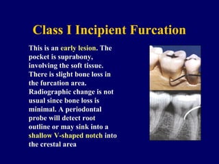
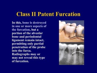
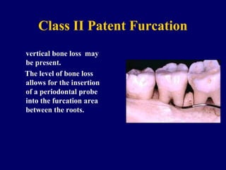
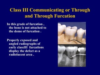
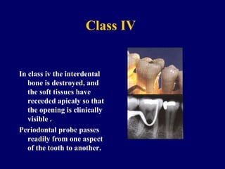
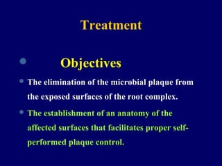
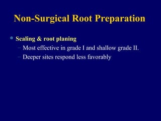
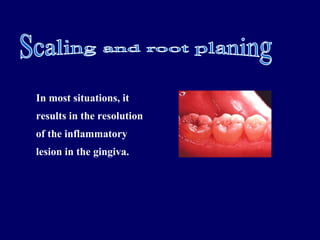
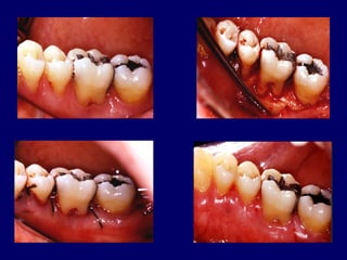
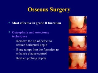
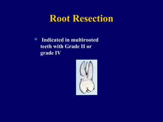
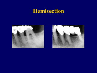
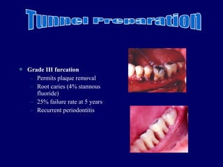
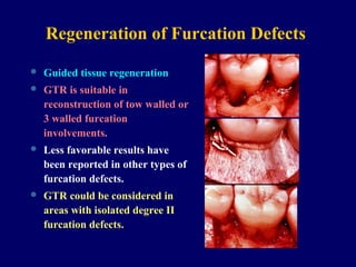
Predented by
Ummarah yaqoob
Furcation and Its surgical
treatment
Definition
 It can be defined as: an area of complex
anatomic morphology that may be difficult or
impossible to be debrided by routine
periodontal instrumentation.
Furcation involvement
 Bone loss usually caused as a result of
periodontal disease affecting the base of
root trunk of a tooth where 2 or more roots
meet.
Glickman`s Classification(1953)
Classification
Class I Incipient Furcation
This is an early lesion. The
pocket is suprabony,
involving the soft tissue.
There is slight bone loss in
the furcation area.
Radiographic change is not
usual since bone loss is
minimal. A periodontal
probe will detect root
outline or may sink into a
shallow V-shaped notch into
the crestal area
Class II Patent Furcation
In this, bone is destroyed
in one or more aspects of
the furcation, but a
portion of the alveolar
bone and periodontal
ligament remain intact,
permitting only partial
penetration of the probe
into the furca.
Radiographs may or
may not reveal this type
of furcation.
Class II Patent Furcation
vertical bone loss may
be present.
The level of bone loss
allows for the insertion
of a periodontal probe
into the furcation area
between the roots.
Class III Communicating or Through
and Through Furcation
In this grade of furcation ,
the bone is not attached to
the dome of furcation .
Properly exposed and
angled radiographs of
early classIII furcations
display the defect as a
radiolucent area .
Class IV
In class iv the interdental
bone is destroyed, and
the soft tissues have
receeded apicaly so that
the opening is clinically
visible .
Periodontal probe passes
readily from one aspect
of the tooth to another.
Treatment
 Objectives
 The elimination of the microbial plaque from
the exposed surfaces of the root complex.
 The establishment of an anatomy of the
affected surfaces that facilitates proper self-
performed plaque control.
Non-Surgical Root Preparation
 Scaling & root planing
– Most effective in grade I and shallow grade II.
– Deeper sites respond less favorably
In most situations, it
results in the resolution
of the inflammatory
lesion in the gingiva.
Antimicrobials
 Adjunct to scaling and root planning
– Chlorhexidine
– Tetracycline fibers
 No clinically significant difference in clinical
parameters after irrigation
Open Debridement
 Greater calculus removal than closed
 Ultrasonic
– Narrow furcations
– Dome of furcation
 Surgical access and increased operator
experience significantly enhance calculus
removal in molar furcation.
Surgical treatment
 Osseous resection
 Root resection.
 Hemisection.
 Tunnel prepration.
 Regeneration of furcation defects.
 Extraction.
 Dental implants.
Osseous Surgery
 Most effective in grade II furcation
 Osteoplasty and ostectomy
techniques
– Remove the lip of defect to
reduce horizontal depth
– Bone ramps into the furcation to
enhance plaque control
– Reduce probing depths
Root Resection
 Indicated in multirooted
teeth with Grade II or
grade IV
 May be performed on vital teeth or
endodontically treated teeth.
 It is preferable to perform endodontic therapy
before resectioon.
 Teeth not suitable for resection are….
 Teeth serving as abutment.
 Molars with advanced bone loss in inter
radicular and interproximal areas.


 WHICH ROOT TO REMOVE .
 Remove the root that will elimnate the
furcation .
 Remove the root with greatest amount of
bone loss and attachment loss.
 Remove the root that best contributes to
the elimnation of periodontal problems of
adjacent teeth.
 Remove the root with greatest number of
anatomic problems .
Hemisection
 Hemisection is the spiliting of a 2 rooted tooth
into 2 separate portions .
 Also called as bicuspidisation or seperation.
 Most likely to be performed on mandibular
molars with buccal and lingual classII or
classIII furcation involvement.
 After sectioning of the teeth one or both
roots can be retained .
 Narrow interradicular zones can
complicate the surgical procedure .
Hemisection
 Grade III furcation
– Permits plaque removal
– Root caries (4% stannous
fluoride)
– 25% failure rate at 5 years
– Recurrent periodontitis
Regeneration of Furcation Defects
 Guided tissue regeneration
 GTR is suitable in
reconstruction of tow walled or
3 walled furcation
involvements.
 Less favorable results have
been reported in other types of
furcation defects.
 GTR could be considered in
areas with isolated degree II
furcation defects.
Osseous Grafting
 Autogenous bone
 Allografts
– Freeze dried bone
– Demineralized Freeze dried bone
 Alloplasts
– Hydroxyapatite
 Non-porous
 Porous
– Bioglass
Extraction
 Extraction is preffered when attachment loss is
so extensive that no root can be maintained.
 Teeth with through and through furcation
defects are extracted.
 If tooth/gingival anatomy will not allow proper
plaque control
 For endodontic or restorative reason
 Preffered in patients with socio economic
drawbacks who cant perform adequate
treatments.
Dental implants
 The advent of osseointegrated dental
implants as an alternative abutment
source has a major impact on retention of
teeth with advanced furcation problems .
 The high level of predictibility of
osseointegration may motivate the
therapists and patient to consider removal
of teeth with a poor prognosis and to seek
an implant supported prosthetic
treatment plan.
Thank you

Ummairah

  • 1.
    Predented by Ummarah yaqoob Furcationand Its surgical treatment
  • 2.
    Definition  It canbe defined as: an area of complex anatomic morphology that may be difficult or impossible to be debrided by routine periodontal instrumentation.
  • 3.
    Furcation involvement  Boneloss usually caused as a result of periodontal disease affecting the base of root trunk of a tooth where 2 or more roots meet.
  • 4.
  • 5.
    Class I IncipientFurcation This is an early lesion. The pocket is suprabony, involving the soft tissue. There is slight bone loss in the furcation area. Radiographic change is not usual since bone loss is minimal. A periodontal probe will detect root outline or may sink into a shallow V-shaped notch into the crestal area
  • 6.
    Class II PatentFurcation In this, bone is destroyed in one or more aspects of the furcation, but a portion of the alveolar bone and periodontal ligament remain intact, permitting only partial penetration of the probe into the furca. Radiographs may or may not reveal this type of furcation.
  • 7.
    Class II PatentFurcation vertical bone loss may be present. The level of bone loss allows for the insertion of a periodontal probe into the furcation area between the roots.
  • 8.
    Class III Communicatingor Through and Through Furcation In this grade of furcation , the bone is not attached to the dome of furcation . Properly exposed and angled radiographs of early classIII furcations display the defect as a radiolucent area .
  • 9.
    Class IV In classiv the interdental bone is destroyed, and the soft tissues have receeded apicaly so that the opening is clinically visible . Periodontal probe passes readily from one aspect of the tooth to another.
  • 10.
    Treatment  Objectives  Theelimination of the microbial plaque from the exposed surfaces of the root complex.  The establishment of an anatomy of the affected surfaces that facilitates proper self- performed plaque control.
  • 11.
    Non-Surgical Root Preparation Scaling & root planing – Most effective in grade I and shallow grade II. – Deeper sites respond less favorably
  • 12.
    In most situations,it results in the resolution of the inflammatory lesion in the gingiva.
  • 13.
    Antimicrobials  Adjunct toscaling and root planning – Chlorhexidine – Tetracycline fibers  No clinically significant difference in clinical parameters after irrigation
  • 14.
    Open Debridement  Greatercalculus removal than closed  Ultrasonic – Narrow furcations – Dome of furcation  Surgical access and increased operator experience significantly enhance calculus removal in molar furcation.
  • 16.
    Surgical treatment  Osseousresection  Root resection.  Hemisection.  Tunnel prepration.  Regeneration of furcation defects.  Extraction.  Dental implants.
  • 17.
    Osseous Surgery  Mosteffective in grade II furcation  Osteoplasty and ostectomy techniques – Remove the lip of defect to reduce horizontal depth – Bone ramps into the furcation to enhance plaque control – Reduce probing depths
  • 18.
    Root Resection  Indicatedin multirooted teeth with Grade II or grade IV
  • 19.
     May beperformed on vital teeth or endodontically treated teeth.  It is preferable to perform endodontic therapy before resectioon.  Teeth not suitable for resection are….  Teeth serving as abutment.  Molars with advanced bone loss in inter radicular and interproximal areas. 
  • 20.
      WHICH ROOTTO REMOVE .  Remove the root that will elimnate the furcation .  Remove the root with greatest amount of bone loss and attachment loss.  Remove the root that best contributes to the elimnation of periodontal problems of adjacent teeth.  Remove the root with greatest number of anatomic problems .
  • 21.
    Hemisection  Hemisection isthe spiliting of a 2 rooted tooth into 2 separate portions .  Also called as bicuspidisation or seperation.  Most likely to be performed on mandibular molars with buccal and lingual classII or classIII furcation involvement.
  • 22.
     After sectioningof the teeth one or both roots can be retained .  Narrow interradicular zones can complicate the surgical procedure .
  • 23.
  • 24.
     Grade IIIfurcation – Permits plaque removal – Root caries (4% stannous fluoride) – 25% failure rate at 5 years – Recurrent periodontitis
  • 25.
    Regeneration of FurcationDefects  Guided tissue regeneration  GTR is suitable in reconstruction of tow walled or 3 walled furcation involvements.  Less favorable results have been reported in other types of furcation defects.  GTR could be considered in areas with isolated degree II furcation defects.
  • 27.
    Osseous Grafting  Autogenousbone  Allografts – Freeze dried bone – Demineralized Freeze dried bone  Alloplasts – Hydroxyapatite  Non-porous  Porous – Bioglass
  • 28.
    Extraction  Extraction ispreffered when attachment loss is so extensive that no root can be maintained.  Teeth with through and through furcation defects are extracted.  If tooth/gingival anatomy will not allow proper plaque control  For endodontic or restorative reason
  • 29.
     Preffered inpatients with socio economic drawbacks who cant perform adequate treatments.
  • 30.
    Dental implants  Theadvent of osseointegrated dental implants as an alternative abutment source has a major impact on retention of teeth with advanced furcation problems .  The high level of predictibility of osseointegration may motivate the therapists and patient to consider removal of teeth with a poor prognosis and to seek an implant supported prosthetic treatment plan.
  • 31.