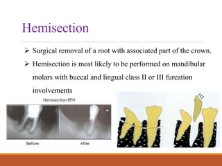
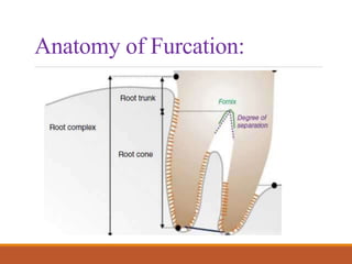
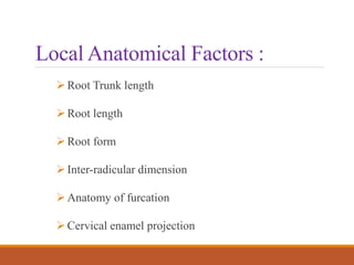
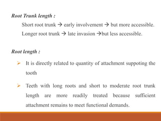
Furcation : Involvement
and Treatment
Furcation is an area or zone of division of roots in a multi
rooted tooth
Furcation involvement refers to the invasion of the
bifurcation and trifurcation of multirooted teeth by
periodontal disease.
Definition :
Anatomy of Furcation:
Etiological factors :
 Long term-bacterial plaque and inflammatory consequenses.
 Local anatomical factors (root trunk length, root morphology
proximity of the furcation to the cemento-enamel junction,
presence of accessory pulpal canals )
 Local developmental anomalies (cervical enamel projections
and enamel pearls),
 Trauma from occlusion
Local Anatomical Factors :
Root Trunk length
Root length
Root form
Inter-radicular dimension
Anatomy of furcation
Cervical enamel projection
Root Trunk length :
Short root trunk  early involvement  but more accessible.
Longer root trunk  late invasion but less accessible.
Root length :
 It is directly related to quantity of attachment suppoting the
tooth
 Teeth with long roots and short to moderate root trunk
length are more readily treated because sufficient
attachment remains to meet functional demands.
Root form :
 The mesial root of most mandibular first and second molars
and the mesiofacial root of the maxillary first molar are
typically curved to the distal side in the apical third and the
distal aspect of this root is usually heavily fluted.
Interradicular dimension :
 Teeth with widely separated roots present more treatment
options and are readily treated
Anatomy of furcation :
 Presence of bifurcation ridges, concavity domes, accessory
canals complicate treatment.
Cervical enamel projections :
 Should be removed because they facilitate plaque
accumulation and complicate treatment.
Classification
GLICKMAN’S CLASSIFICATION (1953)
Grade I-
 Incipient or early lesion.
 Pocket is suprabony with slight bone
loss in loss in furcation area.
 No radiographic changes
Grade II-
 Lesion is cul-de-sac.
 Bone is destroyed in one or more aspects but portion of
PDL and portion of alvolar bone are intact permitting only
partial probe penetration.
 Radiographic changes may or may not be present.
Grade III-
 Interradicular bone is lost completely but occluded by gingival
tissues hence not seen clinically.
 Though and through penetration of probe.
 Radiologically a radiolucency is seen between roots.
Grade IV-
 Complete interadicular bone loss with apical recession of
tissues so clinically visible.
 Radiological changes same as grade III.
TARNOW & FLETCHER (1984)
(Sub-classification based on the degree of vertical involvement)
Subclass A. 1–3 mm
Subclass B. 4–6 mm
Subclass C. >7 mm
Diagnosis :
Probing with the specially designed probes (Nabers
probe) or No.23 explorer.
Transgingival sounding.
Radiographic Appearance
Three diagnostic criteria are
suggested:
1. The slightest radiographic
change in the furcation area
should be investigated
clinically, especially if there is
bone loss on adjacent roots.
2. Diminished radiodensity in the
furcation area in which
outlines of bony trabeculae are
visible suggests furcation
involvement.
3) Whenever there is marked
bone loss in relation to a
single molar root, it may be
assumed that the furcation is
also involved
Management
GRADE I-
 Intial preparation for scaling and root planing.
 Curettage or gigivectomy to expose furcation area.
 Odontoplasty – to reshape or eliminate local factors
which cause plaque accumulation.
GRADE II-
Early (non invasive):
 Scaling, rootplaning, curettage.
 Furcationplasty osteoplasty+odontoplasty
Advanced :
 Tunneling
 Autogenous or allogenous bone grafts can be given
along with GTR
Grade III :
Early - Periodontal regeneration
Advanced - Resective procedures.
Grade IV :
Treated mostly by resective procedures.
Extraction - with advanced attachment loss
Resective procedures :
1) Root resection
2) Hemisection
3) Bicuspidization
Root Resection:
The removal of a root without the removal of any portion of
the crown
Which tooth to remove ?
1) Remove the roots that will eliminate the furcation and allow
the production of a maintainable architecture on the remaining
roots.
2) Remove the root with the greatest amount of bone and
attachment loss.
3) Remove the root that best contributes to the elimination of
periodontal problems on adjacent teeth.
4) Remove the root with the greatest number of anatomic
problems, such as severe curvature, developmental grooves,
root flutings, or accessory and multiple root canals.
5) Remove the root that least complicates future periodontal
maintenance.
Hemisection
 Surgical removal of a root with associated part of the crown.
 Hemisection is most likely to be performed on mandibular
molars with buccal and lingual class II or III furcation
involvements
Bicuspidization
 Sectioning of root complex and
maintainence of all roots.
 Decision is based on the extent and pattern
of bony loss, root trunk and root length,
ability to eliminate the osseous defect, and
endodontic and restorative considerations.
Thank you

Furcation involvement

  • 1.
  • 2.
    Furcation is anarea or zone of division of roots in a multi rooted tooth Furcation involvement refers to the invasion of the bifurcation and trifurcation of multirooted teeth by periodontal disease. Definition :
  • 3.
  • 4.
    Etiological factors : Long term-bacterial plaque and inflammatory consequenses.  Local anatomical factors (root trunk length, root morphology proximity of the furcation to the cemento-enamel junction, presence of accessory pulpal canals )  Local developmental anomalies (cervical enamel projections and enamel pearls),  Trauma from occlusion
  • 5.
    Local Anatomical Factors: Root Trunk length Root length Root form Inter-radicular dimension Anatomy of furcation Cervical enamel projection
  • 6.
    Root Trunk length: Short root trunk  early involvement  but more accessible. Longer root trunk  late invasion but less accessible. Root length :  It is directly related to quantity of attachment suppoting the tooth  Teeth with long roots and short to moderate root trunk length are more readily treated because sufficient attachment remains to meet functional demands.
  • 7.
    Root form : The mesial root of most mandibular first and second molars and the mesiofacial root of the maxillary first molar are typically curved to the distal side in the apical third and the distal aspect of this root is usually heavily fluted. Interradicular dimension :  Teeth with widely separated roots present more treatment options and are readily treated
  • 8.
    Anatomy of furcation:  Presence of bifurcation ridges, concavity domes, accessory canals complicate treatment. Cervical enamel projections :  Should be removed because they facilitate plaque accumulation and complicate treatment.
  • 9.
    Classification GLICKMAN’S CLASSIFICATION (1953) GradeI-  Incipient or early lesion.  Pocket is suprabony with slight bone loss in loss in furcation area.  No radiographic changes
  • 10.
    Grade II-  Lesionis cul-de-sac.  Bone is destroyed in one or more aspects but portion of PDL and portion of alvolar bone are intact permitting only partial probe penetration.  Radiographic changes may or may not be present.
  • 11.
    Grade III-  Interradicularbone is lost completely but occluded by gingival tissues hence not seen clinically.  Though and through penetration of probe.  Radiologically a radiolucency is seen between roots.
  • 12.
    Grade IV-  Completeinteradicular bone loss with apical recession of tissues so clinically visible.  Radiological changes same as grade III.
  • 13.
    TARNOW & FLETCHER(1984) (Sub-classification based on the degree of vertical involvement) Subclass A. 1–3 mm Subclass B. 4–6 mm Subclass C. >7 mm
  • 14.
    Diagnosis : Probing withthe specially designed probes (Nabers probe) or No.23 explorer. Transgingival sounding.
  • 15.
    Radiographic Appearance Three diagnosticcriteria are suggested: 1. The slightest radiographic change in the furcation area should be investigated clinically, especially if there is bone loss on adjacent roots.
  • 16.
    2. Diminished radiodensityin the furcation area in which outlines of bony trabeculae are visible suggests furcation involvement.
  • 17.
    3) Whenever thereis marked bone loss in relation to a single molar root, it may be assumed that the furcation is also involved
  • 18.
    Management GRADE I-  Intialpreparation for scaling and root planing.  Curettage or gigivectomy to expose furcation area.  Odontoplasty – to reshape or eliminate local factors which cause plaque accumulation.
  • 19.
    GRADE II- Early (noninvasive):  Scaling, rootplaning, curettage.  Furcationplasty osteoplasty+odontoplasty Advanced :  Tunneling  Autogenous or allogenous bone grafts can be given along with GTR
  • 20.
    Grade III : Early- Periodontal regeneration Advanced - Resective procedures. Grade IV : Treated mostly by resective procedures. Extraction - with advanced attachment loss
  • 21.
    Resective procedures : 1)Root resection 2) Hemisection 3) Bicuspidization
  • 22.
    Root Resection: The removalof a root without the removal of any portion of the crown
  • 23.
    Which tooth toremove ? 1) Remove the roots that will eliminate the furcation and allow the production of a maintainable architecture on the remaining roots. 2) Remove the root with the greatest amount of bone and attachment loss. 3) Remove the root that best contributes to the elimination of periodontal problems on adjacent teeth.
  • 24.
    4) Remove theroot with the greatest number of anatomic problems, such as severe curvature, developmental grooves, root flutings, or accessory and multiple root canals. 5) Remove the root that least complicates future periodontal maintenance.
  • 25.
    Hemisection  Surgical removalof a root with associated part of the crown.  Hemisection is most likely to be performed on mandibular molars with buccal and lingual class II or III furcation involvements
  • 26.
    Bicuspidization  Sectioning ofroot complex and maintainence of all roots.  Decision is based on the extent and pattern of bony loss, root trunk and root length, ability to eliminate the osseous defect, and endodontic and restorative considerations.
  • 27.