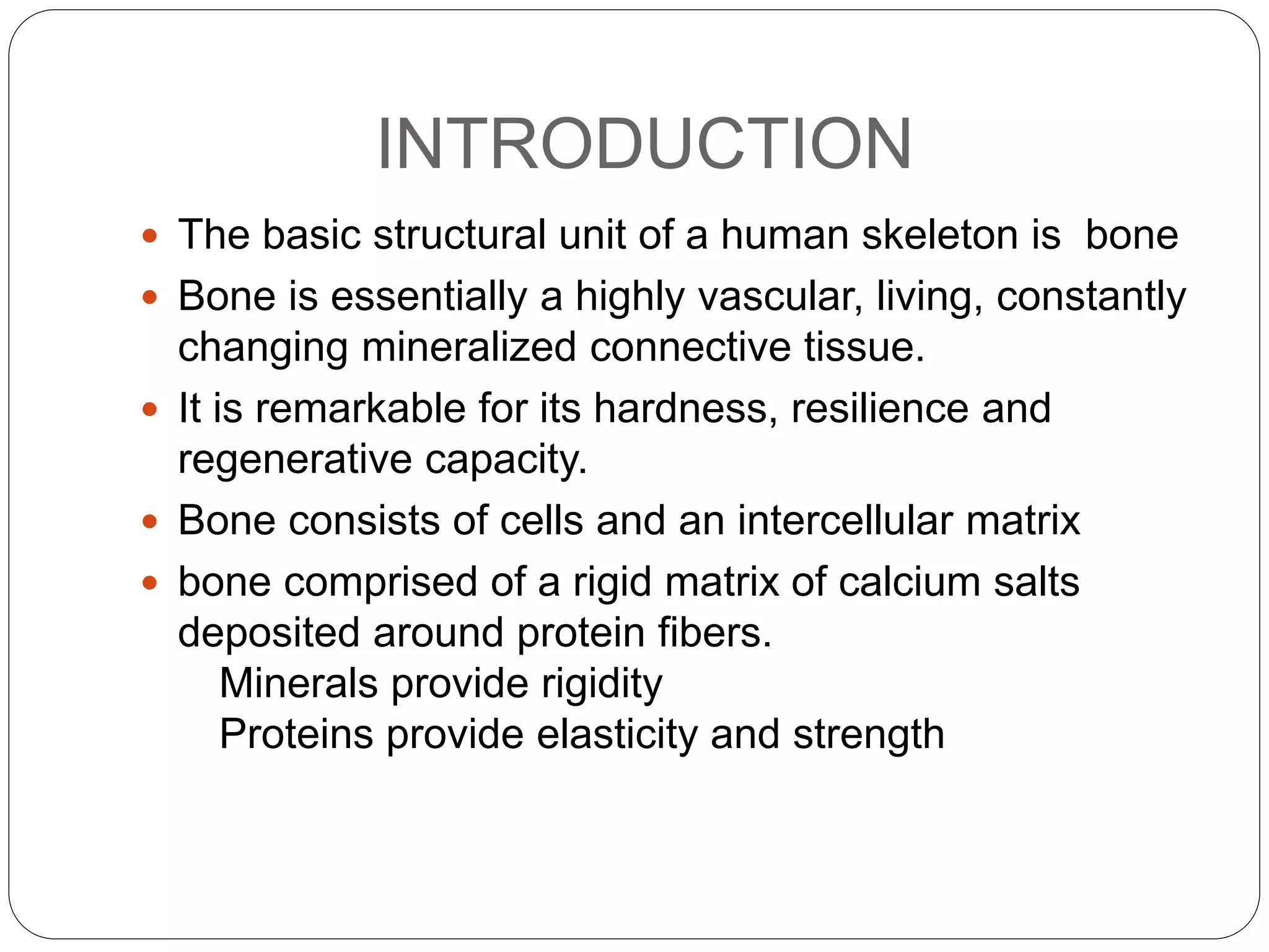
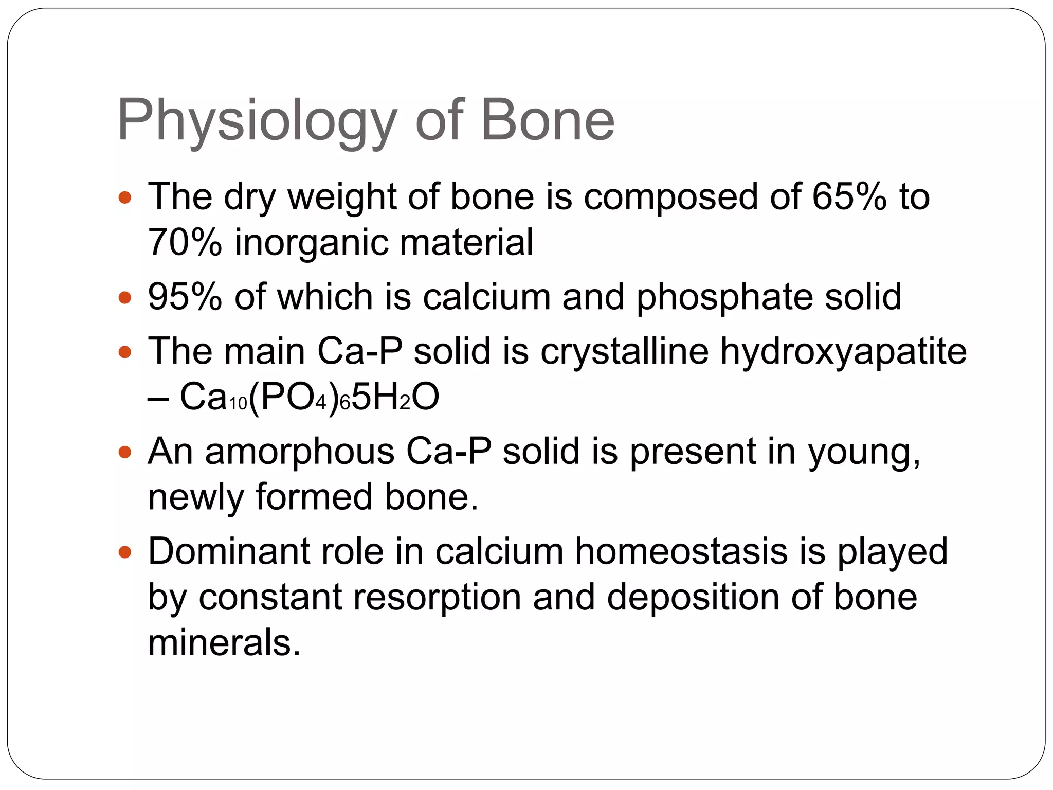
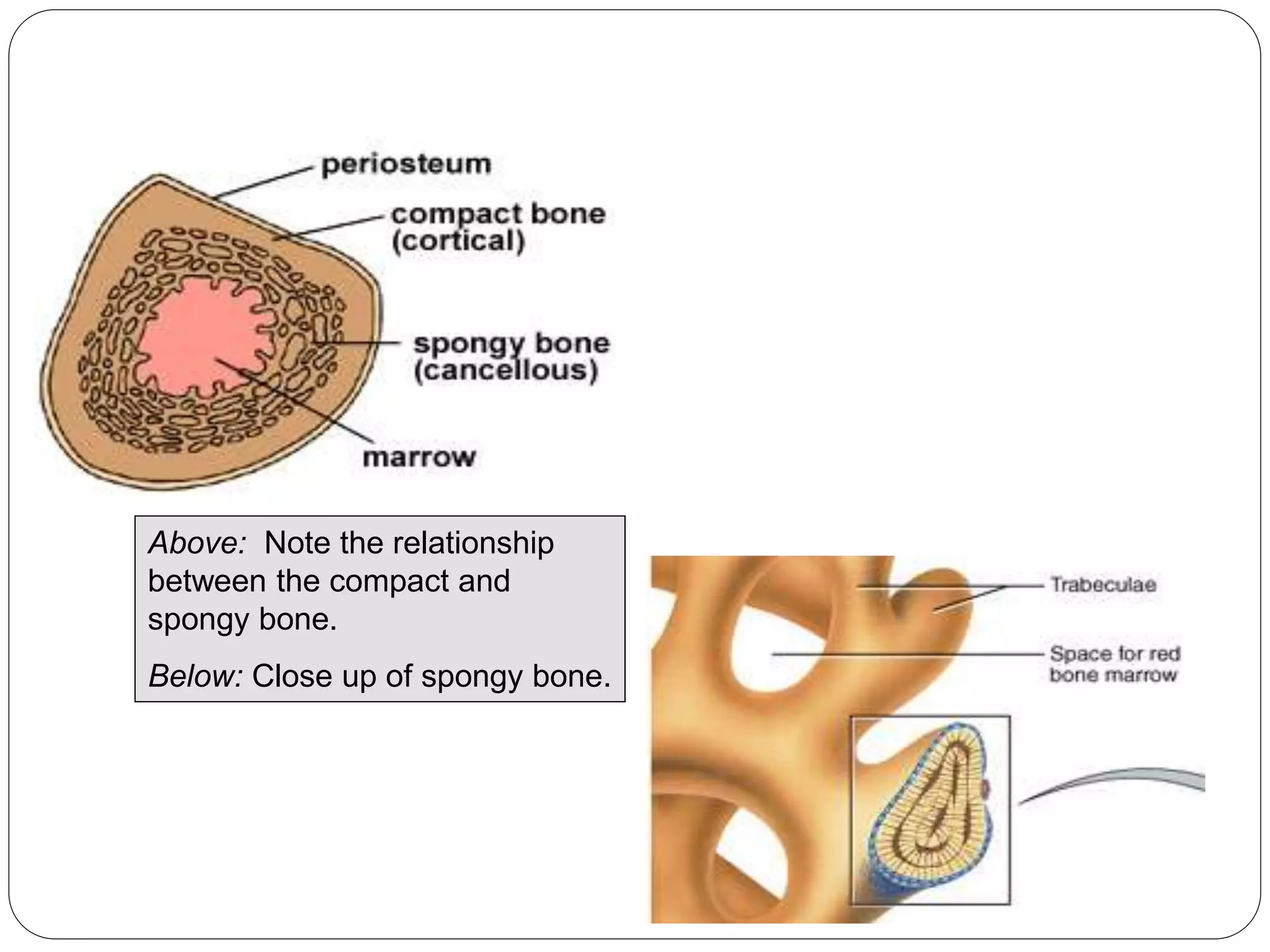
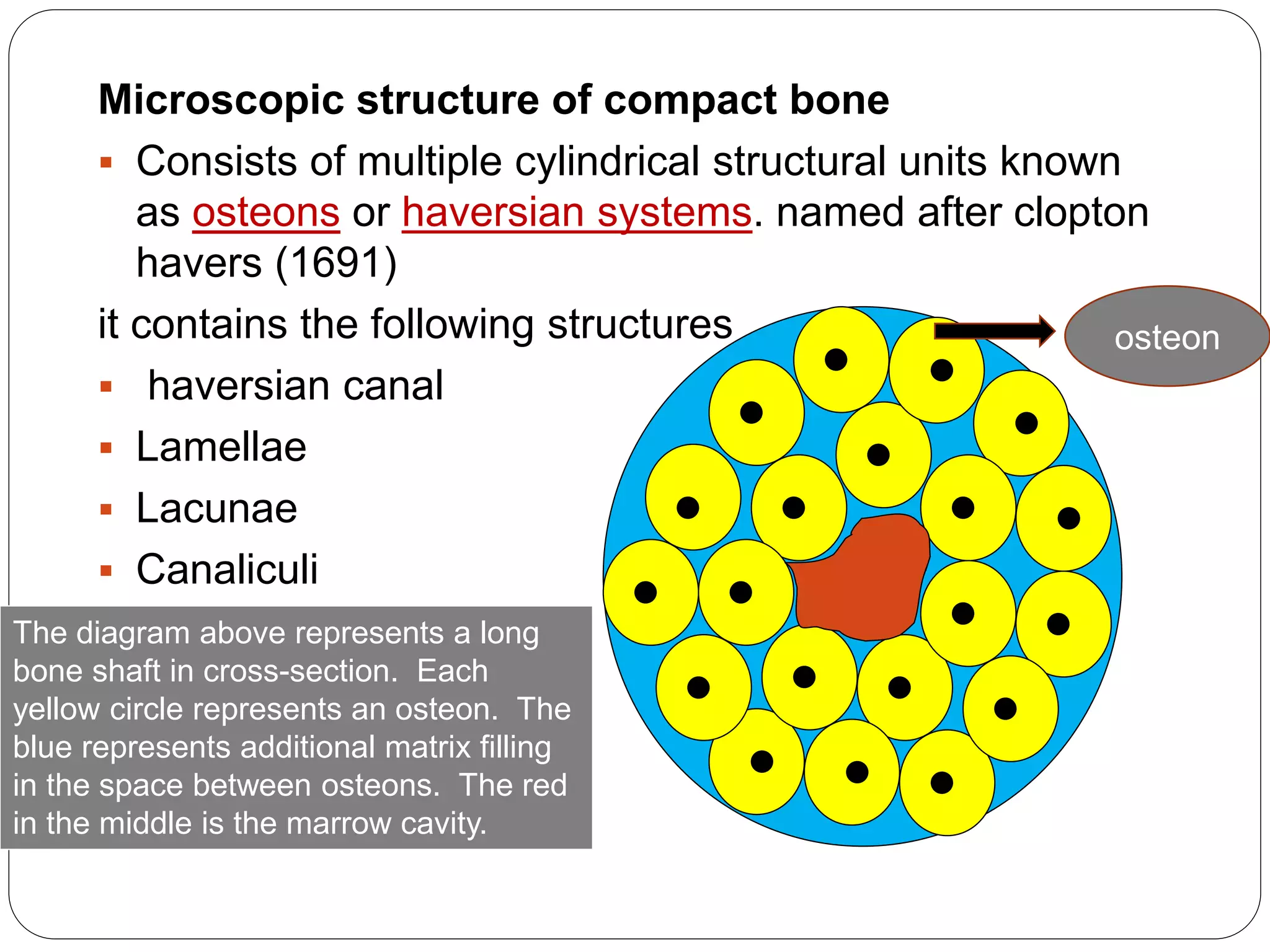
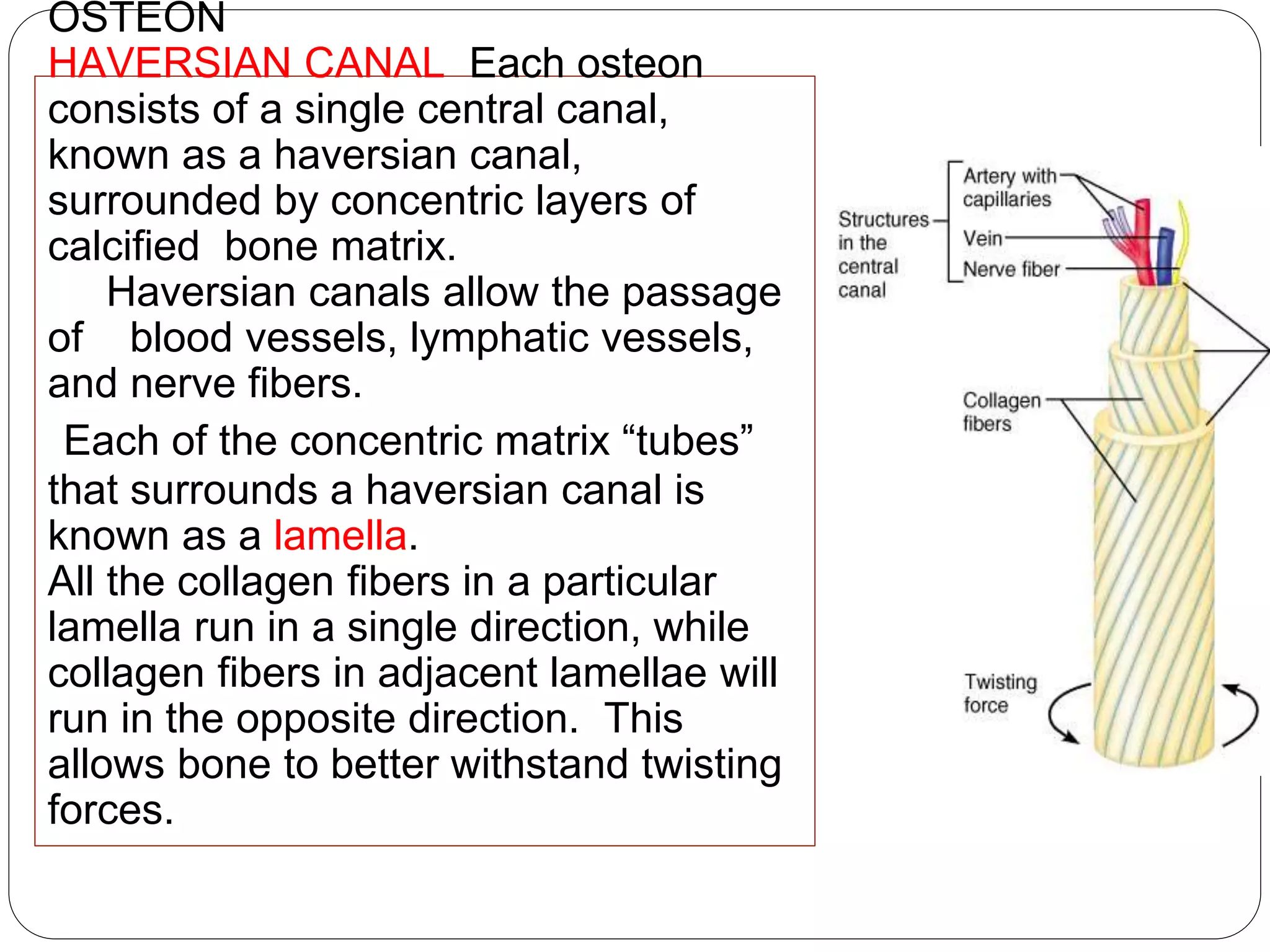
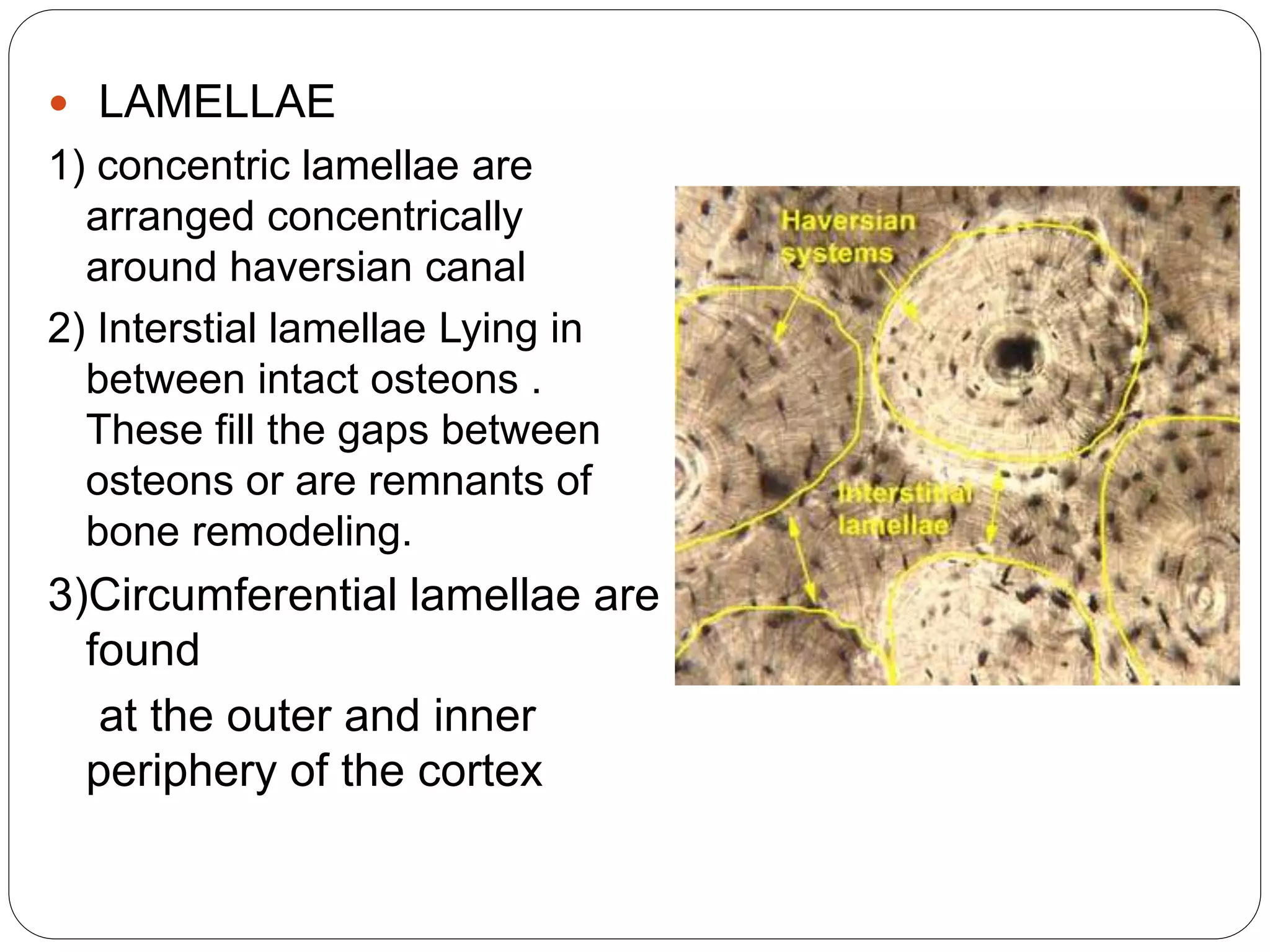
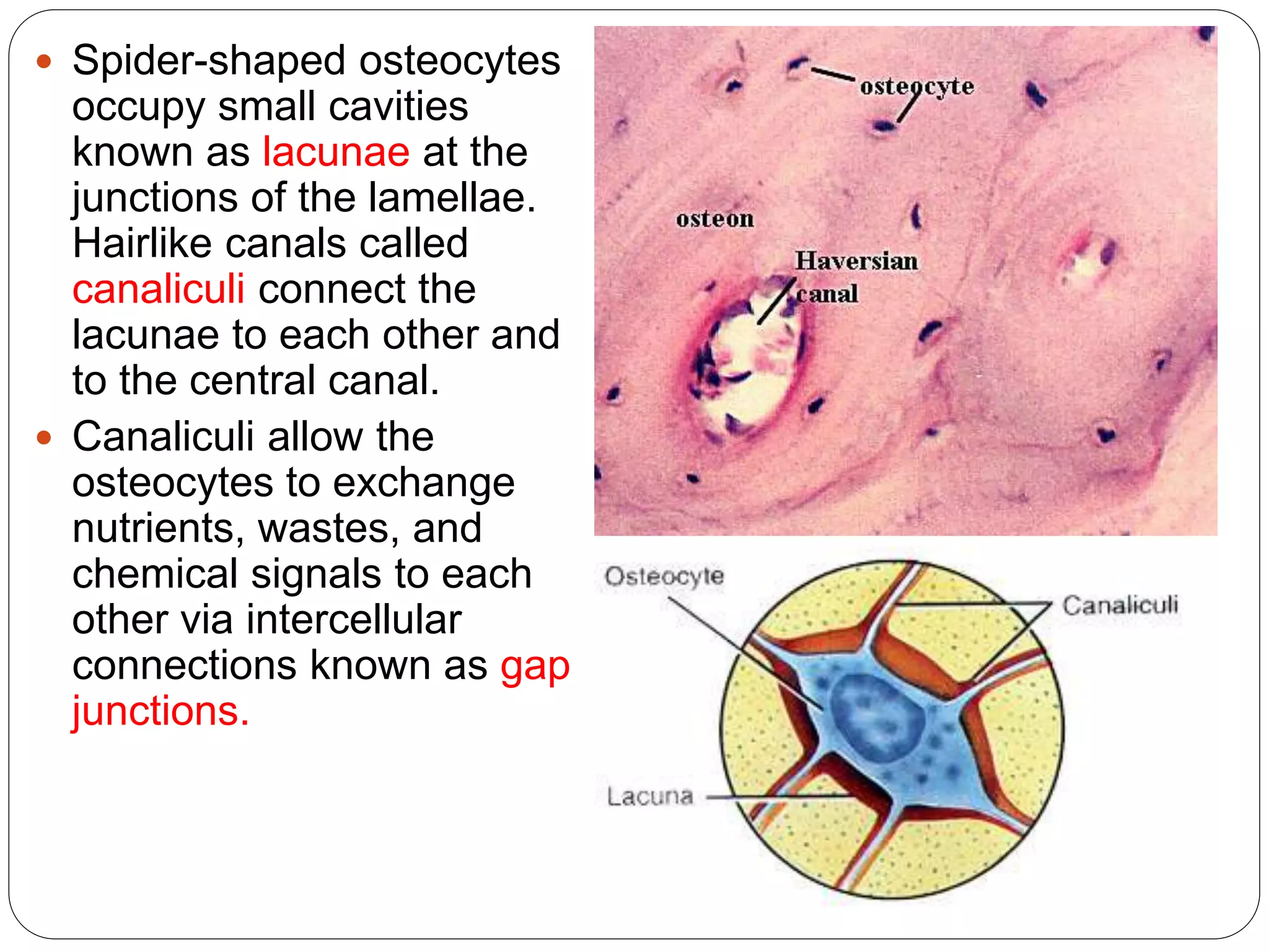
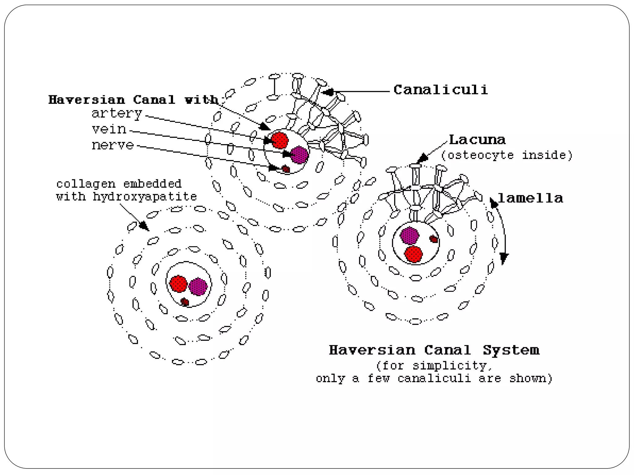
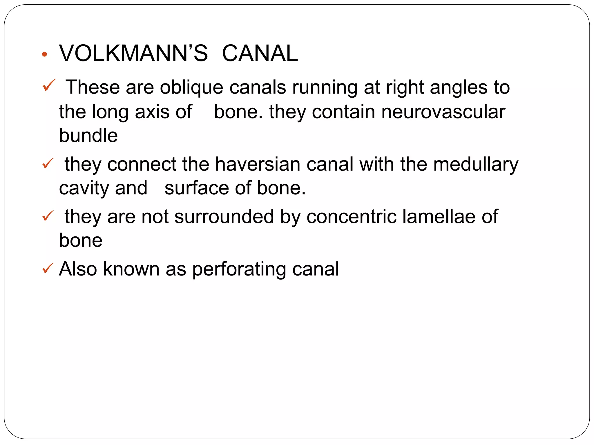
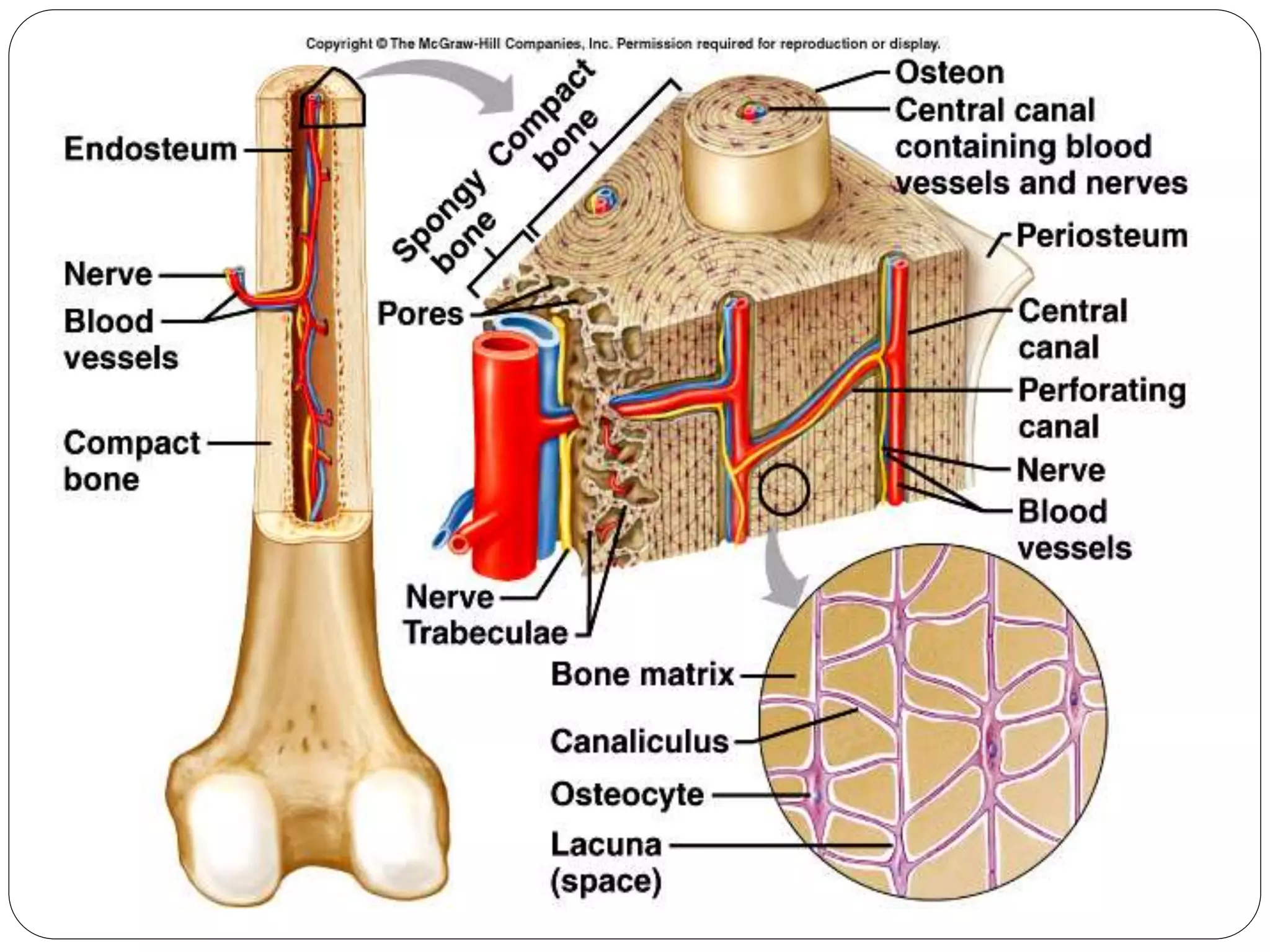
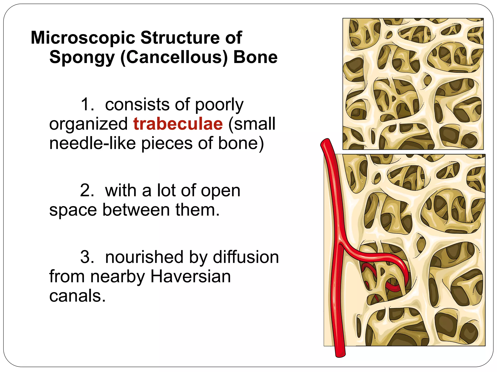
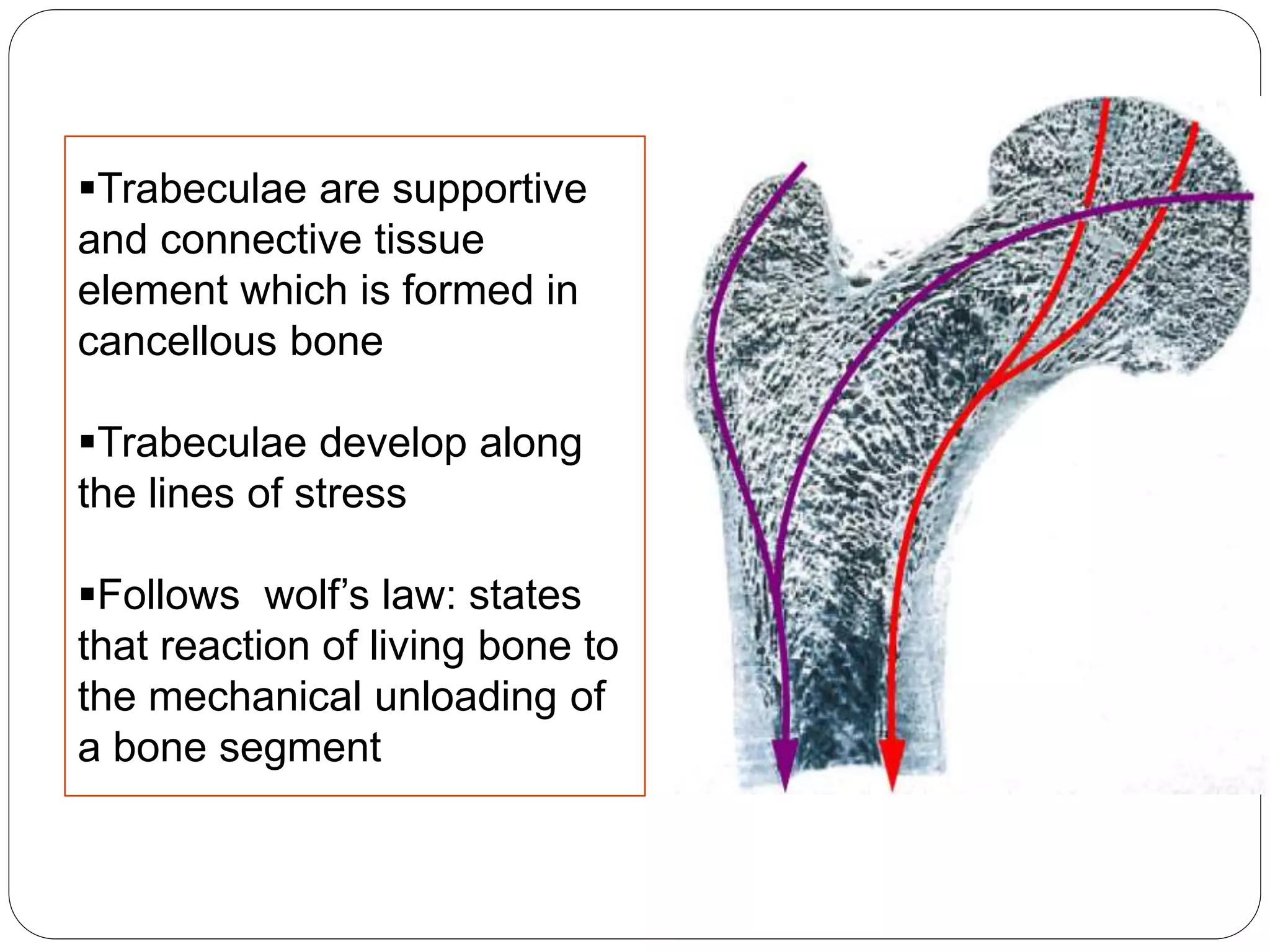
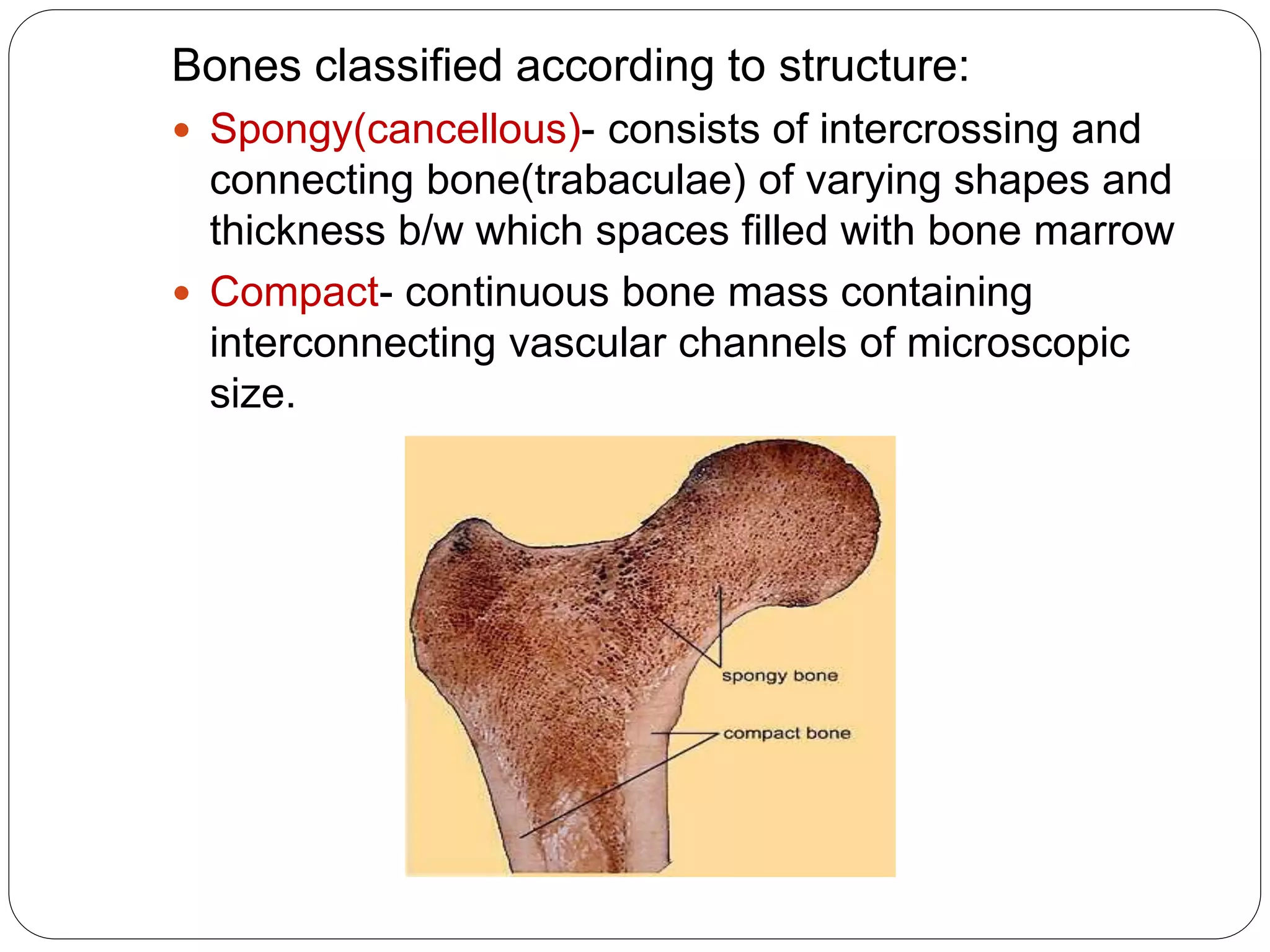
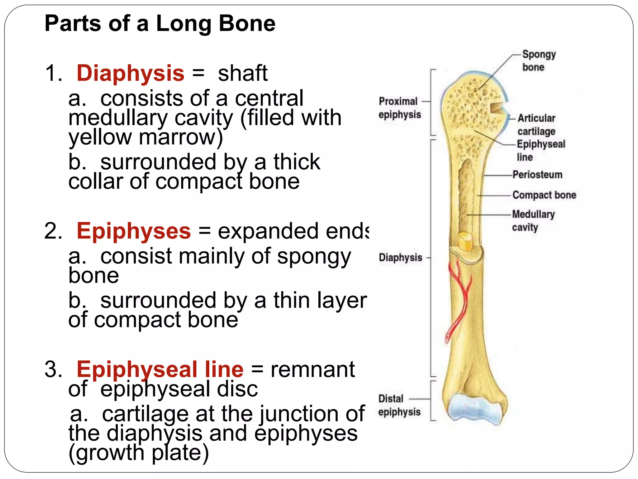
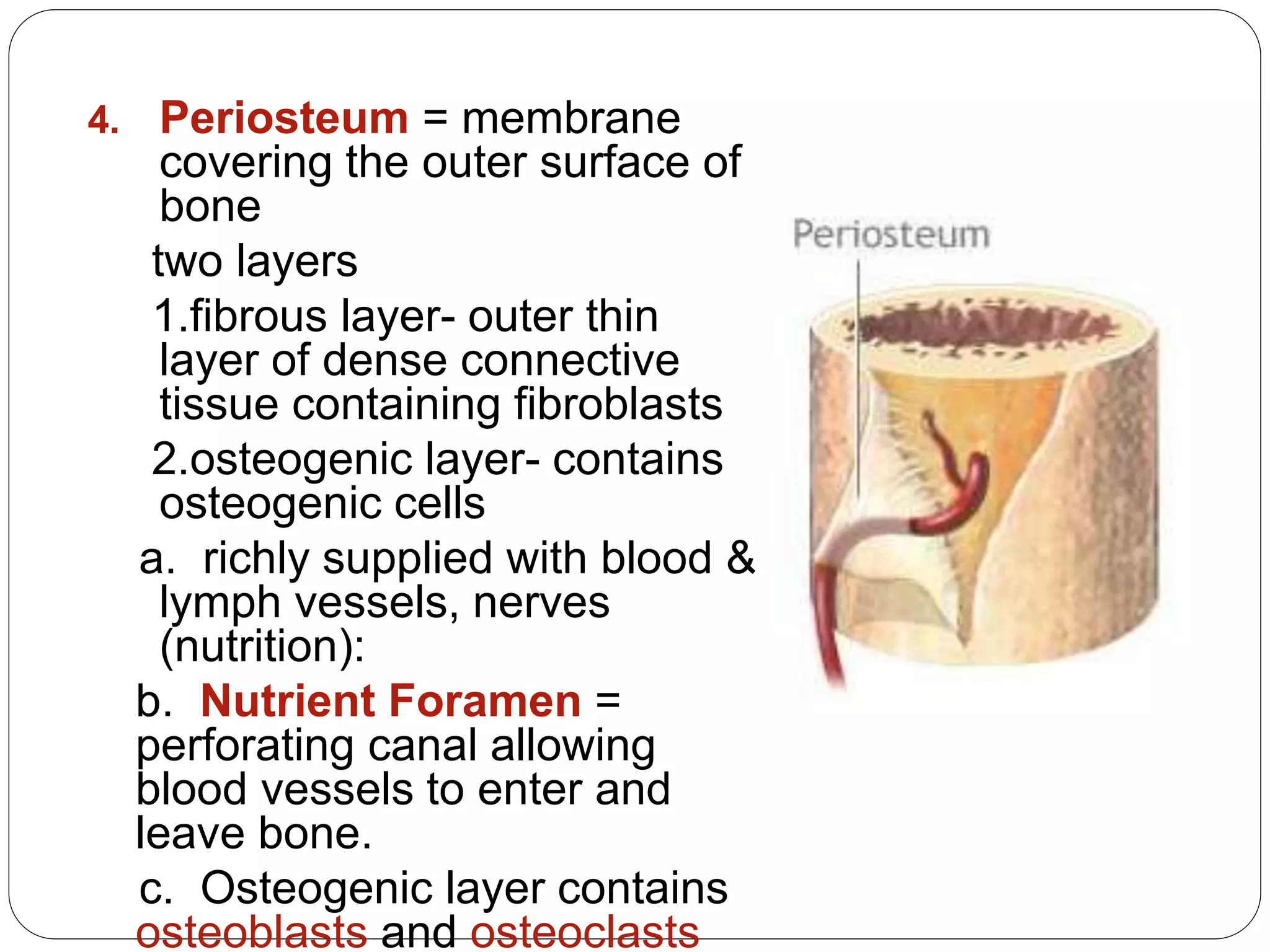
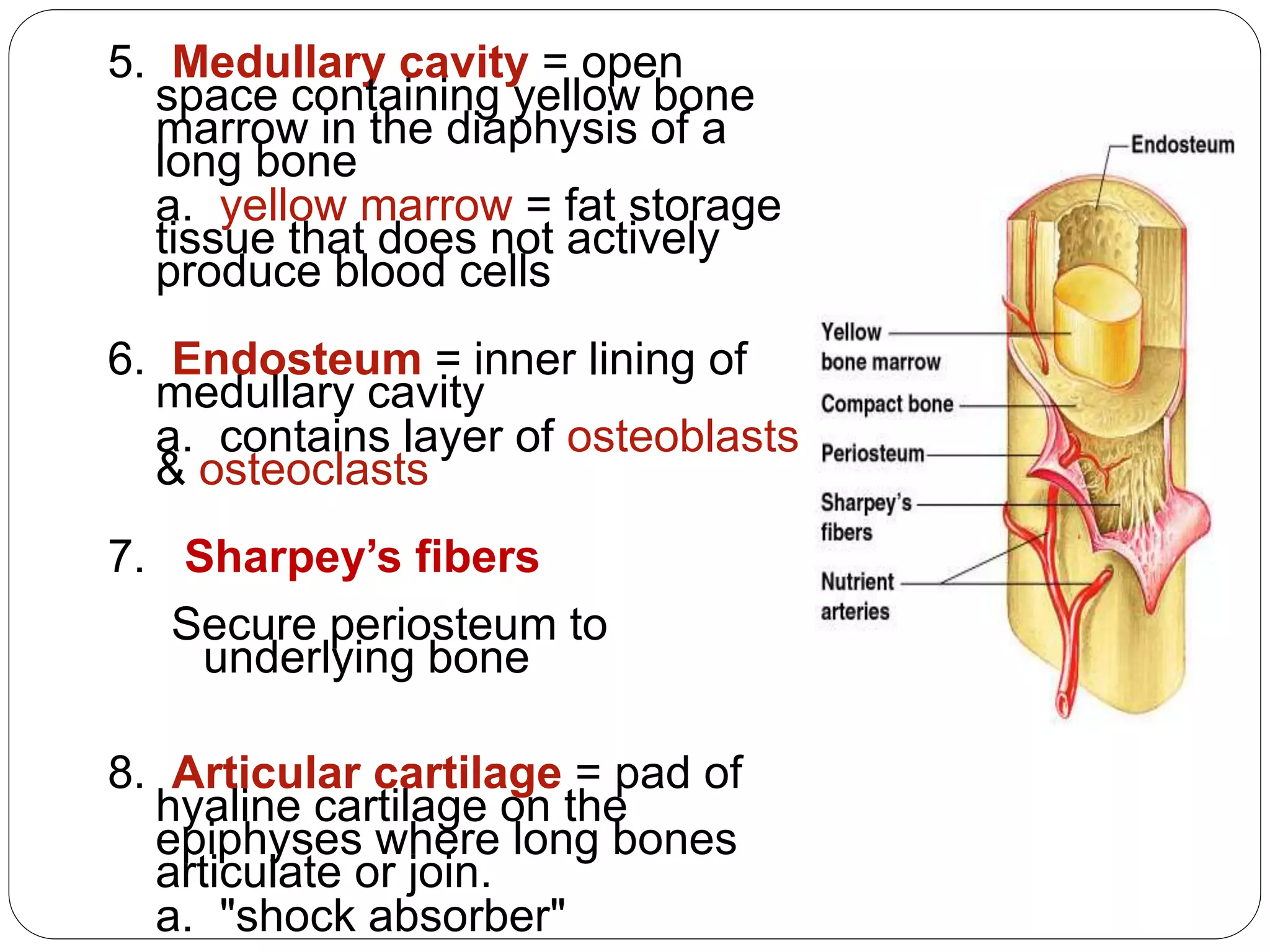
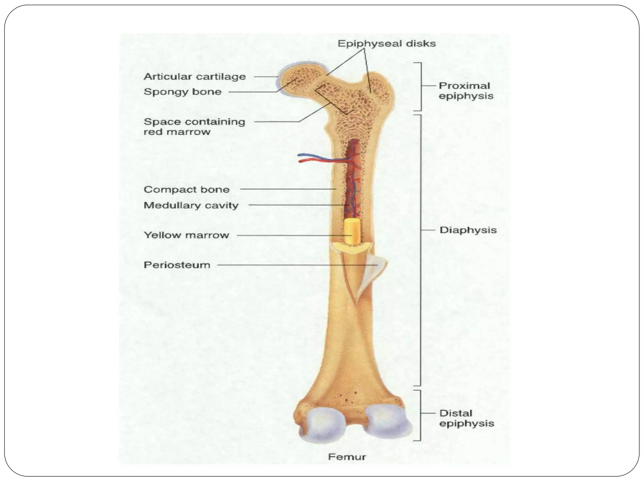
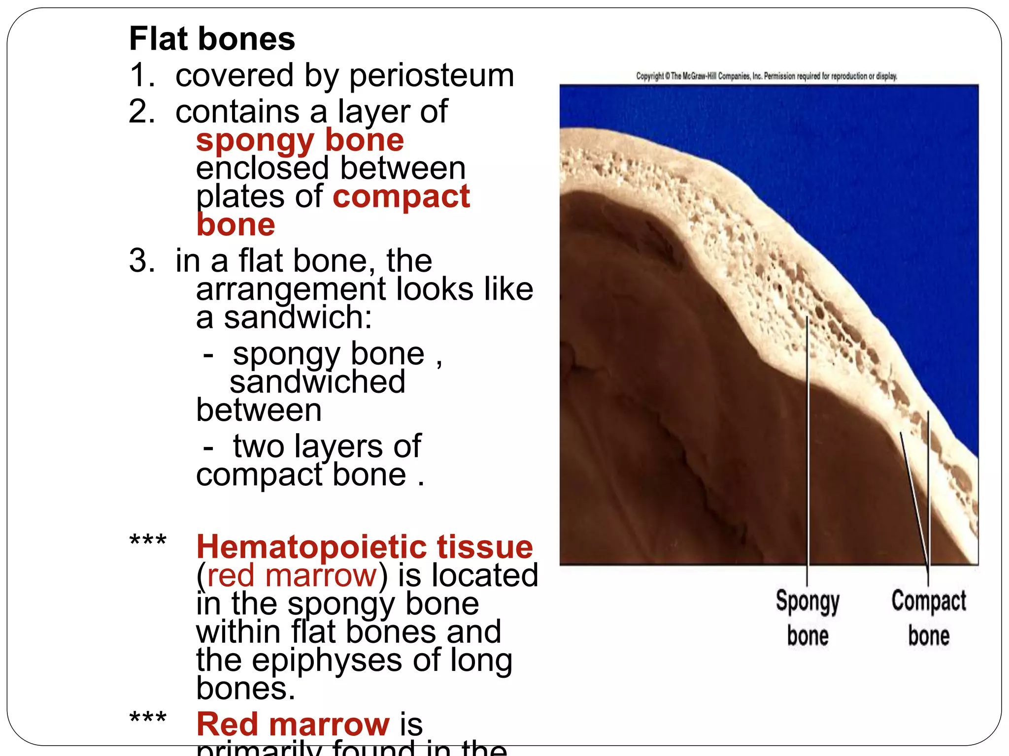
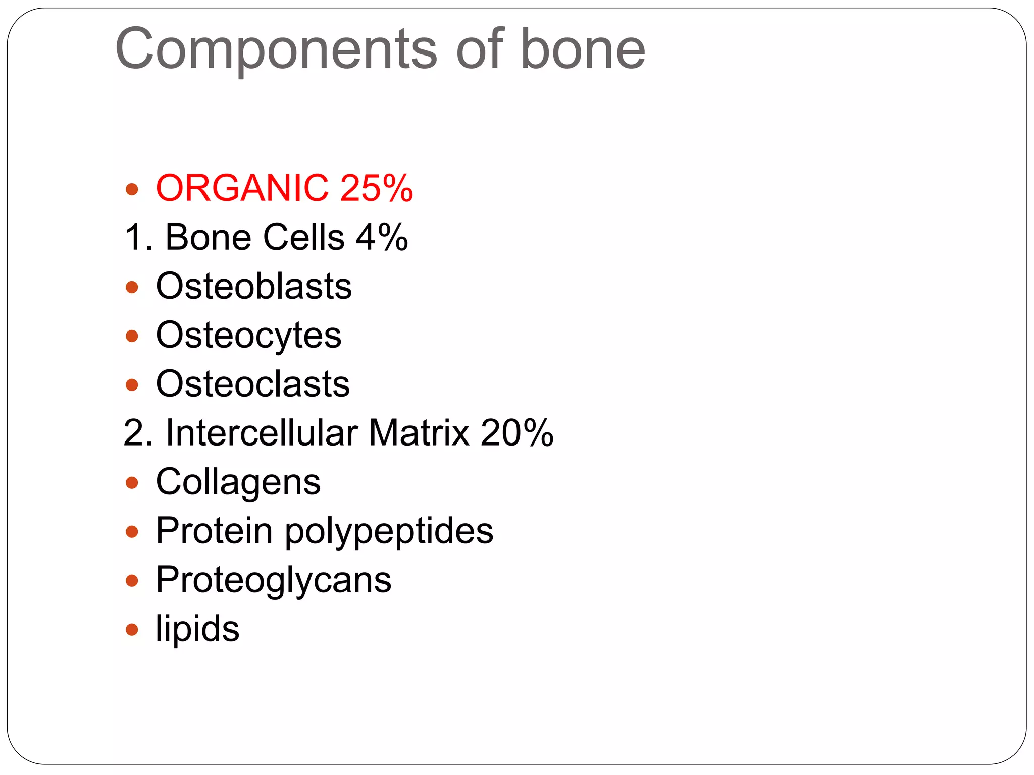
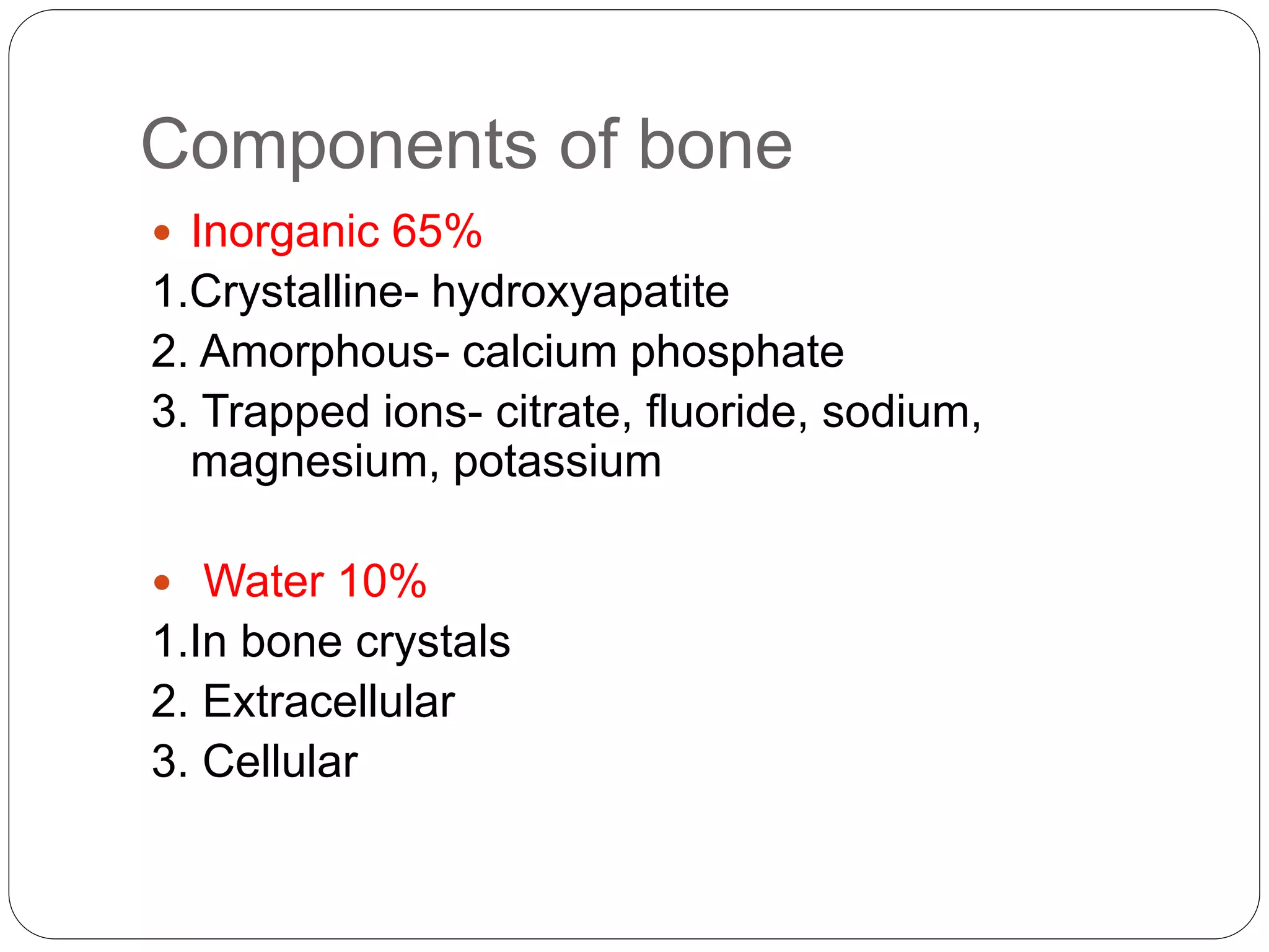
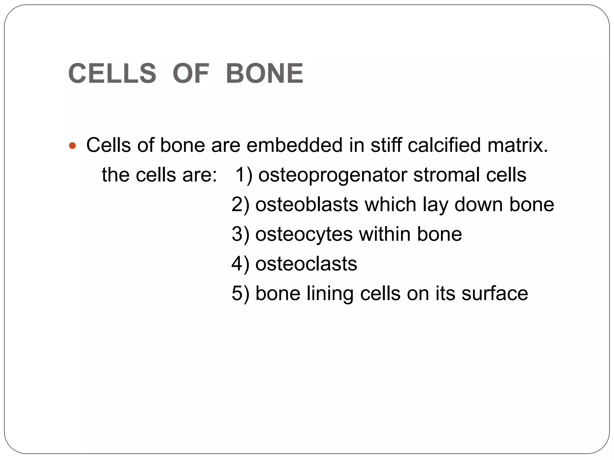
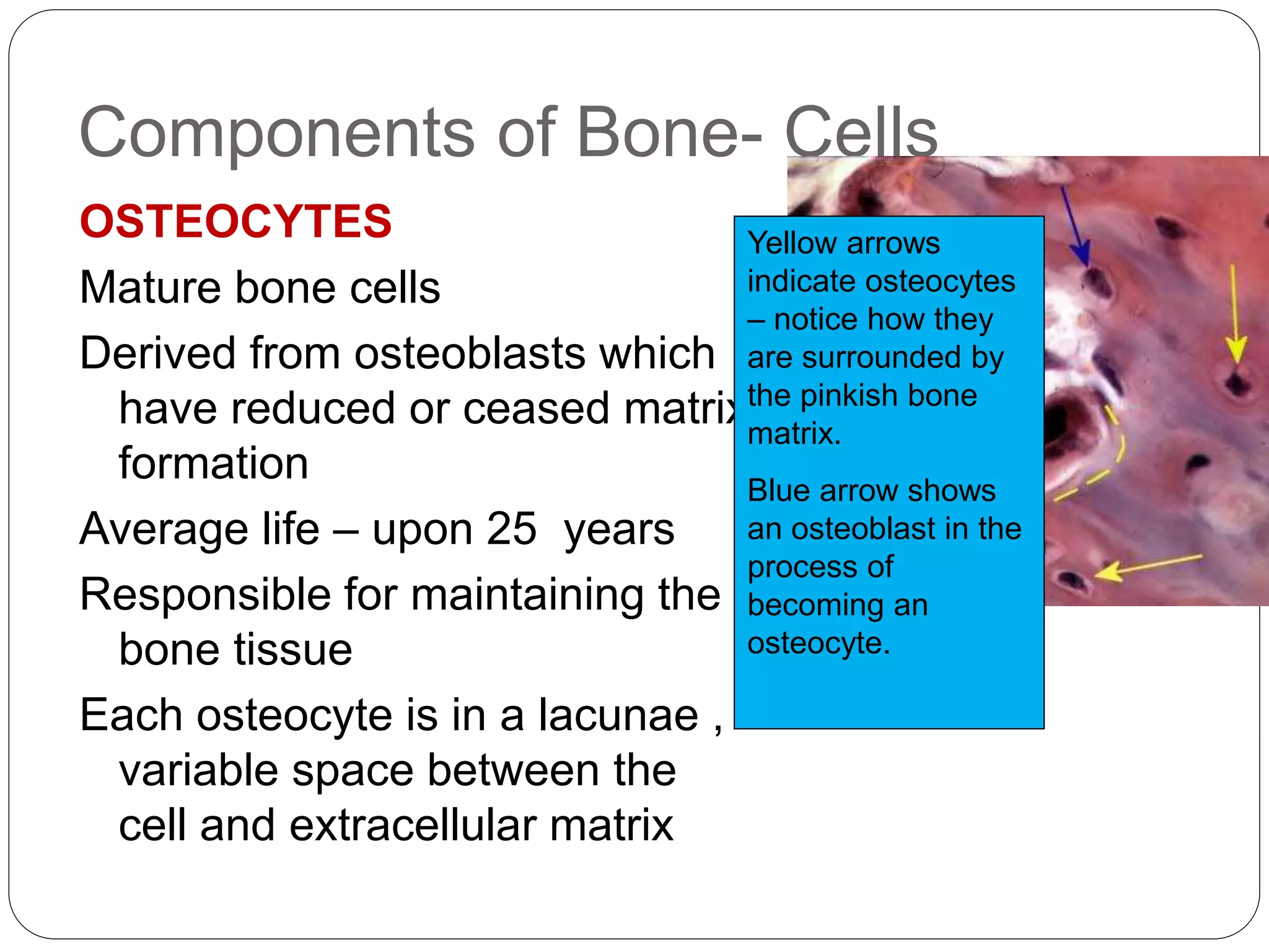
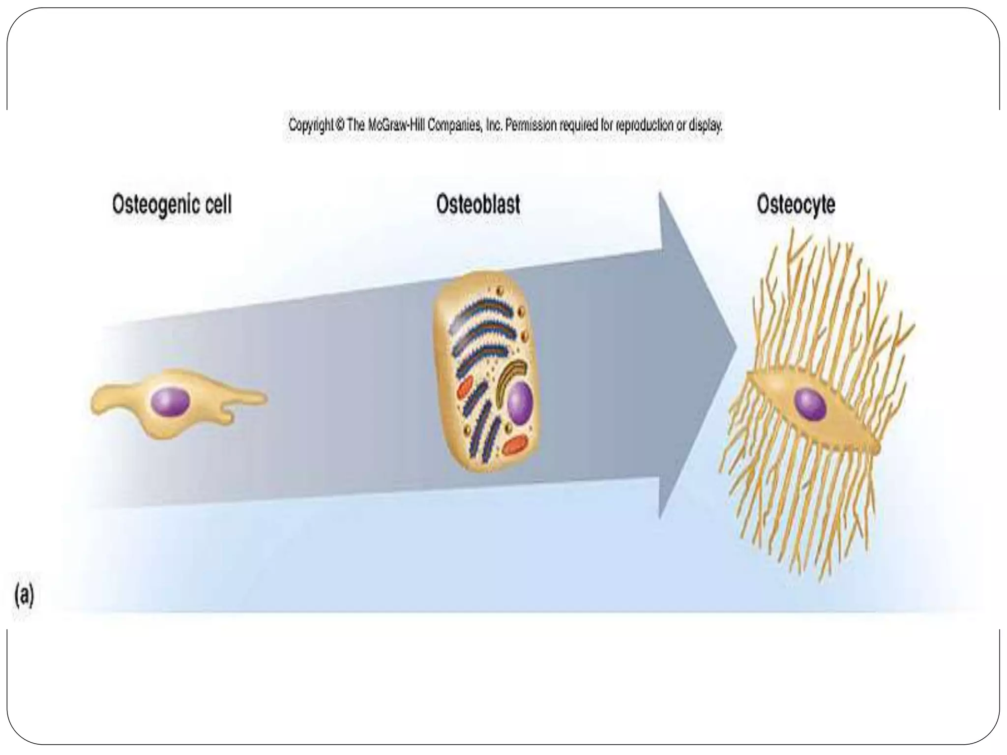
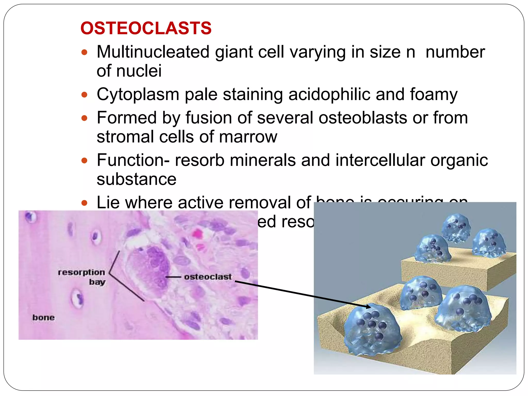
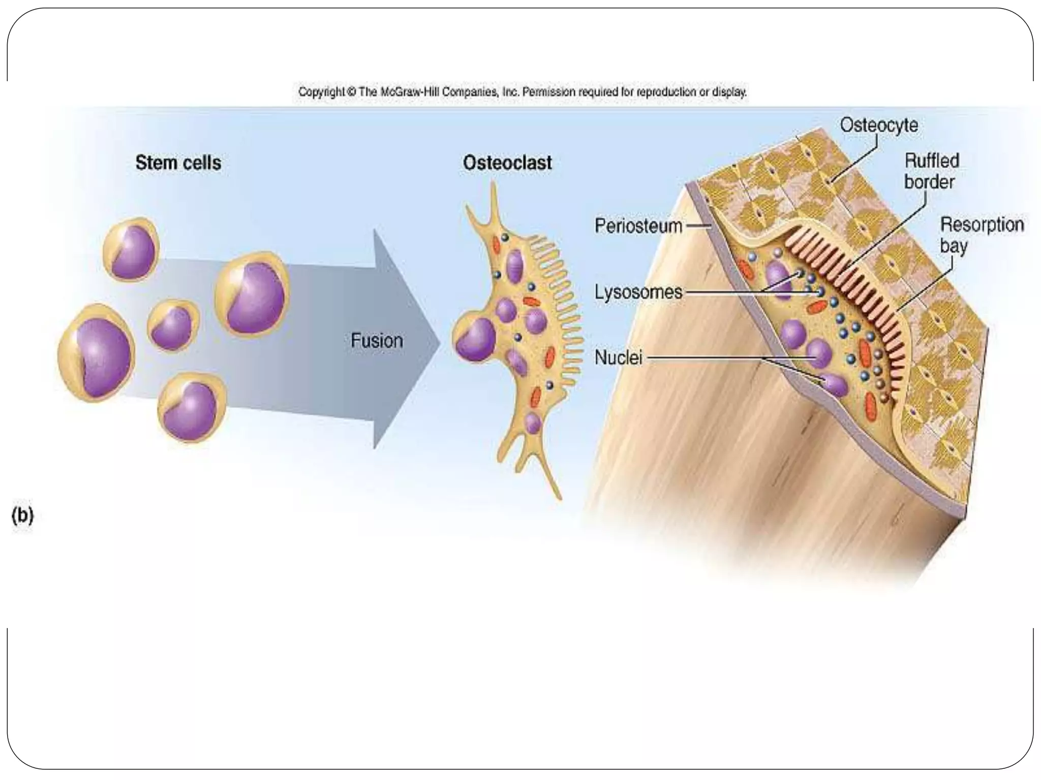
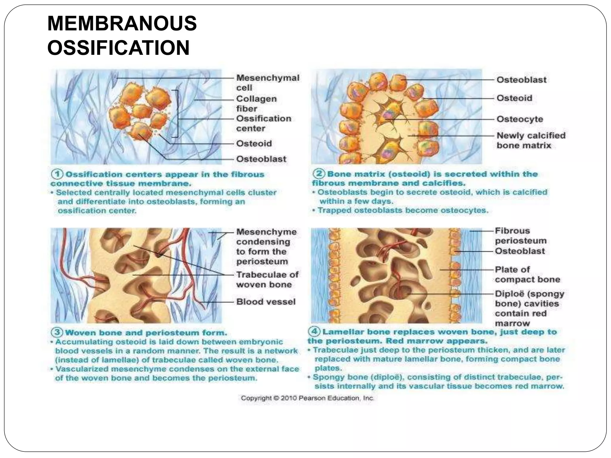
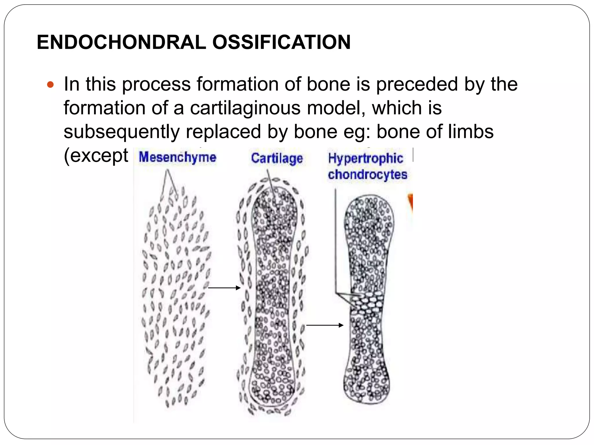
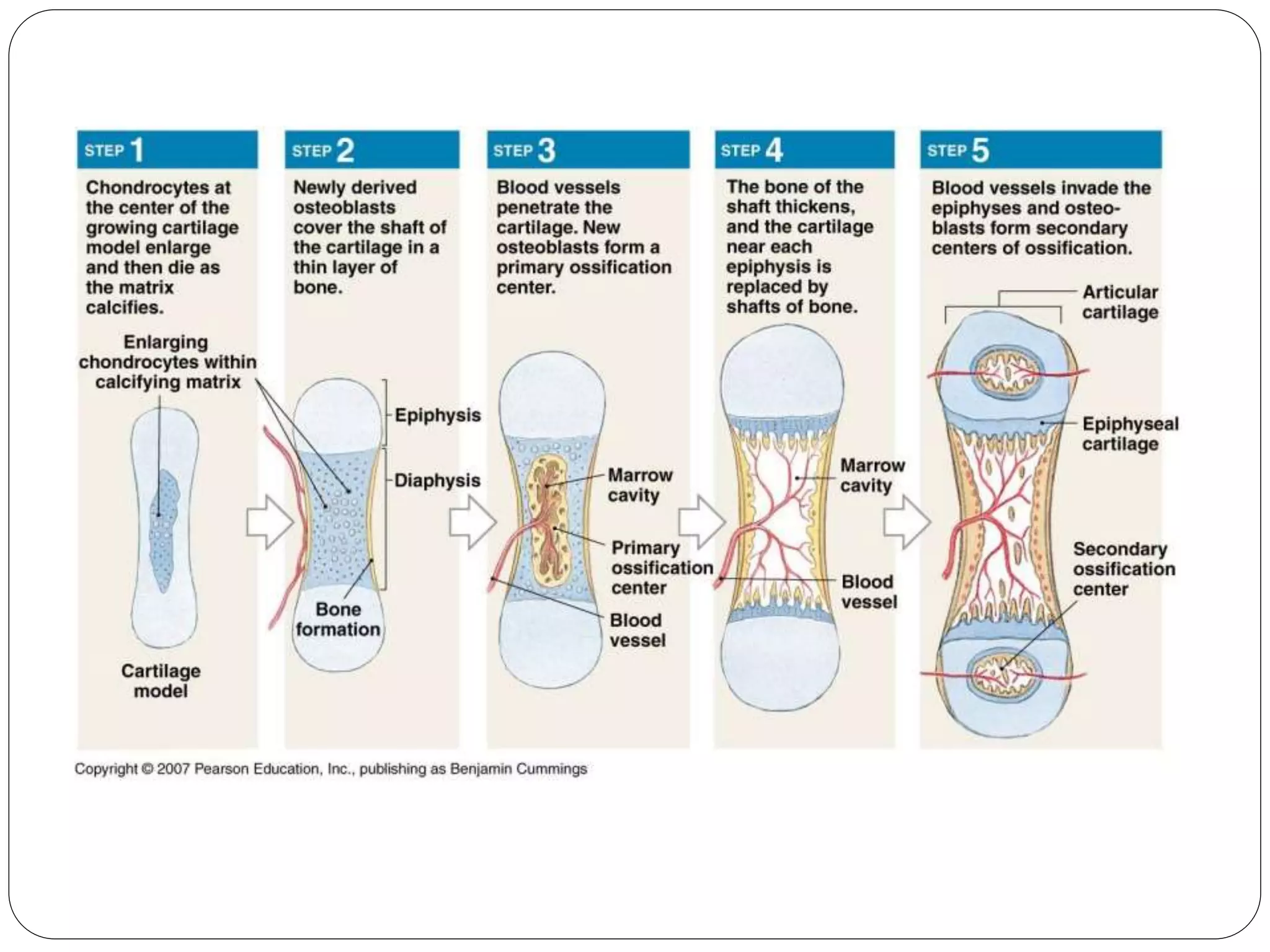
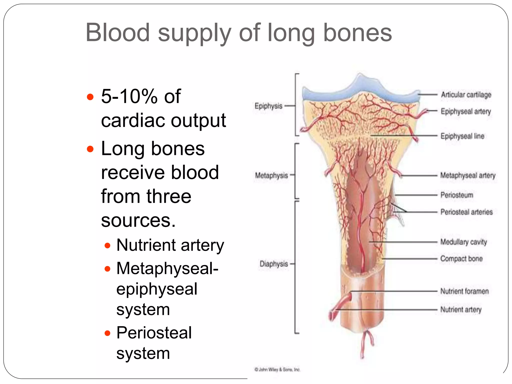
1. Dr. CH.ADITYA (D-ortho)
DEVELOPMENT, GENERAL
STRUCTURE AND ORGANIZATION
AND TYPES OF BONE
MODERATORS :
DR.C. RAGHURAM(PROF&HOD)
DR.B.RAMESH (PROF & HOD)
DR.CH. RAMU (ASSO.PROF)
DR.VENU (ASST.PROF)
DR.VAMSHIDHAR REDDY (ASST.PROF)
DR.K. RAVIKANTH (ASST.PROF)
DR.SURESH (ASST.PROF)
2. INTRODUCTION
The basic structural unit of a human skeleton is bone
Bone is essentially a highly vascular, living, constantly
changing mineralized connective tissue.
It is remarkable for its hardness, resilience and
regenerative capacity.
Bone consists of cells and an intercellular matrix
bone comprised of a rigid matrix of calcium salts
deposited around protein fibers.
Minerals provide rigidity
Proteins provide elasticity and strength
3. Physiology of Bone
The dry weight of bone is composed of 65% to
70% inorganic material
95% of which is calcium and phosphate solid
The main Ca-P solid is crystalline hydroxyapatite
– Ca10(PO4)65H2O
An amorphous Ca-P solid is present in young,
newly formed bone.
Dominant role in calcium homeostasis is played
by constant resorption and deposition of bone
minerals.
4. Other internal factors like hormonal (PTH, calcitonin),
renal (tubular reabsorption), and vitamin D
metabolites help to maintain constant plasma
concentration.
Total Calcium content in body 1 kg
Only 1g is found in plasma and ECF
Remainder in skeleton as phosphates, carbonates
and hydroxides.
Calcium ion necessary for
- Blood coagulation
- Neuromuscular excitability
- Muscular contraction
- Essential ion for enzymes
5. Dietary requirement for normal adult 0.65g/day
Growing children and pregnant- 1g/day
Dietary sources- milk and milk products
About 200-250 mg absorbed rest lost in faeces
Absorption- vit D, PTH, calcitonin
Normal serum ca levels are 8.8 to 10.8 mg/dl
Excretion mainly through kidney- 4oo mg /day
adults
and 4-6mg/kg in children
6. STRUCTURE OF BONE
Macroscopic structure
macroscopically living bone is white, with either a
dense texture like ivory (compact bone), or
honeycombed by large cavities, the bone being
reduced to a latticework of bars and plates
(trabaculae) in which case it is called cancellous,
trabecular or spongy bone
Compact bone is usually limited to the cortices of
mature bones (cortical bone ) function is to
provide strength.
In contrast , cancellous bone lies chiefly in the
interior and particularly in the case of long bones.
Cancellous bones gives additional strength to
cortices and supports the bone marrow
7. Above: Note the relationship
between the compact and
spongy bone.
Below: Close up of spongy bone.
8. Microscopic structure of compact bone
Consists of multiple cylindrical structural units known
as osteons or haversian systems. named after clopton
havers (1691)
it contains the following structures
haversian canal
Lamellae
Lacunae
Canaliculi
volkmann’s canal
osteon
The diagram above represents a long
bone shaft in cross-section. Each
yellow circle represents an osteon. The
blue represents additional matrix filling
in the space between osteons. The red
in the middle is the marrow cavity.
9. OSTEON
HAVERSIAN CANAL Each osteon
consists of a single central canal,
known as a haversian canal,
surrounded by concentric layers of
calcified bone matrix.
Haversian canals allow the passage
of blood vessels, lymphatic vessels,
and nerve fibers.
Each of the concentric matrix “tubes”
that surrounds a haversian canal is
known as a lamella.
All the collagen fibers in a particular
lamella run in a single direction, while
collagen fibers in adjacent lamellae will
run in the opposite direction. This
allows bone to better withstand twisting
forces.
10. LAMELLAE
1) concentric lamellae are
arranged concentrically
around haversian canal
2) Interstial lamellae Lying in
between intact osteons .
These fill the gaps between
osteons or are remnants of
bone remodeling.
3)Circumferential lamellae are
found
at the outer and inner
periphery of the cortex
11. Spider-shaped osteocytes
occupy small cavities
known as lacunae at the
junctions of the lamellae.
Hairlike canals called
canaliculi connect the
lacunae to each other and
to the central canal.
Canaliculi allow the
osteocytes to exchange
nutrients, wastes, and
chemical signals to each
other via intercellular
connections known as gap
junctions.
12.
13. • VOLKMANN’S CANAL
These are oblique canals running at right angles to
the long axis of bone. they contain neurovascular
bundle
they connect the haversian canal with the medullary
cavity and surface of bone.
they are not surrounded by concentric lamellae of
bone
Also known as perforating canal
14.
15. Microscopic Structure of
Spongy (Cancellous) Bone
1. consists of poorly
organized trabeculae (small
needle-like pieces of bone)
2. with a lot of open
space between them.
3. nourished by diffusion
from nearby Haversian
canals.
16. Trabeculae are supportive
and connective tissue
element which is formed in
cancellous bone
Trabeculae develop along
the lines of stress
Follows wolf’s law: states
that reaction of living bone to
the mechanical unloading of
a bone segment
17. CLASSIFICATION OF BONES
Bones classified according to their shape:
A. Long bones consist of a shaft with two ends
1. Examples include:
a. thigh bone = femur
b. upper arm bone = humerus
B. Short bones are cube-like.
1. Examples include:
a. wrist bones = carpals
b. ankle bones = tarsals
18. C. Flat bones are thin and usually curved.
1. Examples include:
a. most skull bones,
b. breast bone = sternum,
c. shoulder blades = scapulae,
d. ribs.
D. Irregular bones are not long, short, or flat.
1. Examples include:
a. vertebrae,
b. auditory ossicles.
19. E. Sesamoid bones develop within a tendon.
1. The patella is a human sesamoid bone.
F. Wormian bones (or sutural bones) are tiny bones
within the skull that lie between major skull bones.
20. Bones classified according to structure:
Spongy(cancellous)- consists of intercrossing and
connecting bone(trabaculae) of varying shapes and
thickness b/w which spaces filled with bone marrow
Compact- continuous bone mass containing
interconnecting vascular channels of microscopic
size.
21. Parts of a Long Bone
1. Diaphysis = shaft
a. consists of a central
medullary cavity (filled with
yellow marrow)
b. surrounded by a thick
collar of compact bone
2. Epiphyses = expanded ends
a. consist mainly of spongy
bone
b. surrounded by a thin layer
of compact bone
3. Epiphyseal line = remnant
of epiphyseal disc
a. cartilage at the junction of
the diaphysis and epiphyses
(growth plate)
22. 4. Periosteum = membrane
covering the outer surface of
bone
two layers
1.fibrous layer- outer thin
layer of dense connective
tissue containing fibroblasts
2.osteogenic layer- contains
osteogenic cells
a. richly supplied with blood &
lymph vessels, nerves
(nutrition):
b. Nutrient Foramen =
perforating canal allowing
blood vessels to enter and
leave bone.
c. Osteogenic layer contains
osteoblasts and osteoclasts
23. 5. Medullary cavity = open
space containing yellow bone
marrow in the diaphysis of a
long bone
a. yellow marrow = fat storage
tissue that does not actively
produce blood cells
6. Endosteum = inner lining of
medullary cavity
a. contains layer of osteoblasts
& osteoclasts
7. Sharpey’s fibers
Secure periosteum to
underlying bone
8. Articular cartilage = pad of
hyaline cartilage on the
epiphyses where long bones
articulate or join.
a. "shock absorber"
24.
25. Flat bones
1. covered by periosteum
2. contains a layer of
spongy bone
enclosed between
plates of compact
bone
3. in a flat bone, the
arrangement looks like
a sandwich:
- spongy bone ,
sandwiched
between
- two layers of
compact bone .
*** Hematopoietic tissue
(red marrow) is located
in the spongy bone
within flat bones and
the epiphyses of long
bones.
*** Red marrow is
26. Components of bone
ORGANIC 25%
1. Bone Cells 4%
Osteoblasts
Osteocytes
Osteoclasts
2. Intercellular Matrix 20%
Collagens
Protein polypeptides
Proteoglycans
lipids
27. Components of bone
Inorganic 65%
1.Crystalline- hydroxyapatite
2. Amorphous- calcium phosphate
3. Trapped ions- citrate, fluoride, sodium,
magnesium, potassium
Water 10%
1.In bone crystals
2. Extracellular
3. Cellular
28. CELLS OF BONE
Cells of bone are embedded in stiff calcified matrix.
the cells are: 1) osteoprogenator stromal cells
2) osteoblasts which lay down bone
3) osteocytes within bone
4) osteoclasts
5) bone lining cells on its surface
29. osteoprogenator stromal cells
from pluripotent stromal stem cells from bone
marrow and other connective tissue
resembles fibroblasts (mesenchymal origin)
Usually differentiate to osteoblasts
2 types –committed (usual)
--inducible-forms ectopic calcification
Depending of nature of induction these may
differentiate into fibroblasts, myoblasts, adipose
cells, chondroblasts
30. Components of bone- Cells
OSTEOBLASTS
bone building cells
15-30 microns,basophilic cuboidal mononuclear
cells
found in both the periosteum and endosteum
Large, roughly fusiform cells characterized by
abundant cytoplasm staining a deep blue with
H&E beneath the deep layer of periosteum.
Abundant rough ER responsible for synthesis of
organic intercellular substance.
Initiate process of calcification
31. Osteoblasts synthesizes organic matters such as
1) type I and V collagen
2) gamma carboxyglutamic acid (GLA)
containing
proteins osteocalcin and GIA protein
3) osteonectin
4) proteases and growth factor.
bears receptors for vit D3 and 1,25(OH)2
vit.D3,which normally inhibits osteoclasts but in
presence of stimulators such as PTH activates
osteoclasts to remove osseous tissue.
32. The blue arrows indicate the
osteoblasts. The yellow arrows
indicate the bone matrix they’ve just
secreted.
33. Components of Bone- Cells
OSTEOCYTES
Mature bone cells
Derived from osteoblasts which
have reduced or ceased matrix
formation
Average life – upon 25 years
Responsible for maintaining the
bone tissue
Each osteocyte is in a lacunae ,
variable space between the
cell and extracellular matrix
Yellow arrows
indicate osteocytes
– notice how they
are surrounded by
the pinkish bone
matrix.
Blue arrow shows
an osteoblast in the
process of
becoming an
osteocyte.
34.
35. OSTEOCLASTS
Multinucleated giant cell varying in size n number
of nuclei
Cytoplasm pale staining acidophilic and foamy
Formed by fusion of several osteoblasts or from
stromal cells of marrow
Function- resorb minerals and intercellular organic
substance
Lie where active removal of bone is occuring on
surface in parts termed resorption bays or lacunae
of howship
36.
37. Agents stimulating osteoclasts
1) factors from osteoblasts
2) macrophages/lymphocytes
3) decrease in intracellular calcium
4) parathyroid hormone
Survival time approximately 7 weeks
38. BONE LINING CELLS
Are flattened epithelium like cells particularly
evident in adult skeleton, found on resting surface
of bone ie: those not undergoing
deposition/resorption
Lines— endosteal surface of marrow cavity
-On Periosteal surfaces
- Vascular canals within osteons
Play active role in regulating differentiation of
osteoprogenator cells
May secrete collagenase
39. Intercellular matrix
Collagen
90% organic matrix
Provides tensile strength to bone
Primarily type I collagen
Structure
◦ Triple helix fibril
◦ Differs from other types of collagen by amino acid
composition and relative insolubility.
◦ X-linking decreases solubility and increases the
tensile strength.
Proteoglycans
Composed of glycosaminoglycans complexes
Partially responsible for compressive strength of
bone.
40. Matrix Proteins
Promote mineralization and bone formation
Osteocalcin
Produced by osteoblasts
Directly related to regulation of bone density
Most abundant non-collagen matrix protein
Inhibited by PTH
Activated by 1,25 Vitamin D
Can measure in urine or serum as marker of
bone turnover
Osteonectin
◦ Secreted by platelets and osteoblasts
◦ Possible role in regulation of calcium and/or
organization of mineral within matrix.
Osteopontin
◦ Cell binding protein
42. www.academic.pgcc.edu/~aimholtz/AandP/LectureNotes/ANP1_Lec/Skeletal/BoneTiss
Formation of the Bony
Skeleton
Before week 8, the human
embryonic skeleton is made
of fibrous membranes and
hyaline cartilage.
After week 8, bone tissue
begins to replace the fibrous
membranes and hyaline
cartilage.
The development of bone from a
fibrous membrane is called
intramembranous ossification.
The replacement of hyaline
cartilage with bone is known as
endochondral ossification.
44. MEMBRANOUS OSSIFICATION
In this process the bone is laid down directly in
membranous sheets eg: clavicle, bones of face vault
of skull..
The various stages in ossification are as follows:
1) at the site where bone is to be formed the
mesenchymal cells become densely packed and the
region becomes highly vascular.
2) some cells lay down bundle of collagen fibres in
the mesenchymal condensation
3)some more mesenchymal cells come and lie
along the collagen fibres. These cells are called
osteoblasts which secrete gelatinous matrix. the
fibres are swollen up. this mass of swollen fibres
and matrix is called OSTEOID. The location in the
45. 4) Under the influence of osteoblasts calcium salts
are deposited in the osteoid and thus one lamellus
of bone is formed.
5) Over this lamellus , another layer of osteoid is laid
down by osteoblasts . the osteoblasts move away
to line the new layer of osteoid. In this process
some cells are trapped between lamellae and
osteoid and are called osteocytes. The new osteoid
is ossified to form another lamellus.
6) In this way number of lamellae are laid down one
over another and forms trabecular bone.
7) Collagen is organized as longitudinal or spiral
bundles and torns
8) During these stages mesenchyme condenses on
surface to form fibrovascular periosteum
46. www.academic.pgcc.edu/~aimholtz/AandP/LectureNotes/ANP1_Lec/Skeletal/BoneTiss
The developing bone grows outward from the ossification
center in small struts called spicules.
Mesenchymal cell divisions provide additional osteoblasts.
The osteoblasts require a reliable source of oxygen and
nutrients. Blood vessels trapped among the spicules meet
these demands and additional vessels branch into the
area. These vessels will eventually become entrapped
within the growing bone.
47. www.academic.pgcc.edu/~aimholtz/AandP/LectureNotes/ANP1_Lec/Skeletal/BoneTiss
Initially, the intramembranous bone consists only of
spongy bone. Subsequent remodeling around
trapped blood vessels can produce osteons typical of
compact bone.
As the rate of growth slows, the connective tissue
around the bone becomes organized into the fibrous
layer of the periosteum. Osteoblasts close to the
bone surface become the inner cellular layer of the
periosteum.
48. ENDOCHONDRAL OSSIFICATION
In this process formation of bone is preceded by the
formation of a cartilaginous model, which is
subsequently replaced by bone eg: bone of limbs
(except clavicle), trunk and base of skull
49.
50. The steps of endochondral ossification are as
follows:
1) at the site where bone is to be formed the
mesenchymal cells become densely packed
2) some mesenchymal cells become
chondroblasts and lay down hyaline cartilage
.mesenchymal cells on the surface of cartilage
form a membrane called perichondrium, which is
vascular and contains osteogenic cells
3) in the area where bone formation is to begin,
the cells enlarge considerably.
51. 4)The intercellular substance b/w enlarged
chondroblasts ossified, under the influence of alkaline
phosphatase, secreted by cartilage cells. The nutrition
to the cells cut off and they die leaving behind empty
spaces called primary areolae
5) Some blood vessels of the perichondrium invades
cartilaginous matrix. they are accompanied by
osteogenic cells and is called periosteal bud. It eats
the primary areolae and forms large cavities called
secondary areolae
6) The osteoblasts are arranged along the surfaces of
secondary areolae.
7) Osteoblasts lay down a layer of ossein fibrils
embeded in gelatinous matrix (osteoid). The osteoid
is calcified and lamellus of bone is formed. In this
way number of lamellae are laid down one over
52. Bone development begins at the primary center of
ossification and spreads toward both ends of the
cartilaginous model.
While the diameter is small, the entire diaphysis is
filled with spongy bone
The primary ossification center enlarges proximally
and distally, while osteoclasts break down the newly
formed spongy bone and open up a medullary cavity
in the center of the shaft.
As the osteoblasts move towards the epiphyses, the
epiphyseal cartilage is growing as well. Thus, even
though the shaft is getting longer, the epiphyses have
yet to be transformed into bone.
53. Around birth, most long bones have a bony diaphysis
surrounding remnants of spongy bone, a widening
medullary cavity, and 2 cartilaginous epiphyses.
At this time, capillaries and osteoblasts will migrate
into the epiphyses and create secondary
ossification centers. The epiphysis will be
transformed into spongy bone. However, a small
cartilaginous plate, known as the epiphyseal plate,
will remain at the juncture between the epiphysis and
the diaphysis.
55. Epiphysis
is the end
of a long
bone.
Diaphysis
is the
shaft of a
long bone.
Epiphyseal plate is the
site of bone growth.
Diaphysis
Compact
bone
Osteoblast
Directionofgrowth
Chondrocyte
Cartilage
owth
Newly calcified
bone
Bone gr
Dividing
chondrocytes
add length
to bone.
Chondrocytes
produce
cartilage.
Old
chondrocytes
disintegrate.
Osteoblasts lay
down bone on
top of cartilage.
56. Microscopic structure of growth plate
Epiphyseal to
diaphyseal end
4 zones
1.Zone of resting
cartilage
2.Zone of young
proliferating
chondrocytes
3.Zone of maturing
chondrocytes
4.Zone of calcified
cartilage.
57. Blood supply of long bones
5-10% of
cardiac output
Long bones
receive blood
from three
sources.
Nutrient artery
Metaphyseal-
epiphyseal
system
Periosteal
system
58. Nutrient artery
- Divides into ascending and descending branch
- Each branch sends lateral(radial) oriented
arteriolar branches most of which lead to cortex,
others to sinusoids within marrow,30% marrow
70% cortical capillary beds
Terminal branches anastamose with epiphyseal
and metaphyseal vessels to form medullary blood
supply.
- Cortical arterioles originating from main medullary
nutrient artery enter cortex some extend
longitudinally and others radially. These branches
ultimately form capillaries within Haversian
systems.
- Nutrient artery and branches- inner two thirds or
59. All long bones have one or more nutrient arteries
that enter through the nutrient foramen
accompanied by thin walled veins and
myelinated nerve
Humerus- single artery, anteromedially at
junction of middle and lower thirds
Femur- two nutrient arteries from profunda
femoris, linea aspera.
Radius and ulna- nutrient foramen proximally and
directed towards elbow
Tibia- from post tibial artery penetrates
posterolateral cortex just below the oblique line
of tibia
60. Venous Drainage
Long bones possess a large central venous
sinus transport effluent blood from marrow
capillary bed
Central venous sinus emerges from diaphysis as
nutrient vein through nutrient canal
Major venous drainage from long bone is into
periosteal venous complex
Only 5- 10% of effluent blood leaving by the way
of nutrient vein
Most leaves by metaphyseal vessels part of
periosteal venous system
61. Blood flow through the compactum is normally
centrifugal flow , blood entering endosteal aspect
from medullary nutrient system and flowing out
through the periosteal surface.
In event of medullary nutrient system interruption,
periosteal system provides reserve supply and
flow becomes centripetal.
62. Functions of bone
A. Support
1. the bones in legs and pelvis support the trunk,
2. the atlas (1st vertebra) supports the skull, etc.
B. Protection of underlying organs
1. the skull protects the brain,
2. the rib cage protects the heart and lungs, etc.
C. Body Movement
1. skeletal muscles attached to bones by tendon.
2. serve as levers to move bones
D. Hematopoiesis
All blood cells are formed in the red marrow of
certain bones
63. E. Inorganic Salt Storage
1. bone stores many minerals
a. calcium,
b. phosphorus
c. others.
2. also a means of calcium homeostasis
F. Energy Storage
1. yellow marrow in the shaft of long bones
2. serve as an important chemical energy reserve