SlideShare a Scribd company logo
1 of 65
Download to read offline
VOL 2, No 63 (2021)
Sciences of Europe
(Praha, Czech Republic)
ISSN 3162-2364
The journal is registered and published in Czech Republic.
Articles in all spheres of sciences are published in the journal.
Journal is published in Czech, English, Polish, Russian, Chinese, German and French, Ukrainian.
Articles are accepted each month.
Frequency: 24 issues per year.
Format - A4
All articles are reviewed
Free access to the electronic version of journal
All manuscripts are peer reviewed by experts in the respective field. Authors of the manuscripts bear responsibil-
ity for their content, credibility and reliability.
Editorial board doesn’t expect the manuscripts’ authors to always agree with its opinion.
Chief editor: Petr Bohacek
Managing editor: Michal Hudecek
• Jiří Pospíšil (Organic and Medicinal Chemistry) Zentiva
• Jaroslav Fähnrich (Organic Chemistry) Institute of Organic Chemistry and Biochemistry
Academy of Sciences of the Czech Republic
• Smirnova Oksana K., Doctor of Pedagogical Sciences, Professor, Department of History
(Moscow, Russia);
• Rasa Boháček – Ph.D. člen Česká zemědělská univerzita v Praze
• Naumov Jaroslav S., MD, Ph.D., assistant professor of history of medicine and the social
sciences and humanities. (Kiev, Ukraine)
• Viktor Pour – Ph.D. člen Univerzita Pardubice
• Petrenko Svyatoslav, PhD in geography, lecturer in social and economic geography.
(Kharkov, Ukraine)
• Karel Schwaninger – Ph.D. člen Vysoká škola báňská – Technická univerzita Ostrava
• Kozachenko Artem Leonidovich, Doctor of Pedagogical Sciences, Professor, Department
of History (Moscow, Russia);
• Václav Pittner -Ph.D. člen Technická univerzita v Liberci
• Dudnik Oleg Arturovich, Doctor of Physical and Mathematical Sciences, Professor, De-
partment of Physical and Mathematical management methods. (Chernivtsi, Ukraine)
• Konovalov Artem Nikolaevich, Doctor of Psychology, Professor, Chair of General Psy-
chology and Pedagogy. (Minsk, Belarus)
«Sciences of Europe» -
Editorial office: Křižíkova 384/101 Karlín, 186 00 Praha
E-mail: info@european-science.org
Web: www.european-science.org
CONTENT
BIOLOGICAL SCIENCES
Syrovatko K.
THE EFFICIENCY OF USING A FEED ACIDIFIER IN
GROWING BROILER CHICKENS....................................3
CHEMICAL SCIENCES
Morozova L., Novakovska V.
RELEASE OF FEED NUTRIENTS BY EXTRUSION OF
LEGUMES.....................................................................9
MEDICAL SCIENCES
Shepetovskaya N.,
Kalenteva S., Shepetovskaya M.
MODERN VIEW ON ULTRASOUND DIAGNOSTICS IN
OBSTETRICS...............................................................13
Vorobev S., Petrushin M.,
Ushakov D., Mikhralieva B.
FREQUENCY AND ROLE OF ANEMIA IN PATIENTS IN
RESCUE UNITS AND INTENSIVE CARE........................20
Loskutova T., Davydenko N., Chulkov O.
PREDICTION OF RECURRENT MISCARRIAGE .............25
Maksinev D.
GENITOMETRIC CHARACTERISTICS OF MALE
STUDENTS..................................................................31
PHARMACEUTICAL SCIENCES
Hroshovyi T., Dobrynchuk M.,
Pavliuk B., Chubka M.
DRONE BROOD – AS A RAW MATERIAL FOR THE
MANUFACTURE OF MEDICINES AND DIETARY
SUPPLEMENTS...........................................................36
Blazhko І., Hroshovyi T.,
Chubka M., Pavliuk B.
ANALYSIS OF THE RANGE OF SEMISOLID MEDICINES
IN THE FORM OF GELS ON THE PHARMACEUTICAL
MARKET OF UKRAINE AND EXCIPIENTS IN THEIR
COMPOSITIONS.........................................................39
Nazarkina V., Kurylenko Yu.,
Popova I., Teterich N., Podkolzina M.
ANALYSIS OF THE COST OF TREATMENT PATIENTS
AFTER COVID-19 WITH NOOTROPIC MEDICINES IN
UKRAINE....................................................................43
PHYSICS AND MATHEMATICS
Parthentiev N., Partfenteva N.
CONNECTION OF NEWTON'S FORMULA FOR KINETIC
ENERGY AND EINSTEIN FORULA ...............................47
Koshman V.
RELICT RADIATION AND PHYSICAL FEATURES OF THE
QUANTUM BIRTH OF OUR UNIVERSE .......................49
Losovsky B., Losovsky A.
DIAGNOSTIC OF INTERMITTENT RADIO EMISSION
FROM PULSARS .........................................................55
Fedun V.
DYNAMICS OF PLASMA PISTON IN PIPE FILLED BY A
GAS-LIQUID MEDIUM................................................60
Sciences of Europe # 63, (2021) 3
BIOLOGICAL SCIENCES
THE EFFICIENCY OF USING A FEED ACIDIFIER IN GROWING BROILER CHICKENS
Syrovatko K.
Candidate of Agricultural Sciences, Associate Professor
Vinnytsia National Agrarian University
DOI: 10.24412/3162-2364-2021-63-2-3-8
ABSTRACT
The influence of the feed acidifier "Ronocid", made on the basis of organic acids with the inclusion of natural
aluminosilicate and oregano essential oil, on the performance indicators of broiler chickens was investigated. It
has been established that the feeding of compound feeds with the addition of acidifier helps to increase the live
weight and safety of broiler chickens, reduce feed costs, increase slaughter rates: the weight of uncooked, semi-
gutted, gutted carcasses, chest and thigh muscles.
Keywords: broiler chickens, acidifier "Ronocid", productivity, slaughter indicators.
Relevance. Improving consumption and increas-
ing the efficiency of feed use in poultry farming is en-
sured by a high level of balanced feeding with the use
of various feed additives [11]. By their intended pur-
pose, feed additives are divided into protein, energy,
mineral, vitamin, antibiotics, enzyme preparations, pro-
biotics, prebiotics, acidifiers, mold inhibitors, toxin ad-
sorbents and combined additives. In the early postnatal
period, the bird has an underdeveloped digestive sys-
tem that is not able to fully digest food. This negatively
affects muscle building and internal organ develop-
ment. In addition, it is during this period that various
harmful microorganisms can develop in the lumen of
the intestinal tract. It should be borne in mind that di-
gestion in young animals is primarily characterized by
a weak secretion of hydrochloric acid in the stomach
and its low activity. In chickens in the first 7 days, less
than 1 milliliter of hydrochloric acid is released. As a
result of such a completely physiological reaction, not
enough acid is released in order for the feed mass to
swell and its alkaline properties to be completely neu-
tralized. Only in a stable acidic environment in the
stomach with a pH of less than 3.5, the enzymes that
are produced in the body begin to influence digestion
and maximize it. In addition, stomach acid is a decisive
barrier up to a certain point for the development of
pathogenic microorganisms and their penetration into
the lower intestine.
Acidifiers are preparations that contain organic
and inorganic acids and other substances. Of the acids,
acetic, ascorbic, succinic, oily, formic, lactic, malic,
propionic, benzoic, citric, fumaric, lauric and their salts
are most often used. Organic acids promote the devel-
opment of the chicken intestinal microflora after hatch-
ing and thus improve the condition of the gastrointesti-
nal tract, inhibit the growth and development of patho-
genic microflora (Salmonella, E. coli and others), mold
pathogens in feed and feed raw materials. Simultane-
ously with a slowdown in the growth of gram-negative
bacteria, which develop optimally at a pH of 6-7, the
work of proteases improves in the gastrointestinal tract
due to acidification of its content [7,12]. The pH and
microbial load in the gastrointestinal tract of the bird is
reduced, the absorption of nutrients is improved,
weight gain is improved and the incidence of digestive
disorders is reduced. This ultimately leads to an in-
crease in the general resistance of the body of chickens,
increases the growth rate and safety of the bird.
In this regard, the use of acidifiers in poultry farm-
ing is promising.
Analysis of recent researches and publications.
In recent years, a number of studies have been carried
out on the use of organic acids in poultry farming as
feed acidifiers. One of the main conditions for the use
of organic acids is a high degree of electrolytic dissoci-
ation and is typical for the environment of the digestive
tract [3]. Among such acids, lactic acid has a high pos-
itive effect, which has received a wide range of appli-
cations not only as an acidifier, flavor enhancer and
preservative, but also as a natural stimulator of animal
productivity, a promoter of intestinal villi development,
a disinfectant, a treatment agent, and others.
It was revealed that lactic acid, being an important
intermediate product of metabolism in living organisms
and possessing antimicrobial properties, in comparison
with other organic acids, has significant advantages,
since the direct action of the metabolite on the hypo-
thalamus and pituitary gland is not excluded by the type
of hormones and adaptogens.
The introduction of 3 to 5 kilograms of a 4% aque-
ous solution of lactic acid in compound feed for laying
hens in the initial phase of laying significantly stimu-
lates the formation of follicles in them, an increase in
resistance.
It was found that the consumption of compound
feed by young quail in the period from 1 to 49 days with
a lactic acid content of 0.5 milliliters / 100 grams con-
tributes to an increase in live weight by 9% (P≤0.05),
average daily gains by 8.8% (p ≤ 0.05). Feeding com-
pound feed for young quail in the period from 1 to 49
days with a content of 0.3 and 0.5 milliliters / 100 grams
of lactic acid helps to reduce feed costs per 1 kg of live
weight gain by 2.4 and 0.6%, respectively [8].
Today, among the most famous acidifiers used in
the production of compound feeds are Biotronic, Ul-
tracid, Salmo-Mil, CuxAcid, Biatsid, Asid Lak. Bio-
tronic is a line of acidifying products for pig and poultry
farming of the company "Biomin GmbH" (Austria). Bi-
otronic SE Forte, Biotronic SE Multi differ in the con-
tent of acids, salts, specific extracts, organic (oligosac-
charides) and inorganic (silicon and its compounds)
4 Sciences of Europe # 63, (2021)
carriers. Biotronic products maintain the pH in the
small intestine at an optimal level (pH 5.5-6.2), inhibit
the growth of pathogenic bacteria and promote the
growth of beneficial intestinal microflora, that is, they
contribute to ensuring microbial balance in the gastro-
intestinal tract. Biotronic SE Forte is a free-flowing
powder, gray-brown in color with a bulk density of 400
grams / dm3. The pH of a 10% hydrogen solution is 3.6.
The drug is intended for use in compound feed for pigs
and poultry in the amount of 1-5 kilograms per 1 ton.
The shelf life is 18 months [6]. As evidenced by the re-
sults of studies by foreign authors [8, 9], the use of or-
ganic acids in feeding improves the digestibility and as-
similation of nutrients, increases the productivity of an-
imals and reduces feed costs for products. The
antioxidant and neurotropic effect of organic acids in
the body of animals and their normalizing effect on en-
ergy metabolism, biosynthesis processes and general
physiological state have also been proven [10, 11]. The
company "NUTRI-AD" (Belgium) produces acidulants
based on organic and inorganic acids under the trade-
marks Ultrasid, Salmo-Mil. Acidifiers are produced us-
ing various technologies, such as spraying-drying, saw-
ing-cooling (coating), on carriers, in liquid form. Ul-
trasid V and Ultrasid 8Plus Dry are buffer acidifiers
made using the Spray Dry technology and contain 20%
of easily assimilable calcium. They are non-reactive,
therefore, they are introduced into the composition of
feed through premixes. Recently, in poultry feeding,
drugs that are derived from yeast have been used, in
particular the drug NuPro - a nucleoprotein containing
nucleotides - the most important components of DNA,
biotin, inositol (vitamin B8), essential amino acids,
macro- and microelements, increases the energy of
growth and development of the body young animals
and poultry. (Dyachenko).
The research results showed that with the content
of 1-3% of the mass fraction of NuPro in the mixed
feed, the average daily body weight gain for 1 week of
rearing in chickens 2, 3, 4 of the research groups was
6.8; 8.7; 7.3% higher, and for the entire experience,
broilers of the experimental groups in average daily
gains were ahead of the control peers by 3.5-9.2%, and
their feed costs per 1 kilogram of gain were less than
that of the control group by 1.10-2 , 21%.
The purpose of the research and methods. The
purpose of the research was to study the effect of the
acidifier "Ronocid" on the productivity, slaughter and
hematological parameters of broiler chickens. The feed
acidifier Ronocid is a product of a perfect combination
of organic acids and salts of fatty acids, specially se-
lected for effective action in every part of the gastroin-
testinal tract of animals. It is a gray powder. It contains
organic acids: orthophosphoric, formic, lactic, succinic,
fumaric, acetic, benzoic; as well as natural aluminosil-
icate and oregano essential oil. The supplier of this ad-
ditive is a private company "Ukrfid". The product re-
duces the Ph of the digestive tract, has a pronounced
antibacterial activity. Penetrating through the bacterial
cell wall, the active substance disrupts metabolism and
the regulation of internal processes in the cell, as a re-
sult of which the bacterial cell dies. In addition, its
pharmacological properties are adsorption properties
and additional enrichment with minerals, especially
easily assimilated phosphorus.
The object of the research- broiler chickens of the
Ross-308 cross. The subject of the research - produc-
tivity, lethality, broiler blood. The study was carried out
at the Rodina farm in the Ilyinets district. The scientific
and economic experience lasted 42 days and consisted
of two periods: an equalizing one, lasting 7 days, ac-
counting for 35 days (Table 1).
According to the principle of analogous groups,
100 heads of broiler chickens were selected and 2
groups of 50 heads each were created. The bird was
kept in cages under microclimate conditions [11]. Dur-
ing the experiment, broilers were fed complete feeds of
the Multigein trademark. The experimental group with
the main diet - complete feeds consumed the acidifier
"Ronocid" in the amount of 1.0 kilograms per ton of
compound feed.
Table 1
Scheme of scientific - husbandry experience
Group
Duration of the period, days
Quantity chickens. Feeding features
equalizing the main
1- control 7 35 50 OR (complete feed)
2- control 7 35 50
OR + "Ronocid"
1.0 kilograms / ton of feed
During the experiment, the safety of the livestock,
the live weight of broiler chickens, the feed consump-
tion were taken into account, the absolute, average
daily and relative gains in the live weight were calcu-
lated, and the slaughter indicators were determined.
Statistical processing of research data was calcu-
lated on a computer using the Micrsoft Excel software.
At the same time, the results of the average statistical
data were considered reliable at * P <0.05; ** P <0.01;
*** P <0.001.
Research results and their discussion. Accord-
ing to the results of the experiment, it was revealed that
broiler chickens of the 2nd group, which were fed the
acidifier "Ronocid" with mixed fodder, had higher
growth and safety indicators (Table 2).
Thus, it was found that at 35 days of age the bird
of the 2nd research group prevailed over its peers from
the control group in terms of live weight by 82.1 grams
or 4.3% (P <0.01).
With age, the growth rate of broiler chickens in the
experimental group increased relative to the control. In
particular, at the end of rearing (6 weeks of age), the
average weight of broiler chickens receiving the acidi-
fier was 224.8 or 9.1% more (P <0.001).
Sciences of Europe # 63, (2021) 5
Table 2
Dynamics of live weight and safety of broilers, g (M ± m, n = 20)
Chicks age, days
Group
1- control 2- research
1 42,8 ± 0,52 42,7 ± 0,68
7 175,6 ± 2,74 178,4 ± 2,82
14 427,5 ± 5,65 430,6 ± 6,47
21 832,2 ± 8,42 856,8 ± 10,64
28 1284,5 ± 13,53 1322,6 ± 14,72
35 1925,7 ± 19,45 2007,8 ± 18,65**
42 2461,5 ± 21,76 2686,3 ± 19,48***
safety, % 94 98
It should be noted that the safety of the poultry of
the 2 group was higher by 4.0% compared to the con-
trol.
It was revealed that in broiler chickens aged 36-42
days for the consumption of the feed acidifier, the av-
erage daily gain was 96.9 grams, which is 20.4 grams
because 26.7% more (P <0.05) compared to the control
(Table . 3).
Table 3
Average daily gain of broiler chickens, g (M ± m, n = 20)
Chicks age, days
Group
1- control 1- research
1-7 19,1 ± 1,23 19,4 ± 1,34
8-14 36,0 ± 1,56 36,0 ± 1,45
15-21 57,8 ± 2,68 60,9 ± 2,54
22-28 64,6 ± 4,85 66,5 ± 4,72
29-35 71,6 ± 5,43 97,9 ± 5,84
36-42 76,5 ± 5,62 96,9 ± 5,56*
Average 54,3 ± 4,23 62,9 ± 5,45
On average, the average daily growth of poultry of
the 2nd group was more by 8.6 grams or 15.8% relative
to the control. The absolute gain in broiler chickens of
the 2nd group at 15-21 days of age increased versus
control peers by 5.3% (P <0.05) (Table 4).
Consumption of the feed additive "Ronocid" al-
lows increasing the absolute growth of broiler chickens
at the age of 22-28 days - by 3.1%, 29-35 days - by
6.9% (P <0.001), and at the age of 36-42 days - by
26.5% compared to control.
On average, over the entire period of growing re-
search broiler chickens, there is an increase in absolute
growth by 9.3% (P <0.001) relative to control ana-
logues.
Table 4
The absolute growth of broiler chickens, g (M ± m, n = 20)
Chicks age, days
Group
1- control 2- research
1-7 133,0 ± 2,75 136,0 ± 2,63
8-14 252,0 ± 2,52 252,0 ± 2,48
15-21 405,0 ± 5,46 426,6 ± 7,54*
22-28 452,0 ± 8,35 466,0 ± 11,62
29-35 641,2 ± 13,43 685,2 ± 14,85*
36-42 535,8 ± 21,55 678,5 ± 19,20***
Over the entire period of experience 2418,7 ± 28,62 2643,6 ± 25,46***
Young poultry grows unevenly, therefore, the ab-
solute growth rate does not reflect the actual intensity
of growth processes, the degree of relationship between
the amount of body weight that increases and the
growth rate. For this purpose, the relative gain is deter-
mined, which shows the percentage increase in body
weight at the end of the period relative to the body
weight at the beginning of the period.
The additional use of an acidifier in poultry feed-
ing also had a positive effect on the relative gains of
broiler chickens (Table 5).
6 Sciences of Europe # 63, (2021)
Table 5
The relative growth of broiler chickens,% (M ± m, n = 20)
Chicks age, days
Group
1- control 2- research
1-7 310,3 ± 3,85 317,8 ± 4,62
8-14 143,5 ± 2,56 141,4 ± 2,71
15-21 94,6 ± 2,82 98,9 ± 2,64
22-28 54,3 ± 1,35 54,4 ± 1,52
29-35 49,9 ± 1,63 51,8 ± 1,35
36-42 27,8 ± 1,28 33,8 ± 1,46*
It was revealed that at the age of 36-42 days, the
relative growth of broiler chickens of the 2nd group,
which received the feed acidifier, prevailed in control
analogues by 6.0% (P <0.05). It has been proven that
feeding broiler chickens with the acidifier "Ronocid"
has a positive effect on feed costs per unit of gain (Ta-
ble 6).
Table 6
Efficiency of feed use, kilograms
Group
Feed costs, kilogram
over the period of experience one head per 1 kilogram gain
1- control 225,5 4,51 1,86
2- research 219,5 4,39 1,63
It was revealed that the cost of feed per 1 kilogram
of poultry growth in the group was 0.23 kilograms or
12.4% less than in the control group. According to the
results of the slaughter, it was found that additional
feeding of the biologically active additive "Ronocid"
contributes to an increase in the slaughter performance
of broiler chickens (Table 7).
Table 7
Slaughter qualities of broiler chickens, g (M ± m, n = 4)
Index
Group
1- control 2- research
Pre-slaughter live weight 2450,2 ± 44,15 2680,0 ± 41,15**
The mass of unpeeled carcass 2326,4 ± 37,28 2468,5 ± 34,45*
Weight of half-gutted carcass 1885,1 ± 31,72 2075,4 ± 32,84**
The weight of the gutted carcass 1628,0 ± 29,48 1796,2 ± 30,18**
Chest muscle massage 486,0 ± 18,57 554,0 ± 20,15*
Thigh muscle mass 315,0 ± 15,48 366,0 ± 17,37
The use of an acidifier in feeding broiler chickens
increases their pre-slaughter live weight by 9.3% (P
<0.01), the weight of a whole carcass by 6.1% (P
<0.05), a half-gutted carcass by 10.0% (P <0.01) and
gutted carcasses by 10.3% (P <0.01) relative to control
analogues.
In addition, for the action of the feed additive, the
mass of the chest muscles is 13.9% (P <0.05) and the
mass of the femoral muscles is 16.2% higher, against
the control indicators.
The use of the acidifier "Ronocid" did not affect
the morphological changes in the internal organs of the
research bird (Table 8). No significant difference in the
mass of the muscular and glandular stomachs, heart,
lungs, liver, pancreas, spleen and kidneys was found.
Table 8
Mass of internal organs, g (M ± m, n = 4)
Index
Group
1- control 2- research
Glandular stomach 8,0 ± 0,64 8,1 ± 0,85
Muscular stomach 29,5 ± 1,28 32,2 ± 1,12
Heart 13,2 ± 1,52 13,8 ± 1,74
Lungs 12,1 ± 0,86 12,5 ± 0,92
Liver 37,3 ± 2,18 38,6 ± 4,25
Pancreas 4,6 ± 0,48 4,9 ± 0,52
Spleen 2,3 ± 0,22 2,5 ± 0,28
Kidneys 16,5 ± 1,04 17,1 ± 1,12
The control slaughter, carried out after the com-
pletion of the scientific and husbandry experiment,
showed that the use of the acidifier "Ronocid" influ-
enced the slaughter performance of broiler chickens in
the control and experimental groups (Table 9).
In broiler chickens of the experimental group, the
yield of semi-gutted and gutted carcasses was greater
relative to the control group by 0.5 and 0.6%, however,
no statistically significant difference was found for this
indicator.
Sciences of Europe # 63, (2021) 7
Table 9
Slaughter products yield,% (M ± m, n = 4)
Index
Group
1- control 1- control
Half-gutted carcasses 76,9 ± 4,92 77,4 ± 6,88
Gutted carcass 66,4 ± 5,84 67,0 ± 4,33
Pectoral muscles 19,8 ± 1,34 20,6 ± 1,45
Thigh muscles 12,9 ± 1,25 13,6 ± 1,32
Glandular stomach 0,3 ± 0,14 0,3 ± 0,15
Muscular stomach 1,2 ± 0,21 1,9 ± 0,92
Liver 1,5 ± 0,18 1,4 ± 0,54
Heart 0,53 ± 0,042 0,51 ± 0,028
When analyzing the mass of edible parts, there
was a tendency to an increase in the mass of the pectoral
and femoral muscles and the glandular stomach in the
experimental group. In broiler chickens of the 2 group,
the weight of edible parts prevailed in their weight in
the control analogues, but there was no significant dif-
ference in these indicators.
One of the main indicators of metabolism and the
physiological state of the body is the morphological and
biochemical blood test. Therefore, during the experi-
ment, the hematological parameters of the bird were
studied (Table 10).
It was found that in broiler chickens of the 2 group,
which received the acidifier "Ronocid" with the mixed
feed, the number of erythrocytes significantly increased
by 10.0% (P <0.05) and hemoglobin by 7.5 (P <0.05)
compared with the control. ... An increase in the hemo-
globin content is positive and indicates an increase in
redox processes and an intensification of metabolic
processes in the body of chickens.
In terms of the content of total protein, albumin
and globulins, there was no significant difference be-
tween the groups. In the blood of group 2 broilers, there
was only a tendency to increase the total protein content
by 4.9%, albumin by 7.8% and globulins by 2.3%. The
biochemical parameters of the blood of broiler chickens
were within the physiological norm.
Table 10
Morphological and biochemical parameters of blood (M ± m, n = 4)
Index
Group
1- control 1- control
Erythrocytes (t / l) 43,0 ± 0,05 43,3 ± 0,04*
Leukocytes (g / l) 420,2 ± 0,76 422,4 ± 1,18
Hemoglobin (g / l) 4110,2 ± 2,42 4118,5 ± 2,25*
Total protein, g / l 432,5 ± 2,54 434,1 ± 2,48
Albumin, g / l 415,3 ± 1,38 416,5 ± 1,25
Globulins, g / l 417,2 ± 1,52 417,6 ± 1,64
ALT, units / L 44,9 ± 1,84 45,1 ± 1,25
AsAT, units / L 4214,2 ± 26,15 4221,2 ± 32,26
Bilirubin, μmol / l 42,8 ± 0,64 43,0 ± 0,82
Cholesterol, mmol / l 42,3 ± 0,27 42,1 ± 0,25
Glucose, mmol / l 47,2 ± 0,68 47,4 ± 0,54
Calcium, mmol / l 42,2 ± 0,34 42,3 ± 0,42
Phosphorus, mmol / l 42,0 ± 0,36 42,2 ± 0,28
The levels of cholesterol, bilirubin, glucose, cal-
cium and phosphorus in the blood were at the level of
values of broiler chickens in the control group.
Conclusions. 1. It was found that when using the
feed acidifier "Ronocid" the live weight of broiler
chickens increased by 9.1% (P <0.001).
2. It was found that for the consumption of the in-
vestigated factor in broiler chickens, the average daily
gain increased by 15.8% (P <0.05), absolute by 9.3%
(P <0.001) relative to control analogues.
3. When using the acidifier "Ronocid" there is a
decrease in feed costs per 1 kg of gain in group 2 by
0.23 kg or 12.4%.
4. The introduction of the acidifier "Ronocid" into
the composition of the broiler chicken feed contributes
to an increase in the pre-slaughter live weight by 9.3%
(P <0.01), the weight of the whole carcass by 6.1% (P
<0.05), half-gutted - by 10.0% (P <0.01), gutted - by
10.3% (P <0.01), pectoral muscle mass - by 13.9% (P
<0.05), femoral muscles - by 16, 2%.
5. The use of the acidifier "Ronocid" increases the
hemoglobin content in the blood of broiler chickens by
7.5%, the number of erythrocytes - by 10.0%. Changes
in blood parameters occurred within the range of phys-
iological values for poultry.
References
1.Demchuschun S. The effectiveness of the use of
acidifiers in the industrial growing of broiler chickensn.
Scientific Messenger LNUVMBT named after S.
Gzhytskyj, 2016. Vol. 18. no 2 (67). P. 82-84.
2.Demchishin A., Kukhtin M. Productivity and
slaughter indicators of broiler chickens for drinking the
8 Sciences of Europe # 63, (2021)
acidifier "Aquasan". Modernization of the national sys-
tem of state development management: challenges and
prospects: materials and II International. scientific and
practical conference the city of Ternopil, November 16,
2018 Ternopil, 2018. Р. 78-80.
3.Jegorov B., Makaryns'ka A. Modern alternative
to feed antibiotics. Cereal products and feed. 2010. №
3. Р. 27 − 34.
4. Ibatullin I.I., Zhurakovsky O. Methodology and
organization scientific research in animal husbandry:
manual. K.: Agricultural science, 2017. 328 pp.
5. Kuz'menko L., Vyslan'ko O., Ban'kovs'ka, I.
The efficiency of a new drug − Acidifiers of feed con-
taining chelated micronutrient compounds − in feeding
young pigs. Journal of Poltava State Agrarian Acad-
emy. 2011. Vol. 4. Р. 81−85.
6. Lokstedt K., Kortil M., Panchenko T. Prospects
for the use of Biotronic ® CE forte to combat salmo-
nella. Efficient poultry farming. - 2007. -No. 11. - Р.
44-47.
7. Nagornaya L., Bondar S., Vorozhbitova A.
Preparations based on organic acids. Our poultry indus-
try. No. 1. Р. 26-27.
8. Nechay N., Otchenashko V. Productivity of
young quail for the use of lactic acid in compound feed.
Bulletin of Agrarian Science. 2015 pp. 31-35.
9. Otchenashko V. Use of lactic acid in animal
husbandry: scientific and practical recommendations.
Kiev, 2012.46 p.
10. Syvachenko, Je.V., Djachenko, L.S. Perfor-
mance and slaughter quality of broiler chickens by
feeding different doses and antibiotics Acidifiers. Sci-
entific and technical bulletin SIC biosafety and envi-
ronmental control resources AIC. 2016. Vol. 4, 1, Р.
244−250
11. Polishchuk A., Bulavkina T., Modern feed ad-
ditives in animal and poultry feeding. Bulletin of the
Poltava State Agrarian Academy. 2010. No. 2. Р. 63-
66.
12. Sivik T., Dyachenko L., Sivachenko E. Com-
position of intestinal microflora, safety and productiv-
ity of broiler chickens when feeding various forms and
doses of acidifier. Production technology and pro-
cessing of livestock products. 2018.No. 2. P. 14-23.
13. Yastrebov K., Krivenok M. Practical use of or-
ganic acids in poultry farming. Cereal products and
compound feed. 2010. No. 4. P.22-24.
14. Samudovska A., Demeterova M. Effect of wa-
ter acidification on performance, carcass characteristic
andsome variables of intermediary metabolism in
chscks . Acta Veterinaria (Beograd). 2010. Vol. 60. (№
4). Р. 363–370.
15. Thyroid Activity, Some Blood Constituents,
Organs Morphology and Performance of Broiler
Chicks Fed Supplemental Organic Acids / S. A. Abdel-
Fattah, M. H. El-Sanhoury, N. M. ElMednay and F. Ab-
del-Azeem. International Journal of Poultry Science.
2008. Vol. 7 (3). P.215–222.
Sciences of Europe # 63, (2021) 9
CHEMICAL SCIENCES
ВИВІЛЬНЕННЯ ПОЖИВНИХ РЕЧОВИН КОРМІВ ШЛЯХОМ ЕКСТРУЗІЇ ЗЕРНОБОБОВОГО
ФУРАЖУ
Морозова Л.П.
Вінницький національний аграрний університет, старший викладач
Новаковська В.Ю.
Інститут кормів та сільського господарства Поділля НААН,
молодший науковий співробітник, Вінниця
RELEASE OF FEED NUTRIENTS BY EXTRUSION OF LEGUMES
Morozova L.
Vinnytsia National Agrarian University, Senior Lecturer
Novakovska V.
Institute of Feed Research and Agriculture of Podillya NAAS, junior researcher
Vinnytsia
DOI: 10.24412/3162-2364-2021-63-2-9-12
АНОТАЦІЯ
Виробництво екструдованих комбікормів для сільськогосподарських тварин є перспективним
напрямком кормовиробництва. Використання такого обладнання економічно доцільно, оскільки забезпе-
чує можливість оперативного зміни рецептур комбікормів з урахуванням наявної сировинної бази.
У роботі досліджено вплив технологічних параметрів процесу екструдування: температури і ступеня
помелу сировини на зміну хімічного складу і руйнування клітковини в бобових культурах: сої, кормових
бобах, горосі і нуті. Згідно з отриманими даними, використання методу екструдування при обробці сої,
кормових бобів, гороху та нуту дозволяє збільшити в них, в порівнянні з натуральним зерном, весь ком-
плекс поживних речовин (кількість обмінної енергії, вміст сухої речовини, сирого і перетравного протеїну,
БЕР, цукру) і, навпаки, знизити вміст сирої клітковини.
ABSTRACT
The production of extruded feed for farm animals is a promising area of feed production. The use of such
equipment is economically feasible, as it provides the ability to quickly change the formulations of feed, taking
into account the available raw material base.
The paper investigates the influence of technological parameters of the extrusion process: temperature and
degree of grinding of raw materials on the change of chemical composition and destruction of fiber in legumes:
soybeans, fodder beans, peas and chickpeas. According to the obtained data, the use of extrusion method in the
processing of soybeans, fodder beans, peas and chickpeas allows them to increase, compared to natural grain, the
whole range of nutrients (metabolic energy, dry matter, crude and digestible protein, NE, sugar ) and, conversely,
reduce the crude fiber content.
Ключові слова: комбікорми, екструдування, екструдер, сирий протеїн, сирий жир, сира клітковина,
безазотисті екстрактивні речовини (БЕР).
Keywords: compound feeds, extrusion, extruder, crude protein, crude fat, crude fiber, nitrogen-free extrac-
tives (NE).
To increase the nutritional value and more rational
use of feed grain, various methods of its processing are
used - grinding, roasting, cooking and steaming, sweet-
ening, extrusion, micronization, flattening, flaking, re-
covery, yeasting.
Extrusion is the processing of grain under the ac-
tion of high pressure and temperature. Pre-cleaned
grain is fed into an extruder in which the pressure is 28
atm and the temperature is 130–150 °C. Grain extrusion
leads to an increase in its composition of sugar, dex-
trins, hemicellulose and a decrease in the content of
starch and cellulose (true fiber). The extrusion process
has a significant effect on the protein complex of the
grain, increases its biological value.
The extrusion process is as follows. In special de-
vices, the feed components are crushed, mixed into a
homogeneous mass, compacted. The resulting high
temperature destroys harmful microorganisms and tox-
ins. Most soybeans are extruded in special screw-type
devices - extruders (Fig. 1). Extruder Insta-Pro 2000 al-
lows to extrude legumes of natural humidity at an outlet
temperature from 140 to 160 ℃.
The value of the pressure on the feed mass inside
the extruder reaches 28-30 atm. This is due to repeated
compression of the soybean mass with the screws of the
auger, the pitch of which is constantly decreasing to-
wards the output of the product. Therefore, the pressure
significantly depends on the design features of the au-
ger, its speed of rotation, the initial humidity of the raw
material. As a result of the interaction of high pressure
and temperature in the feed mass, deep biochemical
processes occur, which significantly improve the as-
similation of feed and neutralize anti-nutritional fac-
tors.
10 Sciences of Europe # 63, (2021)
Fig. 1. Extruder Insta-Pro 2000
Extrusion as a technological process differs favor-
ably from cold pressing, as it allows to increase the nu-
tritional properties of the processed feed components.
As a result of such processing complex structures of
proteins and carbohydrates break down into simpler
ones, fiber into secondary sugars, starch into simple
sugars, and in legumes neutralization of protease inhib-
itors: trypsin and urease. In addition, the temperature
exposure leads to improved hygienic condition of feed
components due to the destruction of unwanted micro-
flora [1].
The use of extrusion has a number of advantages:
- mechanical grinding: the obtained fine feed
structure is quite desirable for optimal digestion. Ex-
pansion at the outlet of the extruder nozzle leads to the
destruction of the internal structure of the material, fa-
cilitating its digestion, as well as to increase the surface
area of the feed, which accelerates the absorption of nu-
trients in the digestive tract;
- destruction of the structure (denaturation) of pro-
teins: short-term heating above 100 ° C with simultane-
ous exposure to high pressure in the extruder very ef-
fectively changes the structure of proteins (coagulation,
denaturation), thereby increasing the energy value of
feed;
- deactivation of unwanted enzymes;
- radical reduction of antinutrients and natural tox-
ins: extrusion very effectively neutralizes a number of
antinutrients. For example, in soybeans after extrusion,
urease activity is clearly reduced. In the feed for mo-
nogastric animals a very positive point is the decrease
in trypsin inhibitor;
- sterilization: temperature and pressure in the ex-
truder reliably destroy all bacteria, fungi and other un-
wanted microorganisms and pests. The growth of mold
and the release of mycotoxins stops, which allows you
to extend the shelf life;
- jelly formation of starch: starch is a very frequent
and important element of feed. In the process of extru-
sion, complex starchy carbohydrates and sugar are con-
verted into simple ones, which simplifies the digestibil-
ity of feed;
- homogenization and the possibility of formation:
in the extruder all the components of the feed are
mixed. Squeezing through the molding matrix, the feed
can be given different shapes. The condition for obtain-
ing and maintaining the required shape is the correct
composition of the extruded raw material - a sufficient
content of binders (most often - starch) [2].
The grain of almost all legumes requires appropri-
ate treatment before feeding, which significantly in-
creases the efficiency of its use by animals.
Soybeans are the leader among grain feeds in
terms of energy, protein and fat nutrition. 1 kg of soy-
beans contains 1.45 feed. units, 14.7 - 15.0 MJ of met-
abolic energy. The content of crude protein is 35-45%,
fat - 16-22%, crude fiber - 7%. Soybeans can be fed to
all species of animals as a protein supplement for defi-
ciencies in feed rations of protein and to balance them
in amino acids.
1 kg of soy contains the following number of
amino acids (g): lysine - 21.1, methionine - 4.6, histi-
dine - 7.6, tryptophan - 4.3, threonine - 12.6, valine -
18.0, arginine - 26.6, leucine - 26.2, isoleucine - 17.6,
phenylalanine - 17.0. The digestibility of soybean or-
ganic matter averages 85–87%.
Assimilation of soy protein reduces the presence
of anti-nutrients in its composition.
The most significant anti-nutritional factors in soy
are inhibitors of proteolytic enzymes: trypsin and chy-
motrypsin. These inhibitors are factors of protein na-
ture, which account for at least 3 - 6% by weight of soy-
bean protein. Protein nature is characterized by anti-nu-
trient enzymes of soy - urease and lipoxidase. The first
enzyme urease - destroys quality proteins and amino
acids in food in the body and converts them into a toxic
substance - ammonia. Lipoxidase destroys the vitamins
of the finished feed, in particular irreversibly breaks
down vitamin A - retinol, which causes typical hypo-
vitaminosis and avitaminosis A. Hematoglutinins and
specific soy protein also have a negative effect on feed
consumption and digestibility [3].
To inactivate anti-nutrient factors, soybeans
should be subjected to heat treatment before feeding:
steaming, autoclaving, extrusion, etc. The best method
of inactivation of soybean anti-nutrients [4] is the pro-
cessing of grain in a barothermal chamber, where it is
heated by contact heat transfer from the surface of the
Sciences of Europe # 63, (2021) 11
heating elements, active mixing of grain mass and
steaming due to moisture of soybean grain. Soy milk
production includes soaking soybeans for 14–16 hours,
homogenizing, and cooking at 103–105 °C for 40–60
minutes.
The composition of compound feeds and feed
mixtures rations processed soybeans can include: for
adult pigs and young animals older than 2 months of
age - up to 15%, pigs for fattening - up to 10%; for cat-
tle - up to 10%.
Peas are one of the best legumes for animals. It has
an advantage over other legumes because it contains
significantly fewer anti-nutrients that adversely affect
digestion, nutrient use and animal health.
1 kg of pea grain contains an average of 1.18 feed.
units, 218 g of digestible protein and 14.2 g of lysine.
The dry matter content is low in fat - 1.9% and fiber -
5.4%. The digestibility of organic matter is 87%.
Peas are fed to all species of animals. Its inclusion
in the diets of dairy cows (1 - 2 kg per day) leads to
increased milk yield and improved milk composition.
In the diets of pigs for fattening peas helps to improve
the quality of meat and the formation of dense granular
fat. Peas are included in the feed mixtures of calves to
reduce the rate of whole milk. Peas should be fed
crushed (in the form of grits) or ground. Cooking or
steaming peas before feeding significantly improves
the use of nutrients by animals.
The norms of inclusion of pea grits in the compo-
sition of feed and feed mixtures of rations are for cattle:
cows and fattening - up to 15%, calves up to 6 months
of age - up to 6%, young cattle - up to 10-15%, breeding
bulls - up to 5 %; for pigs: adults - up to 15-20%, piglets
up to 2 months of age - up to 5%, piglets from 2 to 4
months - up to 10%, during fattening - up to 20%; for
sheep: adults - up to 10%, lambs - up to 5%; for horses
- up to 10%; for poultry: adult chickens, ducks, geese,
turkeys - up to 12%, young - up to 10% (by weight).
Fodder beans have recently become increasingly com-
mon as a source of protein (the content of which is from
25 to 33%). Bean protein contains all the essential
amino acids for animals. Bean protein consists of al-
most 90-95% protein. Digestibility of nutrients by
beans by animals is quite high. For example, in pigs the
digestibility of protein is 84%, fat - 75%, nitrogen-free
extractives - 88%.
1 kg of feed beans contains an average of 1.1 feed.
units, 12.4 MJ of metabolic energy, 227 g of digestible
protein, 16.2 g of lysine.
Beans contain less trypsin inhibitors than soy and
lupine, but their use is limited by tannins, which can
cause digestive disorders in animals. Therefore, when
feeding beans, it is recommended to include wheat bran
and chalk mass, which have a laxative effect, in the diet,
or to carry out their heat treatment.
Due to extrusion, the level of introduction of leg-
umes into compound feeds for growing young pigs up
to 4 months of age reaches 25-30%. During this, the di-
gestibility of protein and the absorption of metabolic
energy increases by 20-25%. Experiments conducted at
the Institute of Agriculture and Animal Husbandry of
the western region showed that the introduction of com-
pound feeds for young pigs 12.5% by weight of ex-
truded and crushed lupine grain improved the physio-
logical condition of pigs, the average daily gain in-
creased by 6.4%. The introduction of extruded
soybeans in the diets of pregnant sows in the amount of
20% by dry matter increases the content of linoleic acid
to 2.9% of dry matter, which increases the fertility, milk
yield and safety of piglets. The addition of extruded and
barothermally treated soybeans in the feed of piglets in-
creases the growth rate by 19.6 - 14.6% with a decrease
in feed costs per 1 kg of growth by 16.3 - 13.1% [4].
The purpose of this work was to investigate the
forage capacity of extruded forage.
Materials and methods
The objects of the study were samples of legumes
used in the production of feed - soybeans, fodder beans,
peas and chickpeas.
Extrusion of feed raw materials was performed on
a single-screw extruder Insta Pro 2000 made in the
USA with a capacity of 600-900 kg / h (2000R). Ex-
truders of this type are "dry" extruders and have a sim-
ple and economical process. At rather small dimensions
they have rather high productivity. Rational parameters
of feed extrusion are established: fineness of grinding
of raw materials with a nozzle diameter of 7,5 mm;
product temperature at the outlet of the extruder – 140
ºC.
Results and discussion
To study the effect of extrusion on the transfor-
mation of nutritional properties, the chemical composi-
tion of feed crops of components before and after ex-
trusion at a temperature of 140 ºC was studied.
Data on changes in the chemical composition of
feed components in natural and completely dry matter
before and after extrusion are given in tables 1 and 2.
Table 1
Chemical composition in natural substance, % in samples of soybeans, fodder beans, peas, chickpeas before
and after extrusion
№ Chemical indicator
The name of the legume
Soybean Fodder beans Pea Chickpea
original extruded original extruded original extruded original extruded
1. Dry matter 92,53 93,19 91,28 92,35 91,93 91,88 93,15 93,51
2. Protein 36,88 38,76 24,24 25,04 20,80 20,68 21,81 21,05
3. Fat 20,09 16,91 0,49 1,15 1,44 1,28 6,70 6,38
4. Cellulose 8,33 5,42 8,53 7,82 6,10 6,06 3,17 2,35
5. Ash 5,64 5,96 3,55 2,64 3,33 3,20 3,60 3,64
6. NЕ 21,59 26,14 54,47 55,70 60,26 60,66 57,87 60,09
12 Sciences of Europe # 63, (2021)
Table 2
Chemical composition in absolutely dry matter,% in samples of soybeans, fodder beans, peas, chickpeas before
and after extrusion
№ Chemical indicator
The name of the legume
Soybean Fodder beans Pea Chickpea
original extruded original extruded original extruded original extruded
1. Protein 39,86 41,59 26,56 27,11 22,63 22,51 23,41 22,51
2. Fat 21,71 18,15 0,54 1,24 1,57 1,39 7,19 6,82
3. Cellulose 9,00 5,82 9,34 8,47 6,64 6,60 3,40 2,51
4. Ash 6,10 6,40 3,89 2,86 3,62 3,48 3,86 3,89
5. NЕ 23,33 28,05 59,67 60,32 65,55 66,02 62,13 64,26
According to the chemical composition of the nu-
trients of the complete zootechnical analysis, the con-
tent of crude protein in extruded samples of soybeans
and fodder beans increases, while in peas and chickpeas
it remains virtually unchanged. The extrusion process
promotes the increase of crude fat content in feed
beans. This is due to the fact that in the process of fric-
tion of the raw material in the extruder was the destruc-
tion of cell walls, which caused the release of fat. The
amount of fiber after extrusion in all tested samples de-
creases. It should also be noted the increase in the share
of nitrogen-free extractives (NE), which combine car-
bohydrates and proteins that significantly increase the
caloric content of the products formed.
The obtained results are consistent with the litera-
ture data [5, 6, 7], according to which during the extru-
sion of grain and oilseeds the crude protein content un-
derwent significant changes, and at high crude fat con-
tent in the seeds there were losses. As a result, there was
a redistribution and increase in ash content, the amount
of protein and carbohydrates in the extruded product.
Сonclusions
1. The use of the extrusion method in the pro-
cessing of legumes allows them to increase, compared
to natural grain, the whole complex of nutrients (meta-
bolic energy, dry matter, crude and digestible protein,
NE) and, conversely, reduce the crude fiber content.
2. The introduction of extruded compound feeds-
concentrates in the diets of farm animals and poultry
increases the intensity of metabolic processes in the
body of animals, helping to improve productivity.
References
1. Щербакова О.Е. Комбикормовое производ-
ство предприятий малой мощности. М.: МГУТУ.
2012. 54 с.
2. Григорьев Д. Ю. Подготовка кормов к
скармливанию. Свиноводство. 2016. № 1. С. 14–20.
3. Подобєд Л.І., Курнаєв О.М. Питання за-
готівлі, зберігання та використання кормів в умовах
інтенсивної технології виробництва молока.
Одесса: Друкарський дім, 2012. 456 с.
4. Петриченко В.Ф. Наукові основи вироб-
ництва і використання зерна сої. Корми і кормови-
робництво. 2012. Вип. 7 . С. 3–11.
5. Маркелова В.Н., Фомичев Ю.П., Никанова
Л.А. Химический состав экструдированного зерна
зерновых, зернобобовых и масличных культур.
Кормопроизводство. 2014. № 9. С. 41–44.
6. Артемов Р.В., Арнаутов М.В., Бочкарев
А.И., Баскакова Ю.А., Артемов А.В., Кокшаров
А.Е., Биндюков С.В.. Обоснование рациональных
параметров экструдирования растительных компо-
нентов на оборудовании малой мощности для полу-
чения комбикормов для аквакультуры. Труды
ВНИРО 2019. т. 176. С. 182–192.
7. Касьянов Р. О., Смоловская О. В., Белова С.
Н. Экструдированный корм в рационах сельскохо-
зяйственных животных. Аграрные проблемы гор-
ного алтая и сопредельных регионов. 2020. С. 176–
183.
8. Попов А. Н. Способы повышения углевод-
ной полноценности концентрированных кормов.
Приоритетные направления регионального разви-
тия. 2020. с. 761–764.
9. Латышевич Е. А., Веселовский Г. В., Анто-
нишин Ю. Т. Эффективность использования пита-
тельных веществ, полученных экструзией. – 2020.
10. Фролов Д. И., Потапов М. А. Увеличение
эффективности работы одношнекового экструдера.
Инновационная техника и технология. 2020. №. 2.
С. 42–47.ї
11. Баитаев М. О., Гаплаев М. Ш. Изменения
химического состава и питательности соевых бобов
в производстве кормов и скармливании их живот-
ным. Инновационная деятельность как фактор раз-
вития агропромышленного комплекса в современ-
ных условиях. 2020. С. 235–239.
Sciences of Europe # 63, (2021) 13
MEDICAL SCIENCES
MODERN VIEW ON ULTRASOUND DIAGNOSTICS IN OBSTETRICS
Shepetovskaya N.
Sechenovsky University, Moscow, Senior Lecturer, Ph.D.
Kalenteva S.
Medical League of Russia, Moscow, Prof., Ph.D., M.D.
Shepetovskaya M.
Sechenovsky University, Moscow, Student, Faculty of Medicine
DOI: 10.24412/3162-2364-2021-63-2-13-19
ABSTRACT
The article provides a review of the modern literature on the use of ultrasound diagnostics in obstetrics and
its effectiveness in different trimesters of pregnancy, during childbirth and the postpartum period.
Ultrasound is a non-invasive method that does not create radiation exposure, which is an ideal imaging
method during pregnancy. Modern technologies and techniques include abdominal and transvaginal 2D scanning,
M-mode imaging, Doppler and color Doppler mapping, energy color Doppler imaging, and 3D ultrasound.
To date, ultrasound diagnostics continues to actively develop and improve.
Keywords: ultrasound diagnostics, dopplerography, trimester of pregnancy, childbirth, the postpartum pe-
riod, obstetrics.
Modern advances in clinical diagnostics are
largely determined by the improvement of research
methods. Significant progress in this area has been
achieved thanks to the development and implementa-
tion of fundamentally new methods of obtaining medi-
cal images, including the ultrasound method.
Ultrasound is a non-invasive method that does not
create radiation exposure, which is an ideal imaging
method during pregnancy. Due to the evolutionary im-
provement of ultrasound technologies in recent years,
there are now a number of unique methods of ultra-
sound examination, the use of which depends on the
duration of pregnancy and the purpose of the study.
Modern technologies and techniques include ab-
dominal and transvaginal 2D scanning, M-mode imag-
ing, Doppler and color Doppler mapping, energy color
Doppler imaging, and 3D ultrasound.
The use of ultrasound diagnostics in obstetrics and
gynecology begins in 1966, when in Europe, the United
States and Japan actively began to apply ultrasound di-
agnostics in this specialty. Both a - and b-modes were
used, including the first "fast b-scanner", the Vidoson
® from Siemens ®, used by D. Hofmann and Hans Hol-
länder at the Wilhelm University in Munster, Germany
[18].
Ultrasound examination in obstetric practice is the
most important diagnostic tool that allows you to iden-
tify a wide range of different pathologies in early preg-
nancy, such as undeveloped and ectopic pregnancy [8],
fetal malformations [2, 21]. To assess the rate of fetal
development, to predict and prevent a number of ob-
stetric syndromes, such as fetal growth retardation [48],
preeclampsia [37], preterm birth [6], antenatal fetal
death [1].
The main tasks of ultrasound examination in ob-
stetrics [3]:
− establishing the fact of pregnancy, monitoring
its course;
− determination of the number of fetal eggs;
− embriometry and fetometry;
− diagnosis of fetal abnormalities;
− evaluation of the functional state of the fetus;
− placentography;
− implementation of control during invasive stud-
ies.
Ultrasound examination in the first trimester
of pregnancy
The purpose of ultrasound in the first trimester of
pregnancy [7, 43]:
− to establish the presence of uterine pregnancy on
the basis of visualization of the fetal egg in the uterine
cavity;
− with the exception of ectopic pregnancy;
− diagnosis of multiple pregnancies;
− evaluation of fetal egg growth (average internal
diameter of the fetal egg, coccygeal-parietal size and
biparietal size of the embryo/fetus);
− assessment of the vital activity of the embryo
(cardiac activity and motor activity);
− study of the anatomy of the embryo/ fetus. De-
tection of chromosomal abnormalities ecomercio;
− study of extraembryonic structures (yolk sac,
amnion, chorion, umbilical cord);
− diagnosis of pregnancy complications (threat-
ened/initiated / complete abortion, cystic drift);
− diagnosis of genital pathology (uterine fibroids,
uterine abnormalities, intrauterine pathology, ovarian
formations).
However, the use of ultrasound screening in the
first trimester of pregnancy up to 10 weeks is impracti-
cal and should be carried out according to strict indica-
tions, since it is impossible to completely exclude the
possible adverse effects of ultrasound on the processes
of embryogenesis and the development of pregnancy in
general [30]. Indications for an emergency immediate
examination in early pregnancy include [3]: • pain in
the lower abdomen, especially in the presence of risk
factors for ectopic pregnancy in patients who have not
yet undergone ultrasound; • massive bleeding; • sys-
temic symptoms of blood loss.
14 Sciences of Europe # 63, (2021)
In the first trimester of pregnancy, transvaginal ul-
trasound is mainly used. One of its advantages is that it
does not require a full bladder when examined. It also
provides a higher image resolution compared to the ab-
dominal one. This is especially important in pregnant
women with retroflexia of the uterus. However, there
are also disadvantages. One of them is that transvaginal
ultrasound shows the pelvic organs in a projection dif-
ferent from the abdominal one. In addition, it has lim-
ited scanning capabilities for higher-placed structures
[29].
The first sign that the patient may be pregnant is
the presence of an eccentrically located small cyst
within one layer of the endometrium. Soon, other signs
also appear, such as the "double bleb sign" symptom,
the presence of a yolk sac, and then the echogenic cyl-
inder, which is the embryonic pole. The echogenic rim
around the fetal egg in the early stages of development
is a decidual reaction [7].
During the first screening ultrasound (10-14
weeks), it is important to diagnose a number of severe
fetal malformations (anencephaly, acrania, some limb
malformations, etc.) [24, 30]. One of the most common
developmental abnormalities is congenital heart dis-
ease (CHD) [23]. However, the sensitivity of echogra-
phy in the diagnosis of congenital heart defects at the
first screening level does not exceed 30% and a high
frequency of false-positive results is noted [7].
Biometrics in the early stages of uncomplicated
pregnancy can be limited to measuring the average in-
ternal diameter of the fetal egg and the coccygeal-pari-
etal size of the embryo. The error in determining the
term of pregnancy in the first trimester can be mini-
mized if the rules of biometrics are strictly followed
[30, 43]. After that, it is necessary to evaluate the signs
of vital activity of the embryo, which include cardiac
activity (recorded from the beginning of week 6) and
motor activity (determined after week 7).
It is important to assess the condition of the inter-
nal pharynx and cervix at an early stage, since isthmic-
cervical insufficiency can be the cause of termination
of pregnancy [30]. Another risk factor for preterm birth
may be the presence of uterine abnormalities in a preg-
nant woman [28] and this is associated with a short
length of the cervix [26].
Predictors of pregnancy pathology in the early
stages of pregnancy are the absence of fetal cardiac ac-
tivity, calcification of the yolk sac, etc. [7].
The absence of a heartbeat of the embryo / fetus is
considered a sign of death, but it should be remembered
that in a very small embryo (<4 mm), heart contractions
may not be detected, so ultrasound should be performed
again after a few days. The value of the embryo length
of 5 mm is a threshold value detected during ultrasound
examination, in which cardiac activity should be visu-
alized in all cases [7].
Ultrasound examination in the second tri-
mester of pregnancy
The purpose of ultrasound in the second trimester
of pregnancy [3, 43]:
− оценка роста плода;
− диагностика пороков развития;
− исследование маркёров хромосомной
патологии;
− диагностика ранних форм задержки разви-
тия плода;
− оценка локализации, толщины и структуры
плаценты;
− определение количества околоплодных вод.
During this time, ultrasound examination makes it
possible to diagnose most of the congenital malfor-
mations of the fetus. However, not all of them. This
may be due to the following reasons. First, some mal-
formations may appear later in pregnancy, or they are
diagnosed only in the third trimester of pregnancy. Sec-
ondly, the quality of prenatal diagnostics is signifi-
cantly affected by the capabilities and resolution of ul-
trasound devices [30].
The spectrum of echomarkers of fetal chromoso-
mal pathology detected in the second trimester of preg-
nancy includes changes in various organs and systems:
ventriculomegaly, cysts of the vascular plexuses of the
lateral ventricles, abnormal forms of the skull and cer-
ebellum ("strawberry", "lemon", "banana"), hyperecho-
genic intestine, pyelectasia, a single umbilical cord ar-
tery, a symmetrical form of fetal development delay
[3].
As part of the diagnosis of intrauterine growth re-
tardation, it is important to determine the size of the fe-
tus 'head, since when the fetus' development is delayed,
its brain continues to grow, despite the fact that the de-
velopment of other parts of the body slows down. To
improve the effectiveness of the diagnosis, it is neces-
sary to use additional parameters when measuring the
fetal head, trunk (the average diameter of the chest), fe-
tal limbs (the length of the radius and ulna, the width of
the scapula, the length of the foot). The determination
of the estimated fetal weight in the diagnosis of intrau-
terine growth retardation is also of great practical im-
portance. The calculation of the expected fetal weight
is carried out on the basis of fetometry. For this pur-
pose, the use of various formulas is proposed [30, 43].
In assessing the nature of the course of pregnancy,
it is essential to determine the volume of amniotic fluid,
which is an integral part of the antenatal ultrasound ex-
amination and serves as a diagnostic test for identifying
pregnant women at high risk for the development of
perinatal pathology [29]. A decrease in amniotic fluid
to 500 ml or less (at the end of pregnancy) is considered
low water, and an increase of more than 1500 ml is con-
sidered polyhydramnios. Low water content occurs in
the second and third trimesters, and high water content
occurs in the third trimester [43]. A significant tortuos-
ity of the amnion indicates an excessive number of ac-
tive-secreting amniocytes. In some observations with
polyhydramnios, thickening and compaction of the
compact layer was revealed, which leads to blocking of
water resorption. In the course of the study, not only the
fact of an abnormal amount of amniotic fluid is rec-
orded, but also the degree of severity of this pathology,
despite the significant element of subjectivity of such a
diagnosis. Estimation of the volume of amniotic fluid
during an echographic examination is possible by de-
termining the average diameter of randomly selected
free spaces ("pockets") filled with water. The volume
Sciences of Europe # 63, (2021) 15
of ocular water is considered normal if the average di-
ameter of the "pockets" is within 2-8 cm. Polyhydram-
nios are characterized by the size of "pockets" of more
than 8 cm, and low-water-less than 2 cm. The presence
of a free "pocket" of less than 1 cm in size is taken as a
pronounced lack of water [30, 43].
With the help of ultrasound in these terms, you can
study the placenta in detail and get the necessary infor-
mation about its localization, thickness, and structure
[29, 30].
The location of the placenta in different periods of
pregnancy changes due to the "migration" from the
lower segment to the bottom of the uterus. If a placenta
previa is detected before 20 weeks of pregnancy, the
ultrasound should be repeated every 4 weeks. The final
conclusion about the location of the placenta should be
made at the end of pregnancy.
Information about the state of the placenta can be
obtained by placentometry [3, 30]. Most often, the
thickness of the placenta is determined, since this meas-
urement is the most accurate and simple. The determi-
nation of the area and volume of the placenta due to the
complexity of the technique is not widely used in the
diagnosis and is used only in individual cases.
During the physiological development of preg-
nancy, the thickness of the placenta continues to in-
crease until the term of 35-36 weeks and then decreases
slightly, amounting to an average of 3.4 cm in the para-
central part at the time of delivery [3]. The size of the
placenta may vary depending on the existing pathology,
the severity and duration of the process. An increase in
the thickness of the placenta is often detected with iso-
serological incompatibility of the blood of the mother
and fetus, diabetes mellitus, non-immune dropsy of the
fetus, the presence of a large fetus. With the threat of
termination of pregnancy, gestosis, delayed intrauterine
development of the fetus, some malformations and low
water content, most often there is a decrease in the size
and thinning of the placenta [29, 30].
During pregnancy, changes in the echographic
structure of the placenta occur, which are mainly due to
involutive-dystrophic processes [30].
With the help of ultrasound examination of the
placenta, it is possible to diagnose such a terrible com-
plication of pregnancy as placental abruption, which is
a consequence of a violation of the mechanisms of its
attachment to the wall of the uterus, followed by bleed-
ing from the vessels of the decidual membrane. Ultra-
sound diagnosis of premature placental abruption is
based on the detection of the echonegative space be-
tween the uterine wall and the placenta. The size of the
hematomas is of great importance. The echographic im-
age of hematomas varies depending on the age of their
existence [30, 43].
Ultrasound is also used to diagnose the con-
sistency of the postoperative scar on the uterus. This
condition is associated with a high risk of uterine rup-
ture with the progression of pregnancy and massive
bleeding [39]. The consistency of the scar is evidenced
by the homogeneous structure of the tissues and the
smooth contours of the lower segment of the uterus, its
thickness is not less than 3-4 mm. The failure of the scar
on the uterus is diagnosed based on the detection of a
defect in the form of a deep niche, thinning in the area
of the alleged scar, the presence of a large number of
hyperechoic inclusions (connective tissue) [30].
Patients with a scar on the uterus after cesarean
section and with low placentation (presentation) of the
placenta in the second trimester of pregnancy are con-
sidered to be at high risk for placental increment [44,
20].
Ultrasound examination in the third trimester
of pregnancy
The purpose of ultrasound in the third trimester of
pregnancy [3, 43]:
− diagnosis of malformations with late manifesta-
tion;
− determination of fetal developmental delay;
− assessment of the functional state of the fetus
(assessment of motor and respiratory activity, Doppler
blood flow in the "mother-placenta-fetus" system).
The main purpose of this screening is to identify
placental dysfunction (intrauterine development delay,
lack of water, pathological form of velocity waves in
the Doppler study) [35, 40].
When studying the growth and development of the
fetus in the second and third trimesters of pregnancy,
fetometry is performed. Fetometry is a required com-
ponent of ultrasound in obstetric practice and allows
you to set according to the size of the fetus in pregnancy
and to assess its growth, to clarify the term of preg-
nancy, to diagnose fetal growth retardation and congen-
ital malformations [29, 30].
The mandatory volume of fetometry includes the
measurement of the biparietal size and circumference
of the head, the diameter or circumference of the abdo-
men, and the length of the femur (the length of the tub-
ular bones is measured on both sides). There are tabular
values of standard gestational indicators of fetometry.
Based on these parameters, it is possible to determine
the estimated weight of the fetus [3, 30].
If one or more of the main fetometric indicators
are found to be inconsistent with the gestational age,
extended fetometry is necessary [30].
Another important indicator is the assessment of
the degree of maturity of the placenta according to the
classification proposed by P. A. Grannum et al. (1979).
Based on the echographic characteristics of the chorial
plate, placental parenchyma, and basal layer, depend-
ing on the gestation period, four degrees of placental
maturity are distinguished: 0, I, II, and III. In the phys-
iological course of pregnancy, structural changes in the
placenta occur in parallel with the development and
maturation of the fetus [29, 30].
A number of other characteristics of the placenta
that may have pathological signs deserve attention: ad-
ditional pathological inclusions in the structure of the
placenta, its thickness and location
Ultrasound scanning reveals pictures of cystic
changes, a sign of which are echonegative formations
of various sizes. However, true placental cysts are rare.
Large cysts can contribute to the development of
atrophic processes in the placenta due to compression
of the surrounding tissues, which adversely affects the
development of the fetus. The presence of multiple
16 Sciences of Europe # 63, (2021)
small cysts can also negatively affect the function of the
placenta [7].
Another important obstetric problem is pathologi-
cal placentation (placenta previa and placenta accreta)
[25]. Antenatal diagnosis is determined by a combina-
tion of anamnestic, clinical, and instrumental data. The
leading role among instrumental research methods is
assigned to ultrasound diagnostics [44, 20]. However,
Z. S. Bowman et al. [9] in their study, they found that
the sensitivity of the method is only 53% and the spec-
ificity is 88%, that is, in every second pregnant woman,
the diagnosis of placental ingrowth is not confirmed by
ultrasound data.
True placental increment, which is extremely rare,
is a serious complication. The echographic picture with
a true increment of the placenta in cases of its presen-
tation is characterized by the presence of multiple hy-
poechoic formations between the area of the internal
pharynx and the placental tissue, which are embedded
in the myometrium. A great help in the diagnosis of this
pathology is provided by the Doppler study, which re-
veals the penetration of placental vessels into the my-
ometrium [12, 30].
With the help of ultrasound, you get valuable in-
formation about the state of the cervix during preg-
nancy and about the risk of premature birth. Transvag-
inal echography has now become an almost non-alter-
native method of investigation used to assess the
condition of the cervical canal and internal pharynx
[16]. It has 100% sensitivity and 80% specificity. Ac-
cording to the 2015 FIGO recommendations [25], the
length of the cervical canal with transvaginal ultra-
sound cervicometry of 35 mm or less indicates a threat
of preterm birth, 25 mm or less-a high risk of direct pre-
term birth. The expansion of the internal pharynx to 5
mm or more, especially to 10 mm, also indicates a high
risk of premature birth.
Using ultrasound cervicometry, the existence of a
latent and active period of labor is clearly confirmed. It
was found that with the onset of labor, there is a trans-
formation of the cervix: its shortening with increasing
thickness. The smoothing process is carried out sequen-
tially in four stages [10]: T-stage (when the cervix is
formed and does not differ from that in the last weeks
of pregnancy), Y -, V-stage and U-stage (corresponding
to the complete smoothing of the cervix). In cases of
induction of labor by oxytocin, cervical smoothing oc-
curred in the same way, but in a shorter time. Echogra-
phy data did not reveal any difference in the mechanism
of cervical smoothing between first-and second-born
women, i.e. smoothing in all cases precedes disclosure,
which differs from the results of a bimanual study.
Ultrasound examinations during childbirth
In classical obstetrics, to assess labor, determine
the type and position of the fetus, the location of its
head, as well as to decide on the timing and method of
surgical intervention, the doctor is traditionally guided
by the data obtained during the vaginal examination.
This approach, of course, remains fundamental, alt-
hough its inherent subjective nature cannot be denied.
The use of echography during labor was previ-
ously insignificant and was mainly used for cephalom-
etry when planning labor. This was due to the high cost
of ultrasound machines and the inability to have an ul-
trasound machine in every delivery room. In recent
years, with the development and cheapening of ultra-
sound technologies in the world, there has been a rapid
introduction of diagnostic ultrasound in childbirth. As
at the time the ultrasound is a revolution in the antenatal
surveillance, as is happening now and in the manage-
ment of labor. This approach is non-invasive, provides
real-time results, and allows you to instantly make a de-
cision in a critical situation [22, 45].
Ultrasound examination in childbirth aims to as-
sess the progress of the fetal head through the bone ring
of the pelvis, to show the possibility of natural delivery
in this particular case, or to give a reliable conclusion
about the need for surgical intervention. During ultra-
sound examination in childbirth, transperineal access is
used. Currently, several methods of transperineal ultra-
sound examination in the second period of labor have
been developed in the world in order to assess the pro-
gress of the fetal head. One of the most accessible and
informative is the progression angle [45]. It represents
the angle between the long axis of the pubic joint and
the line drawn from the lower edge of the symphysis
along the lowest point of the bony structures of the fetal
head. This parameter, first proposed in 2009, allows for
fairly accurate determination of the head's progress in
the pelvic cavity [13, 31, 33].
Extensor options for inserting the fetal head can be
excluded in the longitudinal scan, when the fetal chin is
pressed against the chest. The clinical diagnosis is
made on the basis of palpation of sutures, fontanelles
and determination of bone landmarks of the pelvis of
the mother, but the use of ultrasound improves the di-
agnosis of these abnormalities in childbirth [4]. At the
same time, according to Sherer D. M. (2002) et al.,
when comparing the data of ultrasound and vaginal
studies to determine the position of the fetal head, the
error rate reaches 76% [41, 42].
The combination of transabdominal and transper-
ineal studies allows the most accurate diagnosis of syn-
clitic and asynclitic insertions, flexor and extensor po-
sitions of the fetal head [5].
Transperineal ultrasound examination in the sec-
ond period of labor is highly informative for assessing
the progress of the fetal head. Proper evaluation of the
results allows you to reduce the number of unnecessary
caesarean sections. On the other hand, in the presence
of a birth tumor in the fetus, ultrasound with the meas-
urement of the angle of progression makes it possible
to make the right timely decision in favor of a cesarean
section. The preservation of echograms in electronic
form and on paper allows you to document the deci-
sions made in terms of changing the tactics of labor
management [14].
The use of ultrasound in childbirth improves the
diagnosis of incorrect positions of the fetal head, con-
tributes to the prevention of complications from the
mother and fetus during obstetric operations, but leads
to an increase in the frequency of cesarean sections
when predicting prolonged labor [27]. The use of ultra-
sound examination in childbirth does not allow to diag-
Sciences of Europe # 63, (2021) 17
nose anomalies of labor activity (weakness, discoordi-
nation of labor activity) in the first and second periods
of labor [34].
There are two main situations in which intra-natal
ultrasound examination is advisable [15, 22].
The first situation is a suspicion of weakness of
labor activity in the first or second period of labor, as
well as the need for differential diagnosis of clinical in-
consistency of the fetal head with the pelvis of the de-
livery woman. In this case, you should focus on the an-
gle of progression or the distance from the fetal head to
the perineum during transperineal ultrasound, as well
as on the position of the fetus during transabdominal
ultrasound.
The second situation is predicting the success of
operative vaginal delivery (including manual rotation
of the fetal head from the posterior view of the occipital
insertion to the anterior one). In this case, the practi-
tioner should evaluate the position of the fetal head dur-
ing transabdominal ultrasound and the position of the
head relative to the spinal plane — during transperineal
ultrasound. The most reliable sonographic parameters
for predicting the result of this procedure are the dis-
tance from the head to the perineum and the angle of
progression; an assessment of the median angle and/or
direction of the fetus may also be useful.
The use of ultrasound navigation in childbirth can-
not and should not completely replace vaginal exami-
nation. If, however due to various circumstances (ex-
pressed tribal tumor, asynclitic) to obtain reliable infor-
mation via this is impossible, the echographic pattern
can become a reliable assistant practitioner, especially
in situations when the question about the appropriate-
ness of surgical intervention [19].
Ultrasound examinations in the postpartum
period
The postpartum period is called the first 6 weeks
after delivery. For the first time, ultrasound examina-
tion for the study of postpartum changes was used by
H. P. Robinson in 1972 [36].
Although the postpartum period is a time of phys-
iological involutional processes, events that accompany
delivery through the natural birth canal or by c-section
often lead to complications in a significant number of
patients. These negative consequences are most often
associated with the occurrence of bleeding or an infec-
tious process.
When assessing postpartum bleeding, the ultra-
sound method is used as a source of diagnostic infor-
mation, which is due to its exceptional diagnostic value,
mobility and safety of modern devices [29].
During ultrasound examination, it may be difficult
to make a differential diagnosis between clots, which
are generally more homogeneous, and placental frag-
ments, which are characterized by areas of increased
echogenicity in their structure. B. S. Hertzberg and J.
D. Bowie [17] proposed to distinguish 5 types of echo-
graphic picture of the uterine cavity in patients with
suspected placental tissue remnants. According to their
data, the most characteristic sign of the presence of res-
idues was the detection of a volumetric hyperechoic
formation in the uterine cavity.
Another problem of the postpartum period in mod-
ern obstetrics is infectious and inflammatory diseases,
which is due to their high frequency - 5-26% and their
role in the structure of maternal mortality. Subinvolu-
tion of the uterus and hematolochiometry are character-
ized by an increase in the quantitative indicators of
three-dimensional Dopplerography in all parts of the
uterine vasculature due to pronounced vascular dilata-
tion, and subclinical and clinical forms of endometritis
are characterized by a decrease in the quantitative indi-
cators of three-dimensional Dopplerography in all parts
of the uterine vasculature due to pronounced discoordi-
nated vascular disorders [46].
The incidence of uterine scar failure after c-section
is 10-15%, while the proportion of abdominal delivery
has been steadily increasing in recent years. Thus, rup-
tures of the uterus along the scar after the previous c-
section occur in 0.62-9.0% of cases and often cause an-
tenatal and intranatal fetal death, and life-threatening
bleeding for a pregnant woman [38].
One of the main methods of assessing the condi-
tion of the scar on the uterus during pregnancy is ultra-
sound. However, the information about the diagnostic
value and possibilities of transvaginal and trans-
abdominal ultrasound examination differs significantly
between domestic and foreign authors, and tactical ap-
proaches remain ambiguous [11, 32, 47].
Conclusion
When I. Donald, J. MacVicar, and T. G. Brown
published the first obstetric ultrasound image in the
Lancet in 1958, they could not have imagined the enor-
mous progress and extensive use that ultrasound would
achieve in prenatal diagnostics over the following dec-
ades. Ultrasound examination has become an integral
part of diagnostics in obstetrics and gynecology. In just
a few decades, the use of ultrasound in medicine has
undergone significant changes: from diagnosing the
presence of life in the uterine cavity to measuring the
size of the fetus, from determining the morphology of
the fetus to assessing its blood flow and development
dynamics.
In terms of the absolute number of ultrasound
studies, they were close to X-ray studies, and at the
same time, the boundaries of its use were significantly
expanded. First, it was used to study those objects that
were previously considered inaccessible for ultrasound
evaluation (lungs, stomach, intestines, skeleton), so that
now almost all organs and anatomical structures can be
studied sonographically. Secondly, the practice in-
cludes intracorporeal studies carried out by introducing
special micro-sensors into various cavities of the body
through natural openings, by puncture into blood ves-
sels and the heart, or through surgical wounds. This re-
sulted in a significant increase in the accuracy of ultra-
sound diagnostics. Third, there are new directions of
using the ultrasound method. Along with routine rou-
tine examinations, it is widely used for emergency di-
agnosis, monitoring, screening, and monitoring of di-
agnostic and therapeutic punctures.
To date, ultrasound diagnostics continues to ac-
tively develop and improve.
18 Sciences of Europe # 63, (2021)
References
1.Akoiekar R. Prediction of stillbirth from mater-
nal factors, fetal biometry and uterine artery Doppler at
19-24 weeks / R. Akoiekar, M. Tokunaka, N. Ortega,
A. Syngeiaki, K.H. Nicolaides // Ultrasound Obstet.
Gynecol. 2016; 48(5): 624-30.
2.Bartagova M.N. Prenatal counseling for con-
genital heart disease in the fetus in the aspect of possi-
ble combined damage to the central nervous system.
Bulletin of the Bakulev National Academy of Sciences
of the Russian Academy of Sciences / M.N. Bartagova,
R.M. Gasanova, O.V. Marzoeva // Cardiovascular dis-
eases. 2019 (Appendix 11). P. 182.
3.Baryaeva O.E. Modern principles of conducting
physiological pregnancy: a textbook / O.E. Baryaeva;
GBOU VPO IGMU of the Ministry of Health of Russia.
- Irkutsk: IGMU, 2014. 105 p.
4.Bellussi F. Intrapartum ultrasound to differenti-
ate flexion and deflexion in occipitoposterior rotation /
F. Bellussi, T. Ghi, A. Youssef, I. Cataneo, G. Saisi, G.
Simonazzi et al. // Fetal Diagn. Ther. 2017; 42(4): 249-
56.
5.Bellussi F. The use of intrapartum ultrasound to
diagnose malpositions and cephalic malpresentations /
F. Bellussi, Ghi T., A.Youssef, G. Saisi, F. Giorgetta,
D. Parma et al. // Am. J. Obstet. Gynecol. 2017; 217(6):
633-41.
6.Bergheiia V. Cervical length screening for pre-
vention of preterm birth in singleton pregnancy with
threatened preterm labor: systematic review and meta-
analysis of randomized controlled trials using individ-
ual patient-level data / V. Bergheiia, M. Paiacio, A.
Ness, Z. Aifirevic, K.H. Nicolaides, G. Saccone // Ul-
trasound Obstet. Gynecol. 2017; 49(3): 322-9.
7.Bisset RAL, Durr-e-Sabih, Nigel B. Thomas,
Ali Nawaz Khan. Ultrasound differential diagnostics in
obstetrics and gynecology, trans. from English; under
the general editorship of A. N. Sencha. - M.:
MEDpress-inform, 2018. 344 p.: ill.
8.Bourne T. ISUOG Consensus Statement on ra-
tionalization of early-pregnancy care and provision of
ultrasonography in context of SARS-CoV-2 / T.
Bourne, C. Kyriacou, A. Coomarasamy, M. Ai-Memar
et al. // Ultrasound Obstet. Gynecol. 2020; Apr 8. Ac-
cess mode: https://doi.org/10.1002/uog.22047 , free.
9.Bowman Z.S. Accuracy of ultrasound for the
prediction of placenta accrete / Z.S Bowman, A.G.
Eller, A.M. Kennedy, D.S. Richards et al. // Am. J. Ob-
stet. Gynecol. 2014; 211(2): 177.e1-7.
10. Chehonatskaya M. L. Ultrasound diagnostics
of preterm birth (review) / M. L. Chehonatskaya, L. K.
Vasilevich, N. O. Petrosyan, E. A. Kolesnikova // Sara-
tov Scientific and Medical Journal. 2014; № 10(1): p.
74-79.
11. Clinical recommendations. Obstetrics and gy-
necology. 4th ed. / Ed. V. N. Serov, G. T. Sukhoi. - M.:
GEOTAR-Media, 2014. 1024s.
12. Comstock C.H. Antenatal diagnosis of pla-
centa accreta: a review/ C.H. Comstock // Ultrasound
Obstet. Gynecol. 2005; 26: 89-96.
13. Conversano F. Automatic ultrasound tech-
nique to measure angle of progression during labor / F.
Conversano, M. Peccarisi, P. Pisani et al. // Ultrasound.
Obstet. Gynecol. 2017; 50(6): 766–775.
14. Desurmont S. Assessment of fetal head en-
gagement with transperineal ultrasound: Searching for
the cutoff level / S. Desurmont, A. Houzé de l'Aulnoit,
G. Brabant et al. // J. Gynecol. Obstet. Hum. Reprod.
2018; 47(7): 317-324.
15. Erlik U. Intrapartum sonographic assessment
of labor / U. Erlik, I. Wolman // J. Obstet. Gynaecol.
India. 2013; 63(5): 297-300.
16. Gudicha D.W. Personalized assessment of
cervical length improves prediction of spontaneous pre-
term birth: a standard and a percentile calculator / D.W.
Gudicha, R. Romero, D. Kabiri, E. Hernandez-Andrade
// American Journal of Obstetrics and Gynecology.
2020; 55 (2): 177-188.
17. Hertzberg B.S. Ultrasound of the postpartum
uterus / B.S. Hertzberg, J.D. Bowie // J. Ultrasound
Med. 1991; 10: 451.
18. Hofman D. The importance of placenta local-
ization by means of ultrasonics for amniocentesis / D.
Hofman, H. Mast, HJ. Hollander // Geburtsh Frauen-
heilk. 1967; 27: 1199.
19. Ignatko I. V. Ultrasound examination in child-
birth: opportunities and prospects / I. V. Ignatko, T. E.
Kuzmina, A. N. Strizhakov, T. M. Silaeva, M. A.
Arkhipova / / Obstetrics and Gynecology. 2019; 9: 15-
23.
20. Jauniaux E. FIGO Placenta Accreta Diagnosis
and Management Expert Consensus Panel. FIGO con-
sensus guidelines on placenta accreta spectrum disor-
ders: Epidemiology / E. Jauniaux, F. Chantraine, R.M.
Silver, J. Langhoff-Roos // Int. J. Gynaecol. Obstet.
2018; 140(3): 265-273.
21. Karim J.N. Systematic review of first-tri-
mester ultrasound screening for detection of fetal struc-
tural anomalies and factors that affect screening perfor-
mance / J.N. Karim, N.W. Roberts, L.J. Salomon, A.T.
Papageorghiou // Ultrasound Obstet. Gynecol. 2017;
50(4): 429-41.
22. Koltasheva I.M. Ultrasound in childbirth ul-
trasound navigation in childbirth: the trend of modern
obstetrics / I.M. Koltasheva, S.V. Martirosyan, O.V.
Perevozkina, U.A. Vagushchenko, A.O. Onishchenko
// Statuspraesens. Gynecology, Obstetrics, Infertile
Marriage. 2019; 4 (59): 131-138.
23. Krasuski RA. Congenital heart disease epide-
miology in the United States: blindly feeling for the
charging elephant / R.A. Krasuski, T.M.Bashore // Cir-
culation. 2016; 134(2):110-113.
24. Lebedeva T.V. Analysis of ultrasound studies
of pregnant women at the age of 13-13/6 weeks for
2012 / T. V. Lebedeva// Bulletin of the Chelyabinsk Re-
gional Clinical Hospital. 2013; 2(21): 68-71.
25. Makukhina T.B. Ultrasound diagnostics of
pathological placentation (presentation and increment
of the placenta) / T.B. Makukhina / / Ultrasound and
functional diagnostics. 2019; 4: 48-59.
26. Malanina E.N. Possibilities of transvaginal ul-
trasound evaluation of the cervix in the prognosis of
preterm birth / E.N. Malanina, L.Yu. Davidyan, D.R.
Kasymova, D.T. Khaitova // Modern problems of sci-
ence and education. 2013; 3: 97-100.
Sciences of Europe # 63, (2021) 19
27. Malvasi A. "Can the intrapartum ultrasonog-
raphy reduce the legal liability in distocic labor and de-
livery?" / A. Malvasi, G. Montanari Vergallo, A.
Tinelli, E. Marinelli // J. Matern. Fetal Neonatal Med.
2018; 31(8): 1108-9.
28. Martynenko P.G. Prediction of preterm birth
on the basis of identifying the most significant risk fac-
tors / P.G. Martynenko, V.G. Volkov / / Obstetrics and
Gynecology. 2012; 1: 104-107.
29. Merz E. Ultrasound diagnostics in obstetrics
and gynecology: in 2 vols. / Eberhard Merz; trans. from
English; under the general Ed. Prof. A. I. Gus. - 2nd ed.
- Moscow: MEDpress-inform, 2016; 2018. 720 p.; 360
p.
30. Mitkov V.V. Clinical guidelines for ultra-
sound diagnostics [Electronic resource]. Publishing
house "Vidar-M". Vol. 2. 2005. Access mode:
https://meduniver.com/Medical/Book/40.html, free.
31. Montaguti E., Rizzo N., Pilu G., Youssef A.
Automated 3D ultrasound measurement of the angle of
progression in labor / E. Montaguti, N. Rizzo, G. Pilu,
A. Youssef // J. Matern. Fetal. Neonatal. Med. 2018;
31(2): 141–149.
32. Osser O.V. Cesarean section scar defects:
agreement between transvaginal sonographic findings
with and without saline contrast enhancement / O.V.
Osser, L. Jokubkiene, L. Valentin // Ultrasound Obstet.
Gynecol. 2010; 35(1): 75-83.
33. Perlman S. Correlation between clinical fetal
head station and sonographic angle of progression dur-
ing the second stage of labor / S. Perlman, Z. Kivi-
levitch, O. Moran et al. // J. Matern. Fetal. Neonatal.
Med. 2018; 31(21): 2905–2910.
34. Popowski T. Influence of ultrasound determi-
nation of fetal head position on mode of delivery: a
pragmatic randomized trial / T. Popowski, R. Porcher,
J. Fort, S. Javoise, P. Rozenberg // Ultrasound Obstet.
Gynecol. 2015; 46(5): 520-5.
35. Practical ultrasound diagnostics: a guide for
doctors: in 5 t. t. 4. Ultrasound diagnostics in obstetrics
/ ed. by G. E. Trufanov, D. O. Ivanov, V. V. Ryazanov.
- Moscow: GEOTAR-Media, 2017. 184 p.: ill.
36. Robinson H.P. Sonar in the puerperium / H.P.
Robinson // Scott. Med. J. 1972. 17: 364.
37. Roinik D.L. ASPRE trial: performance of
screening for preterm pre-eclampsia / D.L. Roinik, D.
Wright, L.C.Y. Poon, A. Syngeiaki et al. // Ultrasound
Obstet. Gynecol. 2017; 50(4): 492-5.
38. Savel'eva G. M. The rupture of the operated
uterus during pregnancy and childbirth / G. M.
Savel'eva, I. Yu. Breslav // Questions of gynecology,
obstetrics and perintology. - 2015. - No. 14. - p. 22-27.
39. Seow K.M. Transvaginal sono-guided aspira-
tion of gestational sac concurrent with a local metho-
trexate injection for the treatment of unruptured cesar-
ean scar pregnancy / K.M. Seow, P.H. Wang, L.W.
Huang, J.L. Hwang // Arch. Gynecol. Obstet. 2013; Feb
27: 59-62.
40. Sharma D. Intrauterine growth restriction: an-
tenatal and postnatal aspects / D. Sharma, S. Shastri, P.
Sharma // Clinical Medicine Insights Pediatrics. 2016;
10: 67-83.
41. Sherer D.M. Intrapartum fetal head position I:
comparison between transvaginal digital examination
and transabdominal ultrasound assessment during the
active stage of labor / D.M. Sherer, M. Miodovnik, K.S.
Bradley, O. Langer // Ultrasound Obstet. Gynecol.
2002; 19(3): 258-63.
42. Sherer D.M. Intrapartum fetal head position II:
comparison between transvaginal digital examination
and transabdominal ultrasound assessment during the
second stage of labor / D.M. Sherer, M. Miodovnik,
K.S. Bradley, O. Langer // Ultrasound Obstet. Gynecol.
2002; 19(3): 264-8.
43. Smith N. Ch. Ultrasound diagnostics in obstet-
rics and gynecology in an understandable language. ed-
ited by A. I. Gus. - M.: Practical medicine, 2015. 304
p.: ill.
44. Society of Gynecologic Oncology; American
College of Obstetricians and Gynecologists and the So-
ciety for Maternal-Fetal Medicine; Cahill A.G., Beigi
R., Heine R.P., Silver R.M., Wax J.R. Placenta accreta
spectrum // Am. J. Obstet. Gynecol. 2018; 219(6): B2-
B16.
45. Teregulova L.E. Ultrasound diagnostics in
childbirth / L.E. Teregulova, D.K. Miftakhutdinova,
E.R. Galimova // SonoAce Ultrasound. 2013; 25: 32-
36.
46. Titchenko Yu. P. The possibilities of using
three-dimensional ultrasound technologies in the diag-
nosis of postpartum complications / Yu.P. Titchenko,
L.I. Titchenko, S.V. Novikova, M.A. Chechneva, et al.
// SonoAce International. 2008; 18: 43-50.
47. Van der Voet L.F. Changes in the uterine scar
during the first year after a Caesarean section: a pro-
spective longitudinal study / L.F. Van der Voet, I.P.M.
Jordans, H.A.M. Brolmann, S. Veersema, J.A.F.
Huirne // Gynecol. Obstet. Invest. 2018; 83(2): 164-70.
48. Wiiiiams M. Fetal growth surveillance - Cur-
rent guidelines, practices and challenges/ M. Wiiiiams,
S. Turner, E. Butier, J. Gardosi // Ultrasound. 2018;
26(2): 69-79.
20 Sciences of Europe # 63, (2021)
ЧАСТОТА И РОЛЬ АНЕМИИ У ПАЦИЕНТОВ В ОТДЕЛЕНИИ РЕАНИМАЦИИ И
ИНТЕНСИВНОЙ ТЕРАПИИ
Воробьев С.А.
ФГБОУ ВО Тверской государственный медицинский университет
Петрушин М.А.
Государственное бюджетное учреждение здравоохранения
Тверской области «Областная клиническая больница»
Ушаков Д.А.
ФГБОУ ВО Тверской государственный медицинский университет
Михралиева Б.В.
ФГБОУ ВО Тверской государственный медицинский университет
FREQUENCY AND ROLE OF ANEMIA IN PATIENTS IN RESCUE UNITS AND INTENSIVE CARE
Vorobev S.
Tver State Medical Universite
Petrushin M.
Regional Clinic Hospital, Tver
Ushakov D.
Tver State Medical Universite
Mikhralieva B.
Tver State Medical Universite
DOI: 10.24412/3162-2364-2021-63-2-20-25
АННОТАЦИЯ
В статье представлены данные о частоте анемии и ее характере (микро-, нормо-, макроцитарная; гипо-
, нормо-, гиперхромная) в отделении интенсивной терапии в зависимости от возраста, пола, особенностей
критической патологии. Показана связь наличия анемии, ее тяжести с исходами госпитализации.
ABSTRACT
The article presents data on the frequency of anemia and its nature (micro-, normo-, macrocytic; hypo-,
normo-, hyperchromic) in the intensive care unit, depending on age, gender, characteristics of critical pathology.
The relationship between the presence of anemia and its severity with the outcomes of hospitalization was shown
Ключевые слова: анемия, критическое состояние, прогноз, интенсивная терапия.
Keywords: anemia, critical condition, prognosis, intensive care.
Актуальность проблемы
Анемия является распространенной пробле-
мой у пациентов в критическом состоянии в отде-
лении реанимации и интенсивной терапии (ОРИТ)
[1, 2]. По данным ряда исследований анемия выяв-
ляется у 97% пациентов к 8-му дню пребывания в
ОРИТ [3]. В связи с этим проводятся небезоснова-
тельные параллели между наличием анемии, тяже-
стью состояния больного, показателями продолжи-
тельности пребывания в стационаре и летальности
[4, 5].
Анемия в ОРИТ является многофакторным со-
стоянием, в развитии которого имеют значение
хроническое воспаление [6, 7], нарушения метабо-
лизма железа [8], кровопотеря (в том числе забор
крови на диагностические исследования) [9, 10],
снижение выработки эритропоэтина [11], снижение
чувствительности костномозговых предшественни-
ков эритропоэза к эритропоэтину [10, 12], дефицит
витамина В12 и фолатов [13, 14], усиление эриптоза
при критическом состоянии [15, 16].
По поводу необходимости коррекции и спосо-
бов лечения анемии в ОРИТ ведутся многочислен-
ные дискуссии [17, 18]. В клинической практике пе-
реливание эритроцитарной массы считается самым
быстрым способом повышения концентрации гемо-
глобина, но, в тоже время, использование данного
метода само по себе повышает риск летальности в
ОРИТ [19]. В основном решение о необходимости
переливания крови принимается на основании по-
казателя концентрации гемоглобина [20]. При этом,
с одним и тем же уровнем гемоглобина может быть
совсем разная клиническая ситуация: от полностью
компенсированной хронической анемии - до острой
постгеморрагической анемии, требующей экстрен-
ных вмешательств [21, 22]. По-видимому, усилия
исследователей должны быть направлены на по-
иски группы пациентов, у которых очевидная
польза от гемотрансфузий будет перевешивать воз-
можные негативные последствия. Эта группа, на
наш взгляд, может быть выделена с учетом пола,
возраста пациентов, тяжести анемии, значений
эритроцитарных индексов (MCV, MCH, MCHC) и
RDW, анализа особенностей критической патоло-
гии.
Цель исследования
Выявить распространенность анемии в ОРИТ,
ее степень тяжести и характер (микро-, нормо-, мак-
роцитарная; гипо-, нормо-, гиперхромная), в зави-
симости от возраста и пола пациентов, особенно-
Sciences of Europe VOL 2, No 63 (2021)

More Related Content

What's hot

Probiotics...Beneficial Microbes
Probiotics...Beneficial MicrobesProbiotics...Beneficial Microbes
Probiotics...Beneficial MicrobesNajja Tariq
 
Probiotics basic introduction
Probiotics basic introductionProbiotics basic introduction
Probiotics basic introductionOssama Motawae
 
Probiotic feed
Probiotic feedProbiotic feed
Probiotic feedsiva ni
 
Novel Protein Foods: Alternative Sources of Protein for Human Consumption
Novel Protein Foods: Alternative Sources of Protein for Human Consumption Novel Protein Foods: Alternative Sources of Protein for Human Consumption
Novel Protein Foods: Alternative Sources of Protein for Human Consumption Neeleshkumar Maurya
 
Probiotics and prebiotics mohit shakya
Probiotics and prebiotics mohit shakyaProbiotics and prebiotics mohit shakya
Probiotics and prebiotics mohit shakyaMohitShakya12
 
Ankylosing Spondylitis the gut and the bugs: an integrative approach to treat...
Ankylosing Spondylitis the gut and the bugs: an integrative approach to treat...Ankylosing Spondylitis the gut and the bugs: an integrative approach to treat...
Ankylosing Spondylitis the gut and the bugs: an integrative approach to treat...IFSMED
 
Use of probiotics in aquaculture: can these additives be useful?
Use of probiotics in aquaculture: can these additives be useful?Use of probiotics in aquaculture: can these additives be useful?
Use of probiotics in aquaculture: can these additives be useful?International Aquafeed
 
Sodium butyrate in broiler
Sodium butyrate in broilerSodium butyrate in broiler
Sodium butyrate in broilerOssama Motawae
 
Probiotics , prebiotics and synbiotics
Probiotics , prebiotics and synbioticsProbiotics , prebiotics and synbiotics
Probiotics , prebiotics and synbioticsMunna Joy
 
An overview of gut microbiota
An overview of gut microbiota An overview of gut microbiota
An overview of gut microbiota Asma AlQahtani
 
Scientific Validation Of Polydextrose As A Fibre And
Scientific Validation Of Polydextrose As A Fibre AndScientific Validation Of Polydextrose As A Fibre And
Scientific Validation Of Polydextrose As A Fibre AndGeoffreyOsullivan
 
Pre and probiotics in colorectal cancer Prevention By Dalia Khamis El-Deeb
Pre and probiotics in colorectal cancer Prevention By Dalia Khamis El-DeebPre and probiotics in colorectal cancer Prevention By Dalia Khamis El-Deeb
Pre and probiotics in colorectal cancer Prevention By Dalia Khamis El-DeebDalia Deeb
 
Effect of Synbiotics on Broiler
Effect of Synbiotics on BroilerEffect of Synbiotics on Broiler
Effect of Synbiotics on Broilerchwaqar08
 
Decreasing urea and creatinine in ckd
Decreasing urea and creatinine in ckdDecreasing urea and creatinine in ckd
Decreasing urea and creatinine in ckdALAA AWN
 

What's hot (20)

13.probiotics
13.probiotics13.probiotics
13.probiotics
 
Probiotics...Beneficial Microbes
Probiotics...Beneficial MicrobesProbiotics...Beneficial Microbes
Probiotics...Beneficial Microbes
 
PROBIOTICS AND ITS ROLE IN HUMAN HEALTH AND NUTRITION-Jogvan Houmann
PROBIOTICS AND ITS ROLE IN HUMAN HEALTH AND NUTRITION-Jogvan HoumannPROBIOTICS AND ITS ROLE IN HUMAN HEALTH AND NUTRITION-Jogvan Houmann
PROBIOTICS AND ITS ROLE IN HUMAN HEALTH AND NUTRITION-Jogvan Houmann
 
Probiotics basic introduction
Probiotics basic introductionProbiotics basic introduction
Probiotics basic introduction
 
Probiotic feed
Probiotic feedProbiotic feed
Probiotic feed
 
Novel Protein Foods: Alternative Sources of Protein for Human Consumption
Novel Protein Foods: Alternative Sources of Protein for Human Consumption Novel Protein Foods: Alternative Sources of Protein for Human Consumption
Novel Protein Foods: Alternative Sources of Protein for Human Consumption
 
Probiotics
ProbioticsProbiotics
Probiotics
 
Probiotics and prebiotics mohit shakya
Probiotics and prebiotics mohit shakyaProbiotics and prebiotics mohit shakya
Probiotics and prebiotics mohit shakya
 
PROBIOTICS
PROBIOTICSPROBIOTICS
PROBIOTICS
 
Ankylosing Spondylitis the gut and the bugs: an integrative approach to treat...
Ankylosing Spondylitis the gut and the bugs: an integrative approach to treat...Ankylosing Spondylitis the gut and the bugs: an integrative approach to treat...
Ankylosing Spondylitis the gut and the bugs: an integrative approach to treat...
 
Use of probiotics in aquaculture: can these additives be useful?
Use of probiotics in aquaculture: can these additives be useful?Use of probiotics in aquaculture: can these additives be useful?
Use of probiotics in aquaculture: can these additives be useful?
 
Sodium butyrate in broiler
Sodium butyrate in broilerSodium butyrate in broiler
Sodium butyrate in broiler
 
Probiotics , prebiotics and synbiotics
Probiotics , prebiotics and synbioticsProbiotics , prebiotics and synbiotics
Probiotics , prebiotics and synbiotics
 
Probiotics and prebiotics
Probiotics and prebioticsProbiotics and prebiotics
Probiotics and prebiotics
 
Gut flora and health
Gut flora and healthGut flora and health
Gut flora and health
 
An overview of gut microbiota
An overview of gut microbiota An overview of gut microbiota
An overview of gut microbiota
 
Scientific Validation Of Polydextrose As A Fibre And
Scientific Validation Of Polydextrose As A Fibre AndScientific Validation Of Polydextrose As A Fibre And
Scientific Validation Of Polydextrose As A Fibre And
 
Pre and probiotics in colorectal cancer Prevention By Dalia Khamis El-Deeb
Pre and probiotics in colorectal cancer Prevention By Dalia Khamis El-DeebPre and probiotics in colorectal cancer Prevention By Dalia Khamis El-Deeb
Pre and probiotics in colorectal cancer Prevention By Dalia Khamis El-Deeb
 
Effect of Synbiotics on Broiler
Effect of Synbiotics on BroilerEffect of Synbiotics on Broiler
Effect of Synbiotics on Broiler
 
Decreasing urea and creatinine in ckd
Decreasing urea and creatinine in ckdDecreasing urea and creatinine in ckd
Decreasing urea and creatinine in ckd
 

Similar to Sciences of Europe VOL 2, No 63 (2021)

Sciences of Europe VOL 1, No 65 (2021)
Sciences of Europe VOL 1, No 65 (2021)Sciences of Europe VOL 1, No 65 (2021)
Sciences of Europe VOL 1, No 65 (2021)Sciences of Europe
 
Food Microbiology
Food Microbiology Food Microbiology
Food Microbiology SoftAndSand
 
Nutrition and the periodontium
Nutrition and the periodontiumNutrition and the periodontium
Nutrition and the periodontiumDR.MD.SHADAB ANWAR
 
Feed additives.pptx
Feed additives.pptxFeed additives.pptx
Feed additives.pptxswarnavet05
 
Microbiological Sources and Nutritional Value of Single Cell Protein (SCP)
Microbiological Sources and Nutritional Value of Single Cell Protein (SCP)Microbiological Sources and Nutritional Value of Single Cell Protein (SCP)
Microbiological Sources and Nutritional Value of Single Cell Protein (SCP)AnuragSingh1049
 
Presentation for first doctoral seminar on Advances in poultry nutrition.pptx
Presentation for first doctoral seminar on Advances in poultry nutrition.pptxPresentation for first doctoral seminar on Advances in poultry nutrition.pptx
Presentation for first doctoral seminar on Advances in poultry nutrition.pptxPallaviMali14
 
Factors affecting microbial growth in Livestock products
Factors affecting microbial growth in Livestock productsFactors affecting microbial growth in Livestock products
Factors affecting microbial growth in Livestock productsRavi Kant Agrawal
 
Soy protein concentrate: a value- added soy product for aquafeeds
Soy protein concentrate: a value- added soy product for aquafeedsSoy protein concentrate: a value- added soy product for aquafeeds
Soy protein concentrate: a value- added soy product for aquafeedsCJBio3
 
1-s2.0-S095816691630266X-main
1-s2.0-S095816691630266X-main1-s2.0-S095816691630266X-main
1-s2.0-S095816691630266X-mainDustin Heeney
 
Microbiology of food
Microbiology of foodMicrobiology of food
Microbiology of foodSuprakash Das
 
Proteomic analysis of the interaction between the plant growth promoting fhiz...
Proteomic analysis of the interaction between the plant growth promoting fhiz...Proteomic analysis of the interaction between the plant growth promoting fhiz...
Proteomic analysis of the interaction between the plant growth promoting fhiz...kys9723331
 
Effect of probiotic (saccharomyces cerevisiae) on performance of broiler
Effect of probiotic (saccharomyces cerevisiae) on performance of broilerEffect of probiotic (saccharomyces cerevisiae) on performance of broiler
Effect of probiotic (saccharomyces cerevisiae) on performance of broilerOssama Motawae
 

Similar to Sciences of Europe VOL 2, No 63 (2021) (20)

POLISH-JOURNAL-OF-SCIENCE-№32-2020-VOL.-1
POLISH-JOURNAL-OF-SCIENCE-№32-2020-VOL.-1POLISH-JOURNAL-OF-SCIENCE-№32-2020-VOL.-1
POLISH-JOURNAL-OF-SCIENCE-№32-2020-VOL.-1
 
Sciences of Europe VOL 1, No 65 (2021)
Sciences of Europe VOL 1, No 65 (2021)Sciences of Europe VOL 1, No 65 (2021)
Sciences of Europe VOL 1, No 65 (2021)
 
Food Processing, Food Spoilage and their Prevention: An Overview
Food Processing, Food Spoilage and their Prevention: An OverviewFood Processing, Food Spoilage and their Prevention: An Overview
Food Processing, Food Spoilage and their Prevention: An Overview
 
Food Microbiology
Food Microbiology Food Microbiology
Food Microbiology
 
Nutrition and the periodontium
Nutrition and the periodontiumNutrition and the periodontium
Nutrition and the periodontium
 
Probiotic
ProbioticProbiotic
Probiotic
 
Feed additives.pptx
Feed additives.pptxFeed additives.pptx
Feed additives.pptx
 
Microbiological Sources and Nutritional Value of Single Cell Protein (SCP)
Microbiological Sources and Nutritional Value of Single Cell Protein (SCP)Microbiological Sources and Nutritional Value of Single Cell Protein (SCP)
Microbiological Sources and Nutritional Value of Single Cell Protein (SCP)
 
Food and Microbes History.pptx
Food and Microbes History.pptxFood and Microbes History.pptx
Food and Microbes History.pptx
 
Presentation for first doctoral seminar on Advances in poultry nutrition.pptx
Presentation for first doctoral seminar on Advances in poultry nutrition.pptxPresentation for first doctoral seminar on Advances in poultry nutrition.pptx
Presentation for first doctoral seminar on Advances in poultry nutrition.pptx
 
Factors affecting microbial growth in Livestock products
Factors affecting microbial growth in Livestock productsFactors affecting microbial growth in Livestock products
Factors affecting microbial growth in Livestock products
 
Toxicological_and_Pathological_Effects_Mycotoxins_Poultry.pdf
Toxicological_and_Pathological_Effects_Mycotoxins_Poultry.pdfToxicological_and_Pathological_Effects_Mycotoxins_Poultry.pdf
Toxicological_and_Pathological_Effects_Mycotoxins_Poultry.pdf
 
Soy protein concentrate: a value- added soy product for aquafeeds
Soy protein concentrate: a value- added soy product for aquafeedsSoy protein concentrate: a value- added soy product for aquafeeds
Soy protein concentrate: a value- added soy product for aquafeeds
 
9
99
9
 
1-s2.0-S095816691630266X-main
1-s2.0-S095816691630266X-main1-s2.0-S095816691630266X-main
1-s2.0-S095816691630266X-main
 
Microbiology of food
Microbiology of foodMicrobiology of food
Microbiology of food
 
GUT HEALTH IN 4 (EASY) STEPS
GUT HEALTH IN 4 (EASY) STEPSGUT HEALTH IN 4 (EASY) STEPS
GUT HEALTH IN 4 (EASY) STEPS
 
Proteomic analysis of the interaction between the plant growth promoting fhiz...
Proteomic analysis of the interaction between the plant growth promoting fhiz...Proteomic analysis of the interaction between the plant growth promoting fhiz...
Proteomic analysis of the interaction between the plant growth promoting fhiz...
 
abbas ali zamini
 abbas ali zamini abbas ali zamini
abbas ali zamini
 
Effect of probiotic (saccharomyces cerevisiae) on performance of broiler
Effect of probiotic (saccharomyces cerevisiae) on performance of broilerEffect of probiotic (saccharomyces cerevisiae) on performance of broiler
Effect of probiotic (saccharomyces cerevisiae) on performance of broiler
 

More from Sciences of Europe

Sciences of Europe jounal No 140 (2024)
Sciences of Europe jounal  No 140 (2024)Sciences of Europe jounal  No 140 (2024)
Sciences of Europe jounal No 140 (2024)Sciences of Europe
 
Sciences of Europe journal No 139 (2024).pdf
Sciences of Europe journal No 139 (2024).pdfSciences of Europe journal No 139 (2024).pdf
Sciences of Europe journal No 139 (2024).pdfSciences of Europe
 
Sciences of Europe journal No 138 (2024)
Sciences of Europe journal No 138 (2024)Sciences of Europe journal No 138 (2024)
Sciences of Europe journal No 138 (2024)Sciences of Europe
 
Sciences of Europe journal No 137 (2024)
Sciences of Europe journal No 137 (2024)Sciences of Europe journal No 137 (2024)
Sciences of Europe journal No 137 (2024)Sciences of Europe
 
Sciences of Europe journal No 136 (2024)
Sciences of Europe journal No 136 (2024)Sciences of Europe journal No 136 (2024)
Sciences of Europe journal No 136 (2024)Sciences of Europe
 
"Sciences of Europe" journal No 135 (2024)
"Sciences of Europe" journal No 135 (2024)"Sciences of Europe" journal No 135 (2024)
"Sciences of Europe" journal No 135 (2024)Sciences of Europe
 
Sciences of Europe No 134 (2024)
Sciences of Europe No 134 (2024)Sciences of Europe No 134 (2024)
Sciences of Europe No 134 (2024)Sciences of Europe
 
Sciences of Europe No 133 (2024)
Sciences of Europe No 133 (2024)Sciences of Europe No 133 (2024)
Sciences of Europe No 133 (2024)Sciences of Europe
 
Sciences of Europe No 132 (2023)
Sciences of Europe No 132 (2023)Sciences of Europe No 132 (2023)
Sciences of Europe No 132 (2023)Sciences of Europe
 
Sciences of Europe No 131 (2023)
Sciences of Europe No 131 (2023)Sciences of Europe No 131 (2023)
Sciences of Europe No 131 (2023)Sciences of Europe
 
Sciences of Europe No 130 (2023)
Sciences of Europe No 130 (2023)Sciences of Europe No 130 (2023)
Sciences of Europe No 130 (2023)Sciences of Europe
 
Sciences of Europe No 129 (2023)
Sciences of Europe No 129 (2023)Sciences of Europe No 129 (2023)
Sciences of Europe No 129 (2023)Sciences of Europe
 
Sciences of Europe No 128 (2023)
Sciences of Europe No 128 (2023)Sciences of Europe No 128 (2023)
Sciences of Europe No 128 (2023)Sciences of Europe
 
Sciences of Europe No 127 (2023)
Sciences of Europe No 127 (2023)Sciences of Europe No 127 (2023)
Sciences of Europe No 127 (2023)Sciences of Europe
 
Sciences of Europe No 126 (2023)
Sciences of Europe No 126 (2023)Sciences of Europe No 126 (2023)
Sciences of Europe No 126 (2023)Sciences of Europe
 
Sciences of Europe No 125 (2023)
Sciences of Europe No 125 (2023)Sciences of Europe No 125 (2023)
Sciences of Europe No 125 (2023)Sciences of Europe
 
Sciences of Europe No 124 (2023)
Sciences of Europe No 124 (2023)Sciences of Europe No 124 (2023)
Sciences of Europe No 124 (2023)Sciences of Europe
 
Sciences of Europe No 123 (2023)
Sciences of Europe No 123 (2023)Sciences of Europe No 123 (2023)
Sciences of Europe No 123 (2023)Sciences of Europe
 
Sciences of Europe No 122 (2023)
Sciences of Europe No 122 (2023)Sciences of Europe No 122 (2023)
Sciences of Europe No 122 (2023)Sciences of Europe
 
Sciences of Europe No 121 (2023)
Sciences of Europe No 121 (2023)Sciences of Europe No 121 (2023)
Sciences of Europe No 121 (2023)Sciences of Europe
 

More from Sciences of Europe (20)

Sciences of Europe jounal No 140 (2024)
Sciences of Europe jounal  No 140 (2024)Sciences of Europe jounal  No 140 (2024)
Sciences of Europe jounal No 140 (2024)
 
Sciences of Europe journal No 139 (2024).pdf
Sciences of Europe journal No 139 (2024).pdfSciences of Europe journal No 139 (2024).pdf
Sciences of Europe journal No 139 (2024).pdf
 
Sciences of Europe journal No 138 (2024)
Sciences of Europe journal No 138 (2024)Sciences of Europe journal No 138 (2024)
Sciences of Europe journal No 138 (2024)
 
Sciences of Europe journal No 137 (2024)
Sciences of Europe journal No 137 (2024)Sciences of Europe journal No 137 (2024)
Sciences of Europe journal No 137 (2024)
 
Sciences of Europe journal No 136 (2024)
Sciences of Europe journal No 136 (2024)Sciences of Europe journal No 136 (2024)
Sciences of Europe journal No 136 (2024)
 
"Sciences of Europe" journal No 135 (2024)
"Sciences of Europe" journal No 135 (2024)"Sciences of Europe" journal No 135 (2024)
"Sciences of Europe" journal No 135 (2024)
 
Sciences of Europe No 134 (2024)
Sciences of Europe No 134 (2024)Sciences of Europe No 134 (2024)
Sciences of Europe No 134 (2024)
 
Sciences of Europe No 133 (2024)
Sciences of Europe No 133 (2024)Sciences of Europe No 133 (2024)
Sciences of Europe No 133 (2024)
 
Sciences of Europe No 132 (2023)
Sciences of Europe No 132 (2023)Sciences of Europe No 132 (2023)
Sciences of Europe No 132 (2023)
 
Sciences of Europe No 131 (2023)
Sciences of Europe No 131 (2023)Sciences of Europe No 131 (2023)
Sciences of Europe No 131 (2023)
 
Sciences of Europe No 130 (2023)
Sciences of Europe No 130 (2023)Sciences of Europe No 130 (2023)
Sciences of Europe No 130 (2023)
 
Sciences of Europe No 129 (2023)
Sciences of Europe No 129 (2023)Sciences of Europe No 129 (2023)
Sciences of Europe No 129 (2023)
 
Sciences of Europe No 128 (2023)
Sciences of Europe No 128 (2023)Sciences of Europe No 128 (2023)
Sciences of Europe No 128 (2023)
 
Sciences of Europe No 127 (2023)
Sciences of Europe No 127 (2023)Sciences of Europe No 127 (2023)
Sciences of Europe No 127 (2023)
 
Sciences of Europe No 126 (2023)
Sciences of Europe No 126 (2023)Sciences of Europe No 126 (2023)
Sciences of Europe No 126 (2023)
 
Sciences of Europe No 125 (2023)
Sciences of Europe No 125 (2023)Sciences of Europe No 125 (2023)
Sciences of Europe No 125 (2023)
 
Sciences of Europe No 124 (2023)
Sciences of Europe No 124 (2023)Sciences of Europe No 124 (2023)
Sciences of Europe No 124 (2023)
 
Sciences of Europe No 123 (2023)
Sciences of Europe No 123 (2023)Sciences of Europe No 123 (2023)
Sciences of Europe No 123 (2023)
 
Sciences of Europe No 122 (2023)
Sciences of Europe No 122 (2023)Sciences of Europe No 122 (2023)
Sciences of Europe No 122 (2023)
 
Sciences of Europe No 121 (2023)
Sciences of Europe No 121 (2023)Sciences of Europe No 121 (2023)
Sciences of Europe No 121 (2023)
 

Recently uploaded

FS P2 COMBO MSTA LAST PUSH past exam papers.
FS P2 COMBO MSTA LAST PUSH past exam papers.FS P2 COMBO MSTA LAST PUSH past exam papers.
FS P2 COMBO MSTA LAST PUSH past exam papers.takadzanijustinmaime
 
THE ROLE OF BIOTECHNOLOGY IN THE ECONOMIC UPLIFT.pptx
THE ROLE OF BIOTECHNOLOGY IN THE ECONOMIC UPLIFT.pptxTHE ROLE OF BIOTECHNOLOGY IN THE ECONOMIC UPLIFT.pptx
THE ROLE OF BIOTECHNOLOGY IN THE ECONOMIC UPLIFT.pptxANSARKHAN96
 
Concept of gene and Complementation test.pdf
Concept of gene and Complementation test.pdfConcept of gene and Complementation test.pdf
Concept of gene and Complementation test.pdfCherry
 
Digital Dentistry.Digital Dentistryvv.pptx
Digital Dentistry.Digital Dentistryvv.pptxDigital Dentistry.Digital Dentistryvv.pptx
Digital Dentistry.Digital Dentistryvv.pptxMohamedFarag457087
 
PODOCARPUS...........................pptx
PODOCARPUS...........................pptxPODOCARPUS...........................pptx
PODOCARPUS...........................pptxCherry
 
GBSN - Microbiology (Unit 3)Defense Mechanism of the body
GBSN - Microbiology (Unit 3)Defense Mechanism of the body GBSN - Microbiology (Unit 3)Defense Mechanism of the body
GBSN - Microbiology (Unit 3)Defense Mechanism of the body Areesha Ahmad
 
Climate Change Impacts on Terrestrial and Aquatic Ecosystems.pptx
Climate Change Impacts on Terrestrial and Aquatic Ecosystems.pptxClimate Change Impacts on Terrestrial and Aquatic Ecosystems.pptx
Climate Change Impacts on Terrestrial and Aquatic Ecosystems.pptxDiariAli
 
Porella : features, morphology, anatomy, reproduction etc.
Porella : features, morphology, anatomy, reproduction etc.Porella : features, morphology, anatomy, reproduction etc.
Porella : features, morphology, anatomy, reproduction etc.Cherry
 
Reboulia: features, anatomy, morphology etc.
Reboulia: features, anatomy, morphology etc.Reboulia: features, anatomy, morphology etc.
Reboulia: features, anatomy, morphology etc.Cherry
 
Call Girls Ahmedabad +917728919243 call me Independent Escort Service
Call Girls Ahmedabad +917728919243 call me Independent Escort ServiceCall Girls Ahmedabad +917728919243 call me Independent Escort Service
Call Girls Ahmedabad +917728919243 call me Independent Escort Serviceshivanisharma5244
 
FAIRSpectra - Enabling the FAIRification of Analytical Science
FAIRSpectra - Enabling the FAIRification of Analytical ScienceFAIRSpectra - Enabling the FAIRification of Analytical Science
FAIRSpectra - Enabling the FAIRification of Analytical ScienceAlex Henderson
 
Phenolics: types, biosynthesis and functions.
Phenolics: types, biosynthesis and functions.Phenolics: types, biosynthesis and functions.
Phenolics: types, biosynthesis and functions.Cherry
 
POGONATUM : morphology, anatomy, reproduction etc.
POGONATUM : morphology, anatomy, reproduction etc.POGONATUM : morphology, anatomy, reproduction etc.
POGONATUM : morphology, anatomy, reproduction etc.Cherry
 
Genome sequencing,shotgun sequencing.pptx
Genome sequencing,shotgun sequencing.pptxGenome sequencing,shotgun sequencing.pptx
Genome sequencing,shotgun sequencing.pptxCherry
 
Factory Acceptance Test( FAT).pptx .
Factory Acceptance Test( FAT).pptx       .Factory Acceptance Test( FAT).pptx       .
Factory Acceptance Test( FAT).pptx .Poonam Aher Patil
 
Dr. E. Muralinath_ Blood indices_clinical aspects
Dr. E. Muralinath_ Blood indices_clinical  aspectsDr. E. Muralinath_ Blood indices_clinical  aspects
Dr. E. Muralinath_ Blood indices_clinical aspectsmuralinath2
 
Gwalior ❤CALL GIRL 84099*07087 ❤CALL GIRLS IN Gwalior ESCORT SERVICE❤CALL GIRL
Gwalior ❤CALL GIRL 84099*07087 ❤CALL GIRLS IN Gwalior ESCORT SERVICE❤CALL GIRLGwalior ❤CALL GIRL 84099*07087 ❤CALL GIRLS IN Gwalior ESCORT SERVICE❤CALL GIRL
Gwalior ❤CALL GIRL 84099*07087 ❤CALL GIRLS IN Gwalior ESCORT SERVICE❤CALL GIRLkantirani197
 
Site specific recombination and transposition.........pdf
Site specific recombination and transposition.........pdfSite specific recombination and transposition.........pdf
Site specific recombination and transposition.........pdfCherry
 
Selaginella: features, morphology ,anatomy and reproduction.
Selaginella: features, morphology ,anatomy and reproduction.Selaginella: features, morphology ,anatomy and reproduction.
Selaginella: features, morphology ,anatomy and reproduction.Cherry
 

Recently uploaded (20)

FS P2 COMBO MSTA LAST PUSH past exam papers.
FS P2 COMBO MSTA LAST PUSH past exam papers.FS P2 COMBO MSTA LAST PUSH past exam papers.
FS P2 COMBO MSTA LAST PUSH past exam papers.
 
THE ROLE OF BIOTECHNOLOGY IN THE ECONOMIC UPLIFT.pptx
THE ROLE OF BIOTECHNOLOGY IN THE ECONOMIC UPLIFT.pptxTHE ROLE OF BIOTECHNOLOGY IN THE ECONOMIC UPLIFT.pptx
THE ROLE OF BIOTECHNOLOGY IN THE ECONOMIC UPLIFT.pptx
 
Concept of gene and Complementation test.pdf
Concept of gene and Complementation test.pdfConcept of gene and Complementation test.pdf
Concept of gene and Complementation test.pdf
 
Digital Dentistry.Digital Dentistryvv.pptx
Digital Dentistry.Digital Dentistryvv.pptxDigital Dentistry.Digital Dentistryvv.pptx
Digital Dentistry.Digital Dentistryvv.pptx
 
PODOCARPUS...........................pptx
PODOCARPUS...........................pptxPODOCARPUS...........................pptx
PODOCARPUS...........................pptx
 
GBSN - Microbiology (Unit 3)Defense Mechanism of the body
GBSN - Microbiology (Unit 3)Defense Mechanism of the body GBSN - Microbiology (Unit 3)Defense Mechanism of the body
GBSN - Microbiology (Unit 3)Defense Mechanism of the body
 
Climate Change Impacts on Terrestrial and Aquatic Ecosystems.pptx
Climate Change Impacts on Terrestrial and Aquatic Ecosystems.pptxClimate Change Impacts on Terrestrial and Aquatic Ecosystems.pptx
Climate Change Impacts on Terrestrial and Aquatic Ecosystems.pptx
 
Porella : features, morphology, anatomy, reproduction etc.
Porella : features, morphology, anatomy, reproduction etc.Porella : features, morphology, anatomy, reproduction etc.
Porella : features, morphology, anatomy, reproduction etc.
 
Reboulia: features, anatomy, morphology etc.
Reboulia: features, anatomy, morphology etc.Reboulia: features, anatomy, morphology etc.
Reboulia: features, anatomy, morphology etc.
 
Call Girls Ahmedabad +917728919243 call me Independent Escort Service
Call Girls Ahmedabad +917728919243 call me Independent Escort ServiceCall Girls Ahmedabad +917728919243 call me Independent Escort Service
Call Girls Ahmedabad +917728919243 call me Independent Escort Service
 
FAIRSpectra - Enabling the FAIRification of Analytical Science
FAIRSpectra - Enabling the FAIRification of Analytical ScienceFAIRSpectra - Enabling the FAIRification of Analytical Science
FAIRSpectra - Enabling the FAIRification of Analytical Science
 
Phenolics: types, biosynthesis and functions.
Phenolics: types, biosynthesis and functions.Phenolics: types, biosynthesis and functions.
Phenolics: types, biosynthesis and functions.
 
POGONATUM : morphology, anatomy, reproduction etc.
POGONATUM : morphology, anatomy, reproduction etc.POGONATUM : morphology, anatomy, reproduction etc.
POGONATUM : morphology, anatomy, reproduction etc.
 
Genome sequencing,shotgun sequencing.pptx
Genome sequencing,shotgun sequencing.pptxGenome sequencing,shotgun sequencing.pptx
Genome sequencing,shotgun sequencing.pptx
 
Factory Acceptance Test( FAT).pptx .
Factory Acceptance Test( FAT).pptx       .Factory Acceptance Test( FAT).pptx       .
Factory Acceptance Test( FAT).pptx .
 
Clean In Place(CIP).pptx .
Clean In Place(CIP).pptx                 .Clean In Place(CIP).pptx                 .
Clean In Place(CIP).pptx .
 
Dr. E. Muralinath_ Blood indices_clinical aspects
Dr. E. Muralinath_ Blood indices_clinical  aspectsDr. E. Muralinath_ Blood indices_clinical  aspects
Dr. E. Muralinath_ Blood indices_clinical aspects
 
Gwalior ❤CALL GIRL 84099*07087 ❤CALL GIRLS IN Gwalior ESCORT SERVICE❤CALL GIRL
Gwalior ❤CALL GIRL 84099*07087 ❤CALL GIRLS IN Gwalior ESCORT SERVICE❤CALL GIRLGwalior ❤CALL GIRL 84099*07087 ❤CALL GIRLS IN Gwalior ESCORT SERVICE❤CALL GIRL
Gwalior ❤CALL GIRL 84099*07087 ❤CALL GIRLS IN Gwalior ESCORT SERVICE❤CALL GIRL
 
Site specific recombination and transposition.........pdf
Site specific recombination and transposition.........pdfSite specific recombination and transposition.........pdf
Site specific recombination and transposition.........pdf
 
Selaginella: features, morphology ,anatomy and reproduction.
Selaginella: features, morphology ,anatomy and reproduction.Selaginella: features, morphology ,anatomy and reproduction.
Selaginella: features, morphology ,anatomy and reproduction.
 

Sciences of Europe VOL 2, No 63 (2021)

  • 1. VOL 2, No 63 (2021) Sciences of Europe (Praha, Czech Republic) ISSN 3162-2364 The journal is registered and published in Czech Republic. Articles in all spheres of sciences are published in the journal. Journal is published in Czech, English, Polish, Russian, Chinese, German and French, Ukrainian. Articles are accepted each month. Frequency: 24 issues per year. Format - A4 All articles are reviewed Free access to the electronic version of journal All manuscripts are peer reviewed by experts in the respective field. Authors of the manuscripts bear responsibil- ity for their content, credibility and reliability. Editorial board doesn’t expect the manuscripts’ authors to always agree with its opinion. Chief editor: Petr Bohacek Managing editor: Michal Hudecek • Jiří Pospíšil (Organic and Medicinal Chemistry) Zentiva • Jaroslav Fähnrich (Organic Chemistry) Institute of Organic Chemistry and Biochemistry Academy of Sciences of the Czech Republic • Smirnova Oksana K., Doctor of Pedagogical Sciences, Professor, Department of History (Moscow, Russia); • Rasa Boháček – Ph.D. člen Česká zemědělská univerzita v Praze • Naumov Jaroslav S., MD, Ph.D., assistant professor of history of medicine and the social sciences and humanities. (Kiev, Ukraine) • Viktor Pour – Ph.D. člen Univerzita Pardubice • Petrenko Svyatoslav, PhD in geography, lecturer in social and economic geography. (Kharkov, Ukraine) • Karel Schwaninger – Ph.D. člen Vysoká škola báňská – Technická univerzita Ostrava • Kozachenko Artem Leonidovich, Doctor of Pedagogical Sciences, Professor, Department of History (Moscow, Russia); • Václav Pittner -Ph.D. člen Technická univerzita v Liberci • Dudnik Oleg Arturovich, Doctor of Physical and Mathematical Sciences, Professor, De- partment of Physical and Mathematical management methods. (Chernivtsi, Ukraine) • Konovalov Artem Nikolaevich, Doctor of Psychology, Professor, Chair of General Psy- chology and Pedagogy. (Minsk, Belarus) «Sciences of Europe» - Editorial office: Křižíkova 384/101 Karlín, 186 00 Praha E-mail: info@european-science.org Web: www.european-science.org
  • 2. CONTENT BIOLOGICAL SCIENCES Syrovatko K. THE EFFICIENCY OF USING A FEED ACIDIFIER IN GROWING BROILER CHICKENS....................................3 CHEMICAL SCIENCES Morozova L., Novakovska V. RELEASE OF FEED NUTRIENTS BY EXTRUSION OF LEGUMES.....................................................................9 MEDICAL SCIENCES Shepetovskaya N., Kalenteva S., Shepetovskaya M. MODERN VIEW ON ULTRASOUND DIAGNOSTICS IN OBSTETRICS...............................................................13 Vorobev S., Petrushin M., Ushakov D., Mikhralieva B. FREQUENCY AND ROLE OF ANEMIA IN PATIENTS IN RESCUE UNITS AND INTENSIVE CARE........................20 Loskutova T., Davydenko N., Chulkov O. PREDICTION OF RECURRENT MISCARRIAGE .............25 Maksinev D. GENITOMETRIC CHARACTERISTICS OF MALE STUDENTS..................................................................31 PHARMACEUTICAL SCIENCES Hroshovyi T., Dobrynchuk M., Pavliuk B., Chubka M. DRONE BROOD – AS A RAW MATERIAL FOR THE MANUFACTURE OF MEDICINES AND DIETARY SUPPLEMENTS...........................................................36 Blazhko І., Hroshovyi T., Chubka M., Pavliuk B. ANALYSIS OF THE RANGE OF SEMISOLID MEDICINES IN THE FORM OF GELS ON THE PHARMACEUTICAL MARKET OF UKRAINE AND EXCIPIENTS IN THEIR COMPOSITIONS.........................................................39 Nazarkina V., Kurylenko Yu., Popova I., Teterich N., Podkolzina M. ANALYSIS OF THE COST OF TREATMENT PATIENTS AFTER COVID-19 WITH NOOTROPIC MEDICINES IN UKRAINE....................................................................43 PHYSICS AND MATHEMATICS Parthentiev N., Partfenteva N. CONNECTION OF NEWTON'S FORMULA FOR KINETIC ENERGY AND EINSTEIN FORULA ...............................47 Koshman V. RELICT RADIATION AND PHYSICAL FEATURES OF THE QUANTUM BIRTH OF OUR UNIVERSE .......................49 Losovsky B., Losovsky A. DIAGNOSTIC OF INTERMITTENT RADIO EMISSION FROM PULSARS .........................................................55 Fedun V. DYNAMICS OF PLASMA PISTON IN PIPE FILLED BY A GAS-LIQUID MEDIUM................................................60
  • 3. Sciences of Europe # 63, (2021) 3 BIOLOGICAL SCIENCES THE EFFICIENCY OF USING A FEED ACIDIFIER IN GROWING BROILER CHICKENS Syrovatko K. Candidate of Agricultural Sciences, Associate Professor Vinnytsia National Agrarian University DOI: 10.24412/3162-2364-2021-63-2-3-8 ABSTRACT The influence of the feed acidifier "Ronocid", made on the basis of organic acids with the inclusion of natural aluminosilicate and oregano essential oil, on the performance indicators of broiler chickens was investigated. It has been established that the feeding of compound feeds with the addition of acidifier helps to increase the live weight and safety of broiler chickens, reduce feed costs, increase slaughter rates: the weight of uncooked, semi- gutted, gutted carcasses, chest and thigh muscles. Keywords: broiler chickens, acidifier "Ronocid", productivity, slaughter indicators. Relevance. Improving consumption and increas- ing the efficiency of feed use in poultry farming is en- sured by a high level of balanced feeding with the use of various feed additives [11]. By their intended pur- pose, feed additives are divided into protein, energy, mineral, vitamin, antibiotics, enzyme preparations, pro- biotics, prebiotics, acidifiers, mold inhibitors, toxin ad- sorbents and combined additives. In the early postnatal period, the bird has an underdeveloped digestive sys- tem that is not able to fully digest food. This negatively affects muscle building and internal organ develop- ment. In addition, it is during this period that various harmful microorganisms can develop in the lumen of the intestinal tract. It should be borne in mind that di- gestion in young animals is primarily characterized by a weak secretion of hydrochloric acid in the stomach and its low activity. In chickens in the first 7 days, less than 1 milliliter of hydrochloric acid is released. As a result of such a completely physiological reaction, not enough acid is released in order for the feed mass to swell and its alkaline properties to be completely neu- tralized. Only in a stable acidic environment in the stomach with a pH of less than 3.5, the enzymes that are produced in the body begin to influence digestion and maximize it. In addition, stomach acid is a decisive barrier up to a certain point for the development of pathogenic microorganisms and their penetration into the lower intestine. Acidifiers are preparations that contain organic and inorganic acids and other substances. Of the acids, acetic, ascorbic, succinic, oily, formic, lactic, malic, propionic, benzoic, citric, fumaric, lauric and their salts are most often used. Organic acids promote the devel- opment of the chicken intestinal microflora after hatch- ing and thus improve the condition of the gastrointesti- nal tract, inhibit the growth and development of patho- genic microflora (Salmonella, E. coli and others), mold pathogens in feed and feed raw materials. Simultane- ously with a slowdown in the growth of gram-negative bacteria, which develop optimally at a pH of 6-7, the work of proteases improves in the gastrointestinal tract due to acidification of its content [7,12]. The pH and microbial load in the gastrointestinal tract of the bird is reduced, the absorption of nutrients is improved, weight gain is improved and the incidence of digestive disorders is reduced. This ultimately leads to an in- crease in the general resistance of the body of chickens, increases the growth rate and safety of the bird. In this regard, the use of acidifiers in poultry farm- ing is promising. Analysis of recent researches and publications. In recent years, a number of studies have been carried out on the use of organic acids in poultry farming as feed acidifiers. One of the main conditions for the use of organic acids is a high degree of electrolytic dissoci- ation and is typical for the environment of the digestive tract [3]. Among such acids, lactic acid has a high pos- itive effect, which has received a wide range of appli- cations not only as an acidifier, flavor enhancer and preservative, but also as a natural stimulator of animal productivity, a promoter of intestinal villi development, a disinfectant, a treatment agent, and others. It was revealed that lactic acid, being an important intermediate product of metabolism in living organisms and possessing antimicrobial properties, in comparison with other organic acids, has significant advantages, since the direct action of the metabolite on the hypo- thalamus and pituitary gland is not excluded by the type of hormones and adaptogens. The introduction of 3 to 5 kilograms of a 4% aque- ous solution of lactic acid in compound feed for laying hens in the initial phase of laying significantly stimu- lates the formation of follicles in them, an increase in resistance. It was found that the consumption of compound feed by young quail in the period from 1 to 49 days with a lactic acid content of 0.5 milliliters / 100 grams con- tributes to an increase in live weight by 9% (P≤0.05), average daily gains by 8.8% (p ≤ 0.05). Feeding com- pound feed for young quail in the period from 1 to 49 days with a content of 0.3 and 0.5 milliliters / 100 grams of lactic acid helps to reduce feed costs per 1 kg of live weight gain by 2.4 and 0.6%, respectively [8]. Today, among the most famous acidifiers used in the production of compound feeds are Biotronic, Ul- tracid, Salmo-Mil, CuxAcid, Biatsid, Asid Lak. Bio- tronic is a line of acidifying products for pig and poultry farming of the company "Biomin GmbH" (Austria). Bi- otronic SE Forte, Biotronic SE Multi differ in the con- tent of acids, salts, specific extracts, organic (oligosac- charides) and inorganic (silicon and its compounds)
  • 4. 4 Sciences of Europe # 63, (2021) carriers. Biotronic products maintain the pH in the small intestine at an optimal level (pH 5.5-6.2), inhibit the growth of pathogenic bacteria and promote the growth of beneficial intestinal microflora, that is, they contribute to ensuring microbial balance in the gastro- intestinal tract. Biotronic SE Forte is a free-flowing powder, gray-brown in color with a bulk density of 400 grams / dm3. The pH of a 10% hydrogen solution is 3.6. The drug is intended for use in compound feed for pigs and poultry in the amount of 1-5 kilograms per 1 ton. The shelf life is 18 months [6]. As evidenced by the re- sults of studies by foreign authors [8, 9], the use of or- ganic acids in feeding improves the digestibility and as- similation of nutrients, increases the productivity of an- imals and reduces feed costs for products. The antioxidant and neurotropic effect of organic acids in the body of animals and their normalizing effect on en- ergy metabolism, biosynthesis processes and general physiological state have also been proven [10, 11]. The company "NUTRI-AD" (Belgium) produces acidulants based on organic and inorganic acids under the trade- marks Ultrasid, Salmo-Mil. Acidifiers are produced us- ing various technologies, such as spraying-drying, saw- ing-cooling (coating), on carriers, in liquid form. Ul- trasid V and Ultrasid 8Plus Dry are buffer acidifiers made using the Spray Dry technology and contain 20% of easily assimilable calcium. They are non-reactive, therefore, they are introduced into the composition of feed through premixes. Recently, in poultry feeding, drugs that are derived from yeast have been used, in particular the drug NuPro - a nucleoprotein containing nucleotides - the most important components of DNA, biotin, inositol (vitamin B8), essential amino acids, macro- and microelements, increases the energy of growth and development of the body young animals and poultry. (Dyachenko). The research results showed that with the content of 1-3% of the mass fraction of NuPro in the mixed feed, the average daily body weight gain for 1 week of rearing in chickens 2, 3, 4 of the research groups was 6.8; 8.7; 7.3% higher, and for the entire experience, broilers of the experimental groups in average daily gains were ahead of the control peers by 3.5-9.2%, and their feed costs per 1 kilogram of gain were less than that of the control group by 1.10-2 , 21%. The purpose of the research and methods. The purpose of the research was to study the effect of the acidifier "Ronocid" on the productivity, slaughter and hematological parameters of broiler chickens. The feed acidifier Ronocid is a product of a perfect combination of organic acids and salts of fatty acids, specially se- lected for effective action in every part of the gastroin- testinal tract of animals. It is a gray powder. It contains organic acids: orthophosphoric, formic, lactic, succinic, fumaric, acetic, benzoic; as well as natural aluminosil- icate and oregano essential oil. The supplier of this ad- ditive is a private company "Ukrfid". The product re- duces the Ph of the digestive tract, has a pronounced antibacterial activity. Penetrating through the bacterial cell wall, the active substance disrupts metabolism and the regulation of internal processes in the cell, as a re- sult of which the bacterial cell dies. In addition, its pharmacological properties are adsorption properties and additional enrichment with minerals, especially easily assimilated phosphorus. The object of the research- broiler chickens of the Ross-308 cross. The subject of the research - produc- tivity, lethality, broiler blood. The study was carried out at the Rodina farm in the Ilyinets district. The scientific and economic experience lasted 42 days and consisted of two periods: an equalizing one, lasting 7 days, ac- counting for 35 days (Table 1). According to the principle of analogous groups, 100 heads of broiler chickens were selected and 2 groups of 50 heads each were created. The bird was kept in cages under microclimate conditions [11]. Dur- ing the experiment, broilers were fed complete feeds of the Multigein trademark. The experimental group with the main diet - complete feeds consumed the acidifier "Ronocid" in the amount of 1.0 kilograms per ton of compound feed. Table 1 Scheme of scientific - husbandry experience Group Duration of the period, days Quantity chickens. Feeding features equalizing the main 1- control 7 35 50 OR (complete feed) 2- control 7 35 50 OR + "Ronocid" 1.0 kilograms / ton of feed During the experiment, the safety of the livestock, the live weight of broiler chickens, the feed consump- tion were taken into account, the absolute, average daily and relative gains in the live weight were calcu- lated, and the slaughter indicators were determined. Statistical processing of research data was calcu- lated on a computer using the Micrsoft Excel software. At the same time, the results of the average statistical data were considered reliable at * P <0.05; ** P <0.01; *** P <0.001. Research results and their discussion. Accord- ing to the results of the experiment, it was revealed that broiler chickens of the 2nd group, which were fed the acidifier "Ronocid" with mixed fodder, had higher growth and safety indicators (Table 2). Thus, it was found that at 35 days of age the bird of the 2nd research group prevailed over its peers from the control group in terms of live weight by 82.1 grams or 4.3% (P <0.01). With age, the growth rate of broiler chickens in the experimental group increased relative to the control. In particular, at the end of rearing (6 weeks of age), the average weight of broiler chickens receiving the acidi- fier was 224.8 or 9.1% more (P <0.001).
  • 5. Sciences of Europe # 63, (2021) 5 Table 2 Dynamics of live weight and safety of broilers, g (M ± m, n = 20) Chicks age, days Group 1- control 2- research 1 42,8 ± 0,52 42,7 ± 0,68 7 175,6 ± 2,74 178,4 ± 2,82 14 427,5 ± 5,65 430,6 ± 6,47 21 832,2 ± 8,42 856,8 ± 10,64 28 1284,5 ± 13,53 1322,6 ± 14,72 35 1925,7 ± 19,45 2007,8 ± 18,65** 42 2461,5 ± 21,76 2686,3 ± 19,48*** safety, % 94 98 It should be noted that the safety of the poultry of the 2 group was higher by 4.0% compared to the con- trol. It was revealed that in broiler chickens aged 36-42 days for the consumption of the feed acidifier, the av- erage daily gain was 96.9 grams, which is 20.4 grams because 26.7% more (P <0.05) compared to the control (Table . 3). Table 3 Average daily gain of broiler chickens, g (M ± m, n = 20) Chicks age, days Group 1- control 1- research 1-7 19,1 ± 1,23 19,4 ± 1,34 8-14 36,0 ± 1,56 36,0 ± 1,45 15-21 57,8 ± 2,68 60,9 ± 2,54 22-28 64,6 ± 4,85 66,5 ± 4,72 29-35 71,6 ± 5,43 97,9 ± 5,84 36-42 76,5 ± 5,62 96,9 ± 5,56* Average 54,3 ± 4,23 62,9 ± 5,45 On average, the average daily growth of poultry of the 2nd group was more by 8.6 grams or 15.8% relative to the control. The absolute gain in broiler chickens of the 2nd group at 15-21 days of age increased versus control peers by 5.3% (P <0.05) (Table 4). Consumption of the feed additive "Ronocid" al- lows increasing the absolute growth of broiler chickens at the age of 22-28 days - by 3.1%, 29-35 days - by 6.9% (P <0.001), and at the age of 36-42 days - by 26.5% compared to control. On average, over the entire period of growing re- search broiler chickens, there is an increase in absolute growth by 9.3% (P <0.001) relative to control ana- logues. Table 4 The absolute growth of broiler chickens, g (M ± m, n = 20) Chicks age, days Group 1- control 2- research 1-7 133,0 ± 2,75 136,0 ± 2,63 8-14 252,0 ± 2,52 252,0 ± 2,48 15-21 405,0 ± 5,46 426,6 ± 7,54* 22-28 452,0 ± 8,35 466,0 ± 11,62 29-35 641,2 ± 13,43 685,2 ± 14,85* 36-42 535,8 ± 21,55 678,5 ± 19,20*** Over the entire period of experience 2418,7 ± 28,62 2643,6 ± 25,46*** Young poultry grows unevenly, therefore, the ab- solute growth rate does not reflect the actual intensity of growth processes, the degree of relationship between the amount of body weight that increases and the growth rate. For this purpose, the relative gain is deter- mined, which shows the percentage increase in body weight at the end of the period relative to the body weight at the beginning of the period. The additional use of an acidifier in poultry feed- ing also had a positive effect on the relative gains of broiler chickens (Table 5).
  • 6. 6 Sciences of Europe # 63, (2021) Table 5 The relative growth of broiler chickens,% (M ± m, n = 20) Chicks age, days Group 1- control 2- research 1-7 310,3 ± 3,85 317,8 ± 4,62 8-14 143,5 ± 2,56 141,4 ± 2,71 15-21 94,6 ± 2,82 98,9 ± 2,64 22-28 54,3 ± 1,35 54,4 ± 1,52 29-35 49,9 ± 1,63 51,8 ± 1,35 36-42 27,8 ± 1,28 33,8 ± 1,46* It was revealed that at the age of 36-42 days, the relative growth of broiler chickens of the 2nd group, which received the feed acidifier, prevailed in control analogues by 6.0% (P <0.05). It has been proven that feeding broiler chickens with the acidifier "Ronocid" has a positive effect on feed costs per unit of gain (Ta- ble 6). Table 6 Efficiency of feed use, kilograms Group Feed costs, kilogram over the period of experience one head per 1 kilogram gain 1- control 225,5 4,51 1,86 2- research 219,5 4,39 1,63 It was revealed that the cost of feed per 1 kilogram of poultry growth in the group was 0.23 kilograms or 12.4% less than in the control group. According to the results of the slaughter, it was found that additional feeding of the biologically active additive "Ronocid" contributes to an increase in the slaughter performance of broiler chickens (Table 7). Table 7 Slaughter qualities of broiler chickens, g (M ± m, n = 4) Index Group 1- control 2- research Pre-slaughter live weight 2450,2 ± 44,15 2680,0 ± 41,15** The mass of unpeeled carcass 2326,4 ± 37,28 2468,5 ± 34,45* Weight of half-gutted carcass 1885,1 ± 31,72 2075,4 ± 32,84** The weight of the gutted carcass 1628,0 ± 29,48 1796,2 ± 30,18** Chest muscle massage 486,0 ± 18,57 554,0 ± 20,15* Thigh muscle mass 315,0 ± 15,48 366,0 ± 17,37 The use of an acidifier in feeding broiler chickens increases their pre-slaughter live weight by 9.3% (P <0.01), the weight of a whole carcass by 6.1% (P <0.05), a half-gutted carcass by 10.0% (P <0.01) and gutted carcasses by 10.3% (P <0.01) relative to control analogues. In addition, for the action of the feed additive, the mass of the chest muscles is 13.9% (P <0.05) and the mass of the femoral muscles is 16.2% higher, against the control indicators. The use of the acidifier "Ronocid" did not affect the morphological changes in the internal organs of the research bird (Table 8). No significant difference in the mass of the muscular and glandular stomachs, heart, lungs, liver, pancreas, spleen and kidneys was found. Table 8 Mass of internal organs, g (M ± m, n = 4) Index Group 1- control 2- research Glandular stomach 8,0 ± 0,64 8,1 ± 0,85 Muscular stomach 29,5 ± 1,28 32,2 ± 1,12 Heart 13,2 ± 1,52 13,8 ± 1,74 Lungs 12,1 ± 0,86 12,5 ± 0,92 Liver 37,3 ± 2,18 38,6 ± 4,25 Pancreas 4,6 ± 0,48 4,9 ± 0,52 Spleen 2,3 ± 0,22 2,5 ± 0,28 Kidneys 16,5 ± 1,04 17,1 ± 1,12 The control slaughter, carried out after the com- pletion of the scientific and husbandry experiment, showed that the use of the acidifier "Ronocid" influ- enced the slaughter performance of broiler chickens in the control and experimental groups (Table 9). In broiler chickens of the experimental group, the yield of semi-gutted and gutted carcasses was greater relative to the control group by 0.5 and 0.6%, however, no statistically significant difference was found for this indicator.
  • 7. Sciences of Europe # 63, (2021) 7 Table 9 Slaughter products yield,% (M ± m, n = 4) Index Group 1- control 1- control Half-gutted carcasses 76,9 ± 4,92 77,4 ± 6,88 Gutted carcass 66,4 ± 5,84 67,0 ± 4,33 Pectoral muscles 19,8 ± 1,34 20,6 ± 1,45 Thigh muscles 12,9 ± 1,25 13,6 ± 1,32 Glandular stomach 0,3 ± 0,14 0,3 ± 0,15 Muscular stomach 1,2 ± 0,21 1,9 ± 0,92 Liver 1,5 ± 0,18 1,4 ± 0,54 Heart 0,53 ± 0,042 0,51 ± 0,028 When analyzing the mass of edible parts, there was a tendency to an increase in the mass of the pectoral and femoral muscles and the glandular stomach in the experimental group. In broiler chickens of the 2 group, the weight of edible parts prevailed in their weight in the control analogues, but there was no significant dif- ference in these indicators. One of the main indicators of metabolism and the physiological state of the body is the morphological and biochemical blood test. Therefore, during the experi- ment, the hematological parameters of the bird were studied (Table 10). It was found that in broiler chickens of the 2 group, which received the acidifier "Ronocid" with the mixed feed, the number of erythrocytes significantly increased by 10.0% (P <0.05) and hemoglobin by 7.5 (P <0.05) compared with the control. ... An increase in the hemo- globin content is positive and indicates an increase in redox processes and an intensification of metabolic processes in the body of chickens. In terms of the content of total protein, albumin and globulins, there was no significant difference be- tween the groups. In the blood of group 2 broilers, there was only a tendency to increase the total protein content by 4.9%, albumin by 7.8% and globulins by 2.3%. The biochemical parameters of the blood of broiler chickens were within the physiological norm. Table 10 Morphological and biochemical parameters of blood (M ± m, n = 4) Index Group 1- control 1- control Erythrocytes (t / l) 43,0 ± 0,05 43,3 ± 0,04* Leukocytes (g / l) 420,2 ± 0,76 422,4 ± 1,18 Hemoglobin (g / l) 4110,2 ± 2,42 4118,5 ± 2,25* Total protein, g / l 432,5 ± 2,54 434,1 ± 2,48 Albumin, g / l 415,3 ± 1,38 416,5 ± 1,25 Globulins, g / l 417,2 ± 1,52 417,6 ± 1,64 ALT, units / L 44,9 ± 1,84 45,1 ± 1,25 AsAT, units / L 4214,2 ± 26,15 4221,2 ± 32,26 Bilirubin, μmol / l 42,8 ± 0,64 43,0 ± 0,82 Cholesterol, mmol / l 42,3 ± 0,27 42,1 ± 0,25 Glucose, mmol / l 47,2 ± 0,68 47,4 ± 0,54 Calcium, mmol / l 42,2 ± 0,34 42,3 ± 0,42 Phosphorus, mmol / l 42,0 ± 0,36 42,2 ± 0,28 The levels of cholesterol, bilirubin, glucose, cal- cium and phosphorus in the blood were at the level of values of broiler chickens in the control group. Conclusions. 1. It was found that when using the feed acidifier "Ronocid" the live weight of broiler chickens increased by 9.1% (P <0.001). 2. It was found that for the consumption of the in- vestigated factor in broiler chickens, the average daily gain increased by 15.8% (P <0.05), absolute by 9.3% (P <0.001) relative to control analogues. 3. When using the acidifier "Ronocid" there is a decrease in feed costs per 1 kg of gain in group 2 by 0.23 kg or 12.4%. 4. The introduction of the acidifier "Ronocid" into the composition of the broiler chicken feed contributes to an increase in the pre-slaughter live weight by 9.3% (P <0.01), the weight of the whole carcass by 6.1% (P <0.05), half-gutted - by 10.0% (P <0.01), gutted - by 10.3% (P <0.01), pectoral muscle mass - by 13.9% (P <0.05), femoral muscles - by 16, 2%. 5. The use of the acidifier "Ronocid" increases the hemoglobin content in the blood of broiler chickens by 7.5%, the number of erythrocytes - by 10.0%. Changes in blood parameters occurred within the range of phys- iological values for poultry. References 1.Demchuschun S. The effectiveness of the use of acidifiers in the industrial growing of broiler chickensn. Scientific Messenger LNUVMBT named after S. Gzhytskyj, 2016. Vol. 18. no 2 (67). P. 82-84. 2.Demchishin A., Kukhtin M. Productivity and slaughter indicators of broiler chickens for drinking the
  • 8. 8 Sciences of Europe # 63, (2021) acidifier "Aquasan". Modernization of the national sys- tem of state development management: challenges and prospects: materials and II International. scientific and practical conference the city of Ternopil, November 16, 2018 Ternopil, 2018. Р. 78-80. 3.Jegorov B., Makaryns'ka A. Modern alternative to feed antibiotics. Cereal products and feed. 2010. № 3. Р. 27 − 34. 4. Ibatullin I.I., Zhurakovsky O. Methodology and organization scientific research in animal husbandry: manual. K.: Agricultural science, 2017. 328 pp. 5. Kuz'menko L., Vyslan'ko O., Ban'kovs'ka, I. The efficiency of a new drug − Acidifiers of feed con- taining chelated micronutrient compounds − in feeding young pigs. Journal of Poltava State Agrarian Acad- emy. 2011. Vol. 4. Р. 81−85. 6. Lokstedt K., Kortil M., Panchenko T. Prospects for the use of Biotronic ® CE forte to combat salmo- nella. Efficient poultry farming. - 2007. -No. 11. - Р. 44-47. 7. Nagornaya L., Bondar S., Vorozhbitova A. Preparations based on organic acids. Our poultry indus- try. No. 1. Р. 26-27. 8. Nechay N., Otchenashko V. Productivity of young quail for the use of lactic acid in compound feed. Bulletin of Agrarian Science. 2015 pp. 31-35. 9. Otchenashko V. Use of lactic acid in animal husbandry: scientific and practical recommendations. Kiev, 2012.46 p. 10. Syvachenko, Je.V., Djachenko, L.S. Perfor- mance and slaughter quality of broiler chickens by feeding different doses and antibiotics Acidifiers. Sci- entific and technical bulletin SIC biosafety and envi- ronmental control resources AIC. 2016. Vol. 4, 1, Р. 244−250 11. Polishchuk A., Bulavkina T., Modern feed ad- ditives in animal and poultry feeding. Bulletin of the Poltava State Agrarian Academy. 2010. No. 2. Р. 63- 66. 12. Sivik T., Dyachenko L., Sivachenko E. Com- position of intestinal microflora, safety and productiv- ity of broiler chickens when feeding various forms and doses of acidifier. Production technology and pro- cessing of livestock products. 2018.No. 2. P. 14-23. 13. Yastrebov K., Krivenok M. Practical use of or- ganic acids in poultry farming. Cereal products and compound feed. 2010. No. 4. P.22-24. 14. Samudovska A., Demeterova M. Effect of wa- ter acidification on performance, carcass characteristic andsome variables of intermediary metabolism in chscks . Acta Veterinaria (Beograd). 2010. Vol. 60. (№ 4). Р. 363–370. 15. Thyroid Activity, Some Blood Constituents, Organs Morphology and Performance of Broiler Chicks Fed Supplemental Organic Acids / S. A. Abdel- Fattah, M. H. El-Sanhoury, N. M. ElMednay and F. Ab- del-Azeem. International Journal of Poultry Science. 2008. Vol. 7 (3). P.215–222.
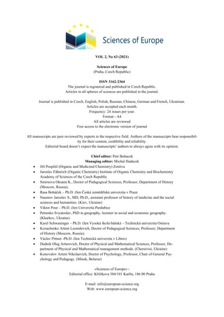
  • 9. Sciences of Europe # 63, (2021) 9 CHEMICAL SCIENCES ВИВІЛЬНЕННЯ ПОЖИВНИХ РЕЧОВИН КОРМІВ ШЛЯХОМ ЕКСТРУЗІЇ ЗЕРНОБОБОВОГО ФУРАЖУ Морозова Л.П. Вінницький національний аграрний університет, старший викладач Новаковська В.Ю. Інститут кормів та сільського господарства Поділля НААН, молодший науковий співробітник, Вінниця RELEASE OF FEED NUTRIENTS BY EXTRUSION OF LEGUMES Morozova L. Vinnytsia National Agrarian University, Senior Lecturer Novakovska V. Institute of Feed Research and Agriculture of Podillya NAAS, junior researcher Vinnytsia DOI: 10.24412/3162-2364-2021-63-2-9-12 АНОТАЦІЯ Виробництво екструдованих комбікормів для сільськогосподарських тварин є перспективним напрямком кормовиробництва. Використання такого обладнання економічно доцільно, оскільки забезпе- чує можливість оперативного зміни рецептур комбікормів з урахуванням наявної сировинної бази. У роботі досліджено вплив технологічних параметрів процесу екструдування: температури і ступеня помелу сировини на зміну хімічного складу і руйнування клітковини в бобових культурах: сої, кормових бобах, горосі і нуті. Згідно з отриманими даними, використання методу екструдування при обробці сої, кормових бобів, гороху та нуту дозволяє збільшити в них, в порівнянні з натуральним зерном, весь ком- плекс поживних речовин (кількість обмінної енергії, вміст сухої речовини, сирого і перетравного протеїну, БЕР, цукру) і, навпаки, знизити вміст сирої клітковини. ABSTRACT The production of extruded feed for farm animals is a promising area of feed production. The use of such equipment is economically feasible, as it provides the ability to quickly change the formulations of feed, taking into account the available raw material base. The paper investigates the influence of technological parameters of the extrusion process: temperature and degree of grinding of raw materials on the change of chemical composition and destruction of fiber in legumes: soybeans, fodder beans, peas and chickpeas. According to the obtained data, the use of extrusion method in the processing of soybeans, fodder beans, peas and chickpeas allows them to increase, compared to natural grain, the whole range of nutrients (metabolic energy, dry matter, crude and digestible protein, NE, sugar ) and, conversely, reduce the crude fiber content. Ключові слова: комбікорми, екструдування, екструдер, сирий протеїн, сирий жир, сира клітковина, безазотисті екстрактивні речовини (БЕР). Keywords: compound feeds, extrusion, extruder, crude protein, crude fat, crude fiber, nitrogen-free extrac- tives (NE). To increase the nutritional value and more rational use of feed grain, various methods of its processing are used - grinding, roasting, cooking and steaming, sweet- ening, extrusion, micronization, flattening, flaking, re- covery, yeasting. Extrusion is the processing of grain under the ac- tion of high pressure and temperature. Pre-cleaned grain is fed into an extruder in which the pressure is 28 atm and the temperature is 130–150 °C. Grain extrusion leads to an increase in its composition of sugar, dex- trins, hemicellulose and a decrease in the content of starch and cellulose (true fiber). The extrusion process has a significant effect on the protein complex of the grain, increases its biological value. The extrusion process is as follows. In special de- vices, the feed components are crushed, mixed into a homogeneous mass, compacted. The resulting high temperature destroys harmful microorganisms and tox- ins. Most soybeans are extruded in special screw-type devices - extruders (Fig. 1). Extruder Insta-Pro 2000 al- lows to extrude legumes of natural humidity at an outlet temperature from 140 to 160 ℃. The value of the pressure on the feed mass inside the extruder reaches 28-30 atm. This is due to repeated compression of the soybean mass with the screws of the auger, the pitch of which is constantly decreasing to- wards the output of the product. Therefore, the pressure significantly depends on the design features of the au- ger, its speed of rotation, the initial humidity of the raw material. As a result of the interaction of high pressure and temperature in the feed mass, deep biochemical processes occur, which significantly improve the as- similation of feed and neutralize anti-nutritional fac- tors.
  • 10. 10 Sciences of Europe # 63, (2021) Fig. 1. Extruder Insta-Pro 2000 Extrusion as a technological process differs favor- ably from cold pressing, as it allows to increase the nu- tritional properties of the processed feed components. As a result of such processing complex structures of proteins and carbohydrates break down into simpler ones, fiber into secondary sugars, starch into simple sugars, and in legumes neutralization of protease inhib- itors: trypsin and urease. In addition, the temperature exposure leads to improved hygienic condition of feed components due to the destruction of unwanted micro- flora [1]. The use of extrusion has a number of advantages: - mechanical grinding: the obtained fine feed structure is quite desirable for optimal digestion. Ex- pansion at the outlet of the extruder nozzle leads to the destruction of the internal structure of the material, fa- cilitating its digestion, as well as to increase the surface area of the feed, which accelerates the absorption of nu- trients in the digestive tract; - destruction of the structure (denaturation) of pro- teins: short-term heating above 100 ° C with simultane- ous exposure to high pressure in the extruder very ef- fectively changes the structure of proteins (coagulation, denaturation), thereby increasing the energy value of feed; - deactivation of unwanted enzymes; - radical reduction of antinutrients and natural tox- ins: extrusion very effectively neutralizes a number of antinutrients. For example, in soybeans after extrusion, urease activity is clearly reduced. In the feed for mo- nogastric animals a very positive point is the decrease in trypsin inhibitor; - sterilization: temperature and pressure in the ex- truder reliably destroy all bacteria, fungi and other un- wanted microorganisms and pests. The growth of mold and the release of mycotoxins stops, which allows you to extend the shelf life; - jelly formation of starch: starch is a very frequent and important element of feed. In the process of extru- sion, complex starchy carbohydrates and sugar are con- verted into simple ones, which simplifies the digestibil- ity of feed; - homogenization and the possibility of formation: in the extruder all the components of the feed are mixed. Squeezing through the molding matrix, the feed can be given different shapes. The condition for obtain- ing and maintaining the required shape is the correct composition of the extruded raw material - a sufficient content of binders (most often - starch) [2]. The grain of almost all legumes requires appropri- ate treatment before feeding, which significantly in- creases the efficiency of its use by animals. Soybeans are the leader among grain feeds in terms of energy, protein and fat nutrition. 1 kg of soy- beans contains 1.45 feed. units, 14.7 - 15.0 MJ of met- abolic energy. The content of crude protein is 35-45%, fat - 16-22%, crude fiber - 7%. Soybeans can be fed to all species of animals as a protein supplement for defi- ciencies in feed rations of protein and to balance them in amino acids. 1 kg of soy contains the following number of amino acids (g): lysine - 21.1, methionine - 4.6, histi- dine - 7.6, tryptophan - 4.3, threonine - 12.6, valine - 18.0, arginine - 26.6, leucine - 26.2, isoleucine - 17.6, phenylalanine - 17.0. The digestibility of soybean or- ganic matter averages 85–87%. Assimilation of soy protein reduces the presence of anti-nutrients in its composition. The most significant anti-nutritional factors in soy are inhibitors of proteolytic enzymes: trypsin and chy- motrypsin. These inhibitors are factors of protein na- ture, which account for at least 3 - 6% by weight of soy- bean protein. Protein nature is characterized by anti-nu- trient enzymes of soy - urease and lipoxidase. The first enzyme urease - destroys quality proteins and amino acids in food in the body and converts them into a toxic substance - ammonia. Lipoxidase destroys the vitamins of the finished feed, in particular irreversibly breaks down vitamin A - retinol, which causes typical hypo- vitaminosis and avitaminosis A. Hematoglutinins and specific soy protein also have a negative effect on feed consumption and digestibility [3]. To inactivate anti-nutrient factors, soybeans should be subjected to heat treatment before feeding: steaming, autoclaving, extrusion, etc. The best method of inactivation of soybean anti-nutrients [4] is the pro- cessing of grain in a barothermal chamber, where it is heated by contact heat transfer from the surface of the
  • 11. Sciences of Europe # 63, (2021) 11 heating elements, active mixing of grain mass and steaming due to moisture of soybean grain. Soy milk production includes soaking soybeans for 14–16 hours, homogenizing, and cooking at 103–105 °C for 40–60 minutes. The composition of compound feeds and feed mixtures rations processed soybeans can include: for adult pigs and young animals older than 2 months of age - up to 15%, pigs for fattening - up to 10%; for cat- tle - up to 10%. Peas are one of the best legumes for animals. It has an advantage over other legumes because it contains significantly fewer anti-nutrients that adversely affect digestion, nutrient use and animal health. 1 kg of pea grain contains an average of 1.18 feed. units, 218 g of digestible protein and 14.2 g of lysine. The dry matter content is low in fat - 1.9% and fiber - 5.4%. The digestibility of organic matter is 87%. Peas are fed to all species of animals. Its inclusion in the diets of dairy cows (1 - 2 kg per day) leads to increased milk yield and improved milk composition. In the diets of pigs for fattening peas helps to improve the quality of meat and the formation of dense granular fat. Peas are included in the feed mixtures of calves to reduce the rate of whole milk. Peas should be fed crushed (in the form of grits) or ground. Cooking or steaming peas before feeding significantly improves the use of nutrients by animals. The norms of inclusion of pea grits in the compo- sition of feed and feed mixtures of rations are for cattle: cows and fattening - up to 15%, calves up to 6 months of age - up to 6%, young cattle - up to 10-15%, breeding bulls - up to 5 %; for pigs: adults - up to 15-20%, piglets up to 2 months of age - up to 5%, piglets from 2 to 4 months - up to 10%, during fattening - up to 20%; for sheep: adults - up to 10%, lambs - up to 5%; for horses - up to 10%; for poultry: adult chickens, ducks, geese, turkeys - up to 12%, young - up to 10% (by weight). Fodder beans have recently become increasingly com- mon as a source of protein (the content of which is from 25 to 33%). Bean protein contains all the essential amino acids for animals. Bean protein consists of al- most 90-95% protein. Digestibility of nutrients by beans by animals is quite high. For example, in pigs the digestibility of protein is 84%, fat - 75%, nitrogen-free extractives - 88%. 1 kg of feed beans contains an average of 1.1 feed. units, 12.4 MJ of metabolic energy, 227 g of digestible protein, 16.2 g of lysine. Beans contain less trypsin inhibitors than soy and lupine, but their use is limited by tannins, which can cause digestive disorders in animals. Therefore, when feeding beans, it is recommended to include wheat bran and chalk mass, which have a laxative effect, in the diet, or to carry out their heat treatment. Due to extrusion, the level of introduction of leg- umes into compound feeds for growing young pigs up to 4 months of age reaches 25-30%. During this, the di- gestibility of protein and the absorption of metabolic energy increases by 20-25%. Experiments conducted at the Institute of Agriculture and Animal Husbandry of the western region showed that the introduction of com- pound feeds for young pigs 12.5% by weight of ex- truded and crushed lupine grain improved the physio- logical condition of pigs, the average daily gain in- creased by 6.4%. The introduction of extruded soybeans in the diets of pregnant sows in the amount of 20% by dry matter increases the content of linoleic acid to 2.9% of dry matter, which increases the fertility, milk yield and safety of piglets. The addition of extruded and barothermally treated soybeans in the feed of piglets in- creases the growth rate by 19.6 - 14.6% with a decrease in feed costs per 1 kg of growth by 16.3 - 13.1% [4]. The purpose of this work was to investigate the forage capacity of extruded forage. Materials and methods The objects of the study were samples of legumes used in the production of feed - soybeans, fodder beans, peas and chickpeas. Extrusion of feed raw materials was performed on a single-screw extruder Insta Pro 2000 made in the USA with a capacity of 600-900 kg / h (2000R). Ex- truders of this type are "dry" extruders and have a sim- ple and economical process. At rather small dimensions they have rather high productivity. Rational parameters of feed extrusion are established: fineness of grinding of raw materials with a nozzle diameter of 7,5 mm; product temperature at the outlet of the extruder – 140 ºC. Results and discussion To study the effect of extrusion on the transfor- mation of nutritional properties, the chemical composi- tion of feed crops of components before and after ex- trusion at a temperature of 140 ºC was studied. Data on changes in the chemical composition of feed components in natural and completely dry matter before and after extrusion are given in tables 1 and 2. Table 1 Chemical composition in natural substance, % in samples of soybeans, fodder beans, peas, chickpeas before and after extrusion № Chemical indicator The name of the legume Soybean Fodder beans Pea Chickpea original extruded original extruded original extruded original extruded 1. Dry matter 92,53 93,19 91,28 92,35 91,93 91,88 93,15 93,51 2. Protein 36,88 38,76 24,24 25,04 20,80 20,68 21,81 21,05 3. Fat 20,09 16,91 0,49 1,15 1,44 1,28 6,70 6,38 4. Cellulose 8,33 5,42 8,53 7,82 6,10 6,06 3,17 2,35 5. Ash 5,64 5,96 3,55 2,64 3,33 3,20 3,60 3,64 6. NЕ 21,59 26,14 54,47 55,70 60,26 60,66 57,87 60,09
  • 12. 12 Sciences of Europe # 63, (2021) Table 2 Chemical composition in absolutely dry matter,% in samples of soybeans, fodder beans, peas, chickpeas before and after extrusion № Chemical indicator The name of the legume Soybean Fodder beans Pea Chickpea original extruded original extruded original extruded original extruded 1. Protein 39,86 41,59 26,56 27,11 22,63 22,51 23,41 22,51 2. Fat 21,71 18,15 0,54 1,24 1,57 1,39 7,19 6,82 3. Cellulose 9,00 5,82 9,34 8,47 6,64 6,60 3,40 2,51 4. Ash 6,10 6,40 3,89 2,86 3,62 3,48 3,86 3,89 5. NЕ 23,33 28,05 59,67 60,32 65,55 66,02 62,13 64,26 According to the chemical composition of the nu- trients of the complete zootechnical analysis, the con- tent of crude protein in extruded samples of soybeans and fodder beans increases, while in peas and chickpeas it remains virtually unchanged. The extrusion process promotes the increase of crude fat content in feed beans. This is due to the fact that in the process of fric- tion of the raw material in the extruder was the destruc- tion of cell walls, which caused the release of fat. The amount of fiber after extrusion in all tested samples de- creases. It should also be noted the increase in the share of nitrogen-free extractives (NE), which combine car- bohydrates and proteins that significantly increase the caloric content of the products formed. The obtained results are consistent with the litera- ture data [5, 6, 7], according to which during the extru- sion of grain and oilseeds the crude protein content un- derwent significant changes, and at high crude fat con- tent in the seeds there were losses. As a result, there was a redistribution and increase in ash content, the amount of protein and carbohydrates in the extruded product. Сonclusions 1. The use of the extrusion method in the pro- cessing of legumes allows them to increase, compared to natural grain, the whole complex of nutrients (meta- bolic energy, dry matter, crude and digestible protein, NE) and, conversely, reduce the crude fiber content. 2. The introduction of extruded compound feeds- concentrates in the diets of farm animals and poultry increases the intensity of metabolic processes in the body of animals, helping to improve productivity. References 1. Щербакова О.Е. Комбикормовое производ- ство предприятий малой мощности. М.: МГУТУ. 2012. 54 с. 2. Григорьев Д. Ю. Подготовка кормов к скармливанию. Свиноводство. 2016. № 1. С. 14–20. 3. Подобєд Л.І., Курнаєв О.М. Питання за- готівлі, зберігання та використання кормів в умовах інтенсивної технології виробництва молока. Одесса: Друкарський дім, 2012. 456 с. 4. Петриченко В.Ф. Наукові основи вироб- ництва і використання зерна сої. Корми і кормови- робництво. 2012. Вип. 7 . С. 3–11. 5. Маркелова В.Н., Фомичев Ю.П., Никанова Л.А. Химический состав экструдированного зерна зерновых, зернобобовых и масличных культур. Кормопроизводство. 2014. № 9. С. 41–44. 6. Артемов Р.В., Арнаутов М.В., Бочкарев А.И., Баскакова Ю.А., Артемов А.В., Кокшаров А.Е., Биндюков С.В.. Обоснование рациональных параметров экструдирования растительных компо- нентов на оборудовании малой мощности для полу- чения комбикормов для аквакультуры. Труды ВНИРО 2019. т. 176. С. 182–192. 7. Касьянов Р. О., Смоловская О. В., Белова С. Н. Экструдированный корм в рационах сельскохо- зяйственных животных. Аграрные проблемы гор- ного алтая и сопредельных регионов. 2020. С. 176– 183. 8. Попов А. Н. Способы повышения углевод- ной полноценности концентрированных кормов. Приоритетные направления регионального разви- тия. 2020. с. 761–764. 9. Латышевич Е. А., Веселовский Г. В., Анто- нишин Ю. Т. Эффективность использования пита- тельных веществ, полученных экструзией. – 2020. 10. Фролов Д. И., Потапов М. А. Увеличение эффективности работы одношнекового экструдера. Инновационная техника и технология. 2020. №. 2. С. 42–47.ї 11. Баитаев М. О., Гаплаев М. Ш. Изменения химического состава и питательности соевых бобов в производстве кормов и скармливании их живот- ным. Инновационная деятельность как фактор раз- вития агропромышленного комплекса в современ- ных условиях. 2020. С. 235–239.
  • 13. Sciences of Europe # 63, (2021) 13 MEDICAL SCIENCES MODERN VIEW ON ULTRASOUND DIAGNOSTICS IN OBSTETRICS Shepetovskaya N. Sechenovsky University, Moscow, Senior Lecturer, Ph.D. Kalenteva S. Medical League of Russia, Moscow, Prof., Ph.D., M.D. Shepetovskaya M. Sechenovsky University, Moscow, Student, Faculty of Medicine DOI: 10.24412/3162-2364-2021-63-2-13-19 ABSTRACT The article provides a review of the modern literature on the use of ultrasound diagnostics in obstetrics and its effectiveness in different trimesters of pregnancy, during childbirth and the postpartum period. Ultrasound is a non-invasive method that does not create radiation exposure, which is an ideal imaging method during pregnancy. Modern technologies and techniques include abdominal and transvaginal 2D scanning, M-mode imaging, Doppler and color Doppler mapping, energy color Doppler imaging, and 3D ultrasound. To date, ultrasound diagnostics continues to actively develop and improve. Keywords: ultrasound diagnostics, dopplerography, trimester of pregnancy, childbirth, the postpartum pe- riod, obstetrics. Modern advances in clinical diagnostics are largely determined by the improvement of research methods. Significant progress in this area has been achieved thanks to the development and implementa- tion of fundamentally new methods of obtaining medi- cal images, including the ultrasound method. Ultrasound is a non-invasive method that does not create radiation exposure, which is an ideal imaging method during pregnancy. Due to the evolutionary im- provement of ultrasound technologies in recent years, there are now a number of unique methods of ultra- sound examination, the use of which depends on the duration of pregnancy and the purpose of the study. Modern technologies and techniques include ab- dominal and transvaginal 2D scanning, M-mode imag- ing, Doppler and color Doppler mapping, energy color Doppler imaging, and 3D ultrasound. The use of ultrasound diagnostics in obstetrics and gynecology begins in 1966, when in Europe, the United States and Japan actively began to apply ultrasound di- agnostics in this specialty. Both a - and b-modes were used, including the first "fast b-scanner", the Vidoson ® from Siemens ®, used by D. Hofmann and Hans Hol- länder at the Wilhelm University in Munster, Germany [18]. Ultrasound examination in obstetric practice is the most important diagnostic tool that allows you to iden- tify a wide range of different pathologies in early preg- nancy, such as undeveloped and ectopic pregnancy [8], fetal malformations [2, 21]. To assess the rate of fetal development, to predict and prevent a number of ob- stetric syndromes, such as fetal growth retardation [48], preeclampsia [37], preterm birth [6], antenatal fetal death [1]. The main tasks of ultrasound examination in ob- stetrics [3]: − establishing the fact of pregnancy, monitoring its course; − determination of the number of fetal eggs; − embriometry and fetometry; − diagnosis of fetal abnormalities; − evaluation of the functional state of the fetus; − placentography; − implementation of control during invasive stud- ies. Ultrasound examination in the first trimester of pregnancy The purpose of ultrasound in the first trimester of pregnancy [7, 43]: − to establish the presence of uterine pregnancy on the basis of visualization of the fetal egg in the uterine cavity; − with the exception of ectopic pregnancy; − diagnosis of multiple pregnancies; − evaluation of fetal egg growth (average internal diameter of the fetal egg, coccygeal-parietal size and biparietal size of the embryo/fetus); − assessment of the vital activity of the embryo (cardiac activity and motor activity); − study of the anatomy of the embryo/ fetus. De- tection of chromosomal abnormalities ecomercio; − study of extraembryonic structures (yolk sac, amnion, chorion, umbilical cord); − diagnosis of pregnancy complications (threat- ened/initiated / complete abortion, cystic drift); − diagnosis of genital pathology (uterine fibroids, uterine abnormalities, intrauterine pathology, ovarian formations). However, the use of ultrasound screening in the first trimester of pregnancy up to 10 weeks is impracti- cal and should be carried out according to strict indica- tions, since it is impossible to completely exclude the possible adverse effects of ultrasound on the processes of embryogenesis and the development of pregnancy in general [30]. Indications for an emergency immediate examination in early pregnancy include [3]: • pain in the lower abdomen, especially in the presence of risk factors for ectopic pregnancy in patients who have not yet undergone ultrasound; • massive bleeding; • sys- temic symptoms of blood loss.
  • 14. 14 Sciences of Europe # 63, (2021) In the first trimester of pregnancy, transvaginal ul- trasound is mainly used. One of its advantages is that it does not require a full bladder when examined. It also provides a higher image resolution compared to the ab- dominal one. This is especially important in pregnant women with retroflexia of the uterus. However, there are also disadvantages. One of them is that transvaginal ultrasound shows the pelvic organs in a projection dif- ferent from the abdominal one. In addition, it has lim- ited scanning capabilities for higher-placed structures [29]. The first sign that the patient may be pregnant is the presence of an eccentrically located small cyst within one layer of the endometrium. Soon, other signs also appear, such as the "double bleb sign" symptom, the presence of a yolk sac, and then the echogenic cyl- inder, which is the embryonic pole. The echogenic rim around the fetal egg in the early stages of development is a decidual reaction [7]. During the first screening ultrasound (10-14 weeks), it is important to diagnose a number of severe fetal malformations (anencephaly, acrania, some limb malformations, etc.) [24, 30]. One of the most common developmental abnormalities is congenital heart dis- ease (CHD) [23]. However, the sensitivity of echogra- phy in the diagnosis of congenital heart defects at the first screening level does not exceed 30% and a high frequency of false-positive results is noted [7]. Biometrics in the early stages of uncomplicated pregnancy can be limited to measuring the average in- ternal diameter of the fetal egg and the coccygeal-pari- etal size of the embryo. The error in determining the term of pregnancy in the first trimester can be mini- mized if the rules of biometrics are strictly followed [30, 43]. After that, it is necessary to evaluate the signs of vital activity of the embryo, which include cardiac activity (recorded from the beginning of week 6) and motor activity (determined after week 7). It is important to assess the condition of the inter- nal pharynx and cervix at an early stage, since isthmic- cervical insufficiency can be the cause of termination of pregnancy [30]. Another risk factor for preterm birth may be the presence of uterine abnormalities in a preg- nant woman [28] and this is associated with a short length of the cervix [26]. Predictors of pregnancy pathology in the early stages of pregnancy are the absence of fetal cardiac ac- tivity, calcification of the yolk sac, etc. [7]. The absence of a heartbeat of the embryo / fetus is considered a sign of death, but it should be remembered that in a very small embryo (<4 mm), heart contractions may not be detected, so ultrasound should be performed again after a few days. The value of the embryo length of 5 mm is a threshold value detected during ultrasound examination, in which cardiac activity should be visu- alized in all cases [7]. Ultrasound examination in the second tri- mester of pregnancy The purpose of ultrasound in the second trimester of pregnancy [3, 43]: − оценка роста плода; − диагностика пороков развития; − исследование маркёров хромосомной патологии; − диагностика ранних форм задержки разви- тия плода; − оценка локализации, толщины и структуры плаценты; − определение количества околоплодных вод. During this time, ultrasound examination makes it possible to diagnose most of the congenital malfor- mations of the fetus. However, not all of them. This may be due to the following reasons. First, some mal- formations may appear later in pregnancy, or they are diagnosed only in the third trimester of pregnancy. Sec- ondly, the quality of prenatal diagnostics is signifi- cantly affected by the capabilities and resolution of ul- trasound devices [30]. The spectrum of echomarkers of fetal chromoso- mal pathology detected in the second trimester of preg- nancy includes changes in various organs and systems: ventriculomegaly, cysts of the vascular plexuses of the lateral ventricles, abnormal forms of the skull and cer- ebellum ("strawberry", "lemon", "banana"), hyperecho- genic intestine, pyelectasia, a single umbilical cord ar- tery, a symmetrical form of fetal development delay [3]. As part of the diagnosis of intrauterine growth re- tardation, it is important to determine the size of the fe- tus 'head, since when the fetus' development is delayed, its brain continues to grow, despite the fact that the de- velopment of other parts of the body slows down. To improve the effectiveness of the diagnosis, it is neces- sary to use additional parameters when measuring the fetal head, trunk (the average diameter of the chest), fe- tal limbs (the length of the radius and ulna, the width of the scapula, the length of the foot). The determination of the estimated fetal weight in the diagnosis of intrau- terine growth retardation is also of great practical im- portance. The calculation of the expected fetal weight is carried out on the basis of fetometry. For this pur- pose, the use of various formulas is proposed [30, 43]. In assessing the nature of the course of pregnancy, it is essential to determine the volume of amniotic fluid, which is an integral part of the antenatal ultrasound ex- amination and serves as a diagnostic test for identifying pregnant women at high risk for the development of perinatal pathology [29]. A decrease in amniotic fluid to 500 ml or less (at the end of pregnancy) is considered low water, and an increase of more than 1500 ml is con- sidered polyhydramnios. Low water content occurs in the second and third trimesters, and high water content occurs in the third trimester [43]. A significant tortuos- ity of the amnion indicates an excessive number of ac- tive-secreting amniocytes. In some observations with polyhydramnios, thickening and compaction of the compact layer was revealed, which leads to blocking of water resorption. In the course of the study, not only the fact of an abnormal amount of amniotic fluid is rec- orded, but also the degree of severity of this pathology, despite the significant element of subjectivity of such a diagnosis. Estimation of the volume of amniotic fluid during an echographic examination is possible by de- termining the average diameter of randomly selected free spaces ("pockets") filled with water. The volume
  • 15. Sciences of Europe # 63, (2021) 15 of ocular water is considered normal if the average di- ameter of the "pockets" is within 2-8 cm. Polyhydram- nios are characterized by the size of "pockets" of more than 8 cm, and low-water-less than 2 cm. The presence of a free "pocket" of less than 1 cm in size is taken as a pronounced lack of water [30, 43]. With the help of ultrasound in these terms, you can study the placenta in detail and get the necessary infor- mation about its localization, thickness, and structure [29, 30]. The location of the placenta in different periods of pregnancy changes due to the "migration" from the lower segment to the bottom of the uterus. If a placenta previa is detected before 20 weeks of pregnancy, the ultrasound should be repeated every 4 weeks. The final conclusion about the location of the placenta should be made at the end of pregnancy. Information about the state of the placenta can be obtained by placentometry [3, 30]. Most often, the thickness of the placenta is determined, since this meas- urement is the most accurate and simple. The determi- nation of the area and volume of the placenta due to the complexity of the technique is not widely used in the diagnosis and is used only in individual cases. During the physiological development of preg- nancy, the thickness of the placenta continues to in- crease until the term of 35-36 weeks and then decreases slightly, amounting to an average of 3.4 cm in the para- central part at the time of delivery [3]. The size of the placenta may vary depending on the existing pathology, the severity and duration of the process. An increase in the thickness of the placenta is often detected with iso- serological incompatibility of the blood of the mother and fetus, diabetes mellitus, non-immune dropsy of the fetus, the presence of a large fetus. With the threat of termination of pregnancy, gestosis, delayed intrauterine development of the fetus, some malformations and low water content, most often there is a decrease in the size and thinning of the placenta [29, 30]. During pregnancy, changes in the echographic structure of the placenta occur, which are mainly due to involutive-dystrophic processes [30]. With the help of ultrasound examination of the placenta, it is possible to diagnose such a terrible com- plication of pregnancy as placental abruption, which is a consequence of a violation of the mechanisms of its attachment to the wall of the uterus, followed by bleed- ing from the vessels of the decidual membrane. Ultra- sound diagnosis of premature placental abruption is based on the detection of the echonegative space be- tween the uterine wall and the placenta. The size of the hematomas is of great importance. The echographic im- age of hematomas varies depending on the age of their existence [30, 43]. Ultrasound is also used to diagnose the con- sistency of the postoperative scar on the uterus. This condition is associated with a high risk of uterine rup- ture with the progression of pregnancy and massive bleeding [39]. The consistency of the scar is evidenced by the homogeneous structure of the tissues and the smooth contours of the lower segment of the uterus, its thickness is not less than 3-4 mm. The failure of the scar on the uterus is diagnosed based on the detection of a defect in the form of a deep niche, thinning in the area of the alleged scar, the presence of a large number of hyperechoic inclusions (connective tissue) [30]. Patients with a scar on the uterus after cesarean section and with low placentation (presentation) of the placenta in the second trimester of pregnancy are con- sidered to be at high risk for placental increment [44, 20]. Ultrasound examination in the third trimester of pregnancy The purpose of ultrasound in the third trimester of pregnancy [3, 43]: − diagnosis of malformations with late manifesta- tion; − determination of fetal developmental delay; − assessment of the functional state of the fetus (assessment of motor and respiratory activity, Doppler blood flow in the "mother-placenta-fetus" system). The main purpose of this screening is to identify placental dysfunction (intrauterine development delay, lack of water, pathological form of velocity waves in the Doppler study) [35, 40]. When studying the growth and development of the fetus in the second and third trimesters of pregnancy, fetometry is performed. Fetometry is a required com- ponent of ultrasound in obstetric practice and allows you to set according to the size of the fetus in pregnancy and to assess its growth, to clarify the term of preg- nancy, to diagnose fetal growth retardation and congen- ital malformations [29, 30]. The mandatory volume of fetometry includes the measurement of the biparietal size and circumference of the head, the diameter or circumference of the abdo- men, and the length of the femur (the length of the tub- ular bones is measured on both sides). There are tabular values of standard gestational indicators of fetometry. Based on these parameters, it is possible to determine the estimated weight of the fetus [3, 30]. If one or more of the main fetometric indicators are found to be inconsistent with the gestational age, extended fetometry is necessary [30]. Another important indicator is the assessment of the degree of maturity of the placenta according to the classification proposed by P. A. Grannum et al. (1979). Based on the echographic characteristics of the chorial plate, placental parenchyma, and basal layer, depend- ing on the gestation period, four degrees of placental maturity are distinguished: 0, I, II, and III. In the phys- iological course of pregnancy, structural changes in the placenta occur in parallel with the development and maturation of the fetus [29, 30]. A number of other characteristics of the placenta that may have pathological signs deserve attention: ad- ditional pathological inclusions in the structure of the placenta, its thickness and location Ultrasound scanning reveals pictures of cystic changes, a sign of which are echonegative formations of various sizes. However, true placental cysts are rare. Large cysts can contribute to the development of atrophic processes in the placenta due to compression of the surrounding tissues, which adversely affects the development of the fetus. The presence of multiple
  • 16. 16 Sciences of Europe # 63, (2021) small cysts can also negatively affect the function of the placenta [7]. Another important obstetric problem is pathologi- cal placentation (placenta previa and placenta accreta) [25]. Antenatal diagnosis is determined by a combina- tion of anamnestic, clinical, and instrumental data. The leading role among instrumental research methods is assigned to ultrasound diagnostics [44, 20]. However, Z. S. Bowman et al. [9] in their study, they found that the sensitivity of the method is only 53% and the spec- ificity is 88%, that is, in every second pregnant woman, the diagnosis of placental ingrowth is not confirmed by ultrasound data. True placental increment, which is extremely rare, is a serious complication. The echographic picture with a true increment of the placenta in cases of its presen- tation is characterized by the presence of multiple hy- poechoic formations between the area of the internal pharynx and the placental tissue, which are embedded in the myometrium. A great help in the diagnosis of this pathology is provided by the Doppler study, which re- veals the penetration of placental vessels into the my- ometrium [12, 30]. With the help of ultrasound, you get valuable in- formation about the state of the cervix during preg- nancy and about the risk of premature birth. Transvag- inal echography has now become an almost non-alter- native method of investigation used to assess the condition of the cervical canal and internal pharynx [16]. It has 100% sensitivity and 80% specificity. Ac- cording to the 2015 FIGO recommendations [25], the length of the cervical canal with transvaginal ultra- sound cervicometry of 35 mm or less indicates a threat of preterm birth, 25 mm or less-a high risk of direct pre- term birth. The expansion of the internal pharynx to 5 mm or more, especially to 10 mm, also indicates a high risk of premature birth. Using ultrasound cervicometry, the existence of a latent and active period of labor is clearly confirmed. It was found that with the onset of labor, there is a trans- formation of the cervix: its shortening with increasing thickness. The smoothing process is carried out sequen- tially in four stages [10]: T-stage (when the cervix is formed and does not differ from that in the last weeks of pregnancy), Y -, V-stage and U-stage (corresponding to the complete smoothing of the cervix). In cases of induction of labor by oxytocin, cervical smoothing oc- curred in the same way, but in a shorter time. Echogra- phy data did not reveal any difference in the mechanism of cervical smoothing between first-and second-born women, i.e. smoothing in all cases precedes disclosure, which differs from the results of a bimanual study. Ultrasound examinations during childbirth In classical obstetrics, to assess labor, determine the type and position of the fetus, the location of its head, as well as to decide on the timing and method of surgical intervention, the doctor is traditionally guided by the data obtained during the vaginal examination. This approach, of course, remains fundamental, alt- hough its inherent subjective nature cannot be denied. The use of echography during labor was previ- ously insignificant and was mainly used for cephalom- etry when planning labor. This was due to the high cost of ultrasound machines and the inability to have an ul- trasound machine in every delivery room. In recent years, with the development and cheapening of ultra- sound technologies in the world, there has been a rapid introduction of diagnostic ultrasound in childbirth. As at the time the ultrasound is a revolution in the antenatal surveillance, as is happening now and in the manage- ment of labor. This approach is non-invasive, provides real-time results, and allows you to instantly make a de- cision in a critical situation [22, 45]. Ultrasound examination in childbirth aims to as- sess the progress of the fetal head through the bone ring of the pelvis, to show the possibility of natural delivery in this particular case, or to give a reliable conclusion about the need for surgical intervention. During ultra- sound examination in childbirth, transperineal access is used. Currently, several methods of transperineal ultra- sound examination in the second period of labor have been developed in the world in order to assess the pro- gress of the fetal head. One of the most accessible and informative is the progression angle [45]. It represents the angle between the long axis of the pubic joint and the line drawn from the lower edge of the symphysis along the lowest point of the bony structures of the fetal head. This parameter, first proposed in 2009, allows for fairly accurate determination of the head's progress in the pelvic cavity [13, 31, 33]. Extensor options for inserting the fetal head can be excluded in the longitudinal scan, when the fetal chin is pressed against the chest. The clinical diagnosis is made on the basis of palpation of sutures, fontanelles and determination of bone landmarks of the pelvis of the mother, but the use of ultrasound improves the di- agnosis of these abnormalities in childbirth [4]. At the same time, according to Sherer D. M. (2002) et al., when comparing the data of ultrasound and vaginal studies to determine the position of the fetal head, the error rate reaches 76% [41, 42]. The combination of transabdominal and transper- ineal studies allows the most accurate diagnosis of syn- clitic and asynclitic insertions, flexor and extensor po- sitions of the fetal head [5]. Transperineal ultrasound examination in the sec- ond period of labor is highly informative for assessing the progress of the fetal head. Proper evaluation of the results allows you to reduce the number of unnecessary caesarean sections. On the other hand, in the presence of a birth tumor in the fetus, ultrasound with the meas- urement of the angle of progression makes it possible to make the right timely decision in favor of a cesarean section. The preservation of echograms in electronic form and on paper allows you to document the deci- sions made in terms of changing the tactics of labor management [14]. The use of ultrasound in childbirth improves the diagnosis of incorrect positions of the fetal head, con- tributes to the prevention of complications from the mother and fetus during obstetric operations, but leads to an increase in the frequency of cesarean sections when predicting prolonged labor [27]. The use of ultra- sound examination in childbirth does not allow to diag-
  • 17. Sciences of Europe # 63, (2021) 17 nose anomalies of labor activity (weakness, discoordi- nation of labor activity) in the first and second periods of labor [34]. There are two main situations in which intra-natal ultrasound examination is advisable [15, 22]. The first situation is a suspicion of weakness of labor activity in the first or second period of labor, as well as the need for differential diagnosis of clinical in- consistency of the fetal head with the pelvis of the de- livery woman. In this case, you should focus on the an- gle of progression or the distance from the fetal head to the perineum during transperineal ultrasound, as well as on the position of the fetus during transabdominal ultrasound. The second situation is predicting the success of operative vaginal delivery (including manual rotation of the fetal head from the posterior view of the occipital insertion to the anterior one). In this case, the practi- tioner should evaluate the position of the fetal head dur- ing transabdominal ultrasound and the position of the head relative to the spinal plane — during transperineal ultrasound. The most reliable sonographic parameters for predicting the result of this procedure are the dis- tance from the head to the perineum and the angle of progression; an assessment of the median angle and/or direction of the fetus may also be useful. The use of ultrasound navigation in childbirth can- not and should not completely replace vaginal exami- nation. If, however due to various circumstances (ex- pressed tribal tumor, asynclitic) to obtain reliable infor- mation via this is impossible, the echographic pattern can become a reliable assistant practitioner, especially in situations when the question about the appropriate- ness of surgical intervention [19]. Ultrasound examinations in the postpartum period The postpartum period is called the first 6 weeks after delivery. For the first time, ultrasound examina- tion for the study of postpartum changes was used by H. P. Robinson in 1972 [36]. Although the postpartum period is a time of phys- iological involutional processes, events that accompany delivery through the natural birth canal or by c-section often lead to complications in a significant number of patients. These negative consequences are most often associated with the occurrence of bleeding or an infec- tious process. When assessing postpartum bleeding, the ultra- sound method is used as a source of diagnostic infor- mation, which is due to its exceptional diagnostic value, mobility and safety of modern devices [29]. During ultrasound examination, it may be difficult to make a differential diagnosis between clots, which are generally more homogeneous, and placental frag- ments, which are characterized by areas of increased echogenicity in their structure. B. S. Hertzberg and J. D. Bowie [17] proposed to distinguish 5 types of echo- graphic picture of the uterine cavity in patients with suspected placental tissue remnants. According to their data, the most characteristic sign of the presence of res- idues was the detection of a volumetric hyperechoic formation in the uterine cavity. Another problem of the postpartum period in mod- ern obstetrics is infectious and inflammatory diseases, which is due to their high frequency - 5-26% and their role in the structure of maternal mortality. Subinvolu- tion of the uterus and hematolochiometry are character- ized by an increase in the quantitative indicators of three-dimensional Dopplerography in all parts of the uterine vasculature due to pronounced vascular dilata- tion, and subclinical and clinical forms of endometritis are characterized by a decrease in the quantitative indi- cators of three-dimensional Dopplerography in all parts of the uterine vasculature due to pronounced discoordi- nated vascular disorders [46]. The incidence of uterine scar failure after c-section is 10-15%, while the proportion of abdominal delivery has been steadily increasing in recent years. Thus, rup- tures of the uterus along the scar after the previous c- section occur in 0.62-9.0% of cases and often cause an- tenatal and intranatal fetal death, and life-threatening bleeding for a pregnant woman [38]. One of the main methods of assessing the condi- tion of the scar on the uterus during pregnancy is ultra- sound. However, the information about the diagnostic value and possibilities of transvaginal and trans- abdominal ultrasound examination differs significantly between domestic and foreign authors, and tactical ap- proaches remain ambiguous [11, 32, 47]. Conclusion When I. Donald, J. MacVicar, and T. G. Brown published the first obstetric ultrasound image in the Lancet in 1958, they could not have imagined the enor- mous progress and extensive use that ultrasound would achieve in prenatal diagnostics over the following dec- ades. Ultrasound examination has become an integral part of diagnostics in obstetrics and gynecology. In just a few decades, the use of ultrasound in medicine has undergone significant changes: from diagnosing the presence of life in the uterine cavity to measuring the size of the fetus, from determining the morphology of the fetus to assessing its blood flow and development dynamics. In terms of the absolute number of ultrasound studies, they were close to X-ray studies, and at the same time, the boundaries of its use were significantly expanded. First, it was used to study those objects that were previously considered inaccessible for ultrasound evaluation (lungs, stomach, intestines, skeleton), so that now almost all organs and anatomical structures can be studied sonographically. Secondly, the practice in- cludes intracorporeal studies carried out by introducing special micro-sensors into various cavities of the body through natural openings, by puncture into blood ves- sels and the heart, or through surgical wounds. This re- sulted in a significant increase in the accuracy of ultra- sound diagnostics. Third, there are new directions of using the ultrasound method. Along with routine rou- tine examinations, it is widely used for emergency di- agnosis, monitoring, screening, and monitoring of di- agnostic and therapeutic punctures. To date, ultrasound diagnostics continues to ac- tively develop and improve.
  • 18. 18 Sciences of Europe # 63, (2021) References 1.Akoiekar R. Prediction of stillbirth from mater- nal factors, fetal biometry and uterine artery Doppler at 19-24 weeks / R. Akoiekar, M. Tokunaka, N. Ortega, A. Syngeiaki, K.H. Nicolaides // Ultrasound Obstet. Gynecol. 2016; 48(5): 624-30. 2.Bartagova M.N. Prenatal counseling for con- genital heart disease in the fetus in the aspect of possi- ble combined damage to the central nervous system. Bulletin of the Bakulev National Academy of Sciences of the Russian Academy of Sciences / M.N. Bartagova, R.M. Gasanova, O.V. Marzoeva // Cardiovascular dis- eases. 2019 (Appendix 11). P. 182. 3.Baryaeva O.E. Modern principles of conducting physiological pregnancy: a textbook / O.E. Baryaeva; GBOU VPO IGMU of the Ministry of Health of Russia. - Irkutsk: IGMU, 2014. 105 p. 4.Bellussi F. Intrapartum ultrasound to differenti- ate flexion and deflexion in occipitoposterior rotation / F. Bellussi, T. Ghi, A. Youssef, I. Cataneo, G. Saisi, G. Simonazzi et al. // Fetal Diagn. Ther. 2017; 42(4): 249- 56. 5.Bellussi F. The use of intrapartum ultrasound to diagnose malpositions and cephalic malpresentations / F. Bellussi, Ghi T., A.Youssef, G. Saisi, F. Giorgetta, D. Parma et al. // Am. J. Obstet. Gynecol. 2017; 217(6): 633-41. 6.Bergheiia V. Cervical length screening for pre- vention of preterm birth in singleton pregnancy with threatened preterm labor: systematic review and meta- analysis of randomized controlled trials using individ- ual patient-level data / V. Bergheiia, M. Paiacio, A. Ness, Z. Aifirevic, K.H. Nicolaides, G. Saccone // Ul- trasound Obstet. Gynecol. 2017; 49(3): 322-9. 7.Bisset RAL, Durr-e-Sabih, Nigel B. Thomas, Ali Nawaz Khan. Ultrasound differential diagnostics in obstetrics and gynecology, trans. from English; under the general editorship of A. N. Sencha. - M.: MEDpress-inform, 2018. 344 p.: ill. 8.Bourne T. ISUOG Consensus Statement on ra- tionalization of early-pregnancy care and provision of ultrasonography in context of SARS-CoV-2 / T. Bourne, C. Kyriacou, A. Coomarasamy, M. Ai-Memar et al. // Ultrasound Obstet. Gynecol. 2020; Apr 8. Ac- cess mode: https://doi.org/10.1002/uog.22047 , free. 9.Bowman Z.S. Accuracy of ultrasound for the prediction of placenta accrete / Z.S Bowman, A.G. Eller, A.M. Kennedy, D.S. Richards et al. // Am. J. Ob- stet. Gynecol. 2014; 211(2): 177.e1-7. 10. Chehonatskaya M. L. Ultrasound diagnostics of preterm birth (review) / M. L. Chehonatskaya, L. K. Vasilevich, N. O. Petrosyan, E. A. Kolesnikova // Sara- tov Scientific and Medical Journal. 2014; № 10(1): p. 74-79. 11. Clinical recommendations. Obstetrics and gy- necology. 4th ed. / Ed. V. N. Serov, G. T. Sukhoi. - M.: GEOTAR-Media, 2014. 1024s. 12. Comstock C.H. Antenatal diagnosis of pla- centa accreta: a review/ C.H. Comstock // Ultrasound Obstet. Gynecol. 2005; 26: 89-96. 13. Conversano F. Automatic ultrasound tech- nique to measure angle of progression during labor / F. Conversano, M. Peccarisi, P. Pisani et al. // Ultrasound. Obstet. Gynecol. 2017; 50(6): 766–775. 14. Desurmont S. Assessment of fetal head en- gagement with transperineal ultrasound: Searching for the cutoff level / S. Desurmont, A. Houzé de l'Aulnoit, G. Brabant et al. // J. Gynecol. Obstet. Hum. Reprod. 2018; 47(7): 317-324. 15. Erlik U. Intrapartum sonographic assessment of labor / U. Erlik, I. Wolman // J. Obstet. Gynaecol. India. 2013; 63(5): 297-300. 16. Gudicha D.W. Personalized assessment of cervical length improves prediction of spontaneous pre- term birth: a standard and a percentile calculator / D.W. Gudicha, R. Romero, D. Kabiri, E. Hernandez-Andrade // American Journal of Obstetrics and Gynecology. 2020; 55 (2): 177-188. 17. Hertzberg B.S. Ultrasound of the postpartum uterus / B.S. Hertzberg, J.D. Bowie // J. Ultrasound Med. 1991; 10: 451. 18. Hofman D. The importance of placenta local- ization by means of ultrasonics for amniocentesis / D. Hofman, H. Mast, HJ. Hollander // Geburtsh Frauen- heilk. 1967; 27: 1199. 19. Ignatko I. V. Ultrasound examination in child- birth: opportunities and prospects / I. V. Ignatko, T. E. Kuzmina, A. N. Strizhakov, T. M. Silaeva, M. A. Arkhipova / / Obstetrics and Gynecology. 2019; 9: 15- 23. 20. Jauniaux E. FIGO Placenta Accreta Diagnosis and Management Expert Consensus Panel. FIGO con- sensus guidelines on placenta accreta spectrum disor- ders: Epidemiology / E. Jauniaux, F. Chantraine, R.M. Silver, J. Langhoff-Roos // Int. J. Gynaecol. Obstet. 2018; 140(3): 265-273. 21. Karim J.N. Systematic review of first-tri- mester ultrasound screening for detection of fetal struc- tural anomalies and factors that affect screening perfor- mance / J.N. Karim, N.W. Roberts, L.J. Salomon, A.T. Papageorghiou // Ultrasound Obstet. Gynecol. 2017; 50(4): 429-41. 22. Koltasheva I.M. Ultrasound in childbirth ul- trasound navigation in childbirth: the trend of modern obstetrics / I.M. Koltasheva, S.V. Martirosyan, O.V. Perevozkina, U.A. Vagushchenko, A.O. Onishchenko // Statuspraesens. Gynecology, Obstetrics, Infertile Marriage. 2019; 4 (59): 131-138. 23. Krasuski RA. Congenital heart disease epide- miology in the United States: blindly feeling for the charging elephant / R.A. Krasuski, T.M.Bashore // Cir- culation. 2016; 134(2):110-113. 24. Lebedeva T.V. Analysis of ultrasound studies of pregnant women at the age of 13-13/6 weeks for 2012 / T. V. Lebedeva// Bulletin of the Chelyabinsk Re- gional Clinical Hospital. 2013; 2(21): 68-71. 25. Makukhina T.B. Ultrasound diagnostics of pathological placentation (presentation and increment of the placenta) / T.B. Makukhina / / Ultrasound and functional diagnostics. 2019; 4: 48-59. 26. Malanina E.N. Possibilities of transvaginal ul- trasound evaluation of the cervix in the prognosis of preterm birth / E.N. Malanina, L.Yu. Davidyan, D.R. Kasymova, D.T. Khaitova // Modern problems of sci- ence and education. 2013; 3: 97-100.
  • 19. Sciences of Europe # 63, (2021) 19 27. Malvasi A. "Can the intrapartum ultrasonog- raphy reduce the legal liability in distocic labor and de- livery?" / A. Malvasi, G. Montanari Vergallo, A. Tinelli, E. Marinelli // J. Matern. Fetal Neonatal Med. 2018; 31(8): 1108-9. 28. Martynenko P.G. Prediction of preterm birth on the basis of identifying the most significant risk fac- tors / P.G. Martynenko, V.G. Volkov / / Obstetrics and Gynecology. 2012; 1: 104-107. 29. Merz E. Ultrasound diagnostics in obstetrics and gynecology: in 2 vols. / Eberhard Merz; trans. from English; under the general Ed. Prof. A. I. Gus. - 2nd ed. - Moscow: MEDpress-inform, 2016; 2018. 720 p.; 360 p. 30. Mitkov V.V. Clinical guidelines for ultra- sound diagnostics [Electronic resource]. Publishing house "Vidar-M". Vol. 2. 2005. Access mode: https://meduniver.com/Medical/Book/40.html, free. 31. Montaguti E., Rizzo N., Pilu G., Youssef A. Automated 3D ultrasound measurement of the angle of progression in labor / E. Montaguti, N. Rizzo, G. Pilu, A. Youssef // J. Matern. Fetal. Neonatal. Med. 2018; 31(2): 141–149. 32. Osser O.V. Cesarean section scar defects: agreement between transvaginal sonographic findings with and without saline contrast enhancement / O.V. Osser, L. Jokubkiene, L. Valentin // Ultrasound Obstet. Gynecol. 2010; 35(1): 75-83. 33. Perlman S. Correlation between clinical fetal head station and sonographic angle of progression dur- ing the second stage of labor / S. Perlman, Z. Kivi- levitch, O. Moran et al. // J. Matern. Fetal. Neonatal. Med. 2018; 31(21): 2905–2910. 34. Popowski T. Influence of ultrasound determi- nation of fetal head position on mode of delivery: a pragmatic randomized trial / T. Popowski, R. Porcher, J. Fort, S. Javoise, P. Rozenberg // Ultrasound Obstet. Gynecol. 2015; 46(5): 520-5. 35. Practical ultrasound diagnostics: a guide for doctors: in 5 t. t. 4. Ultrasound diagnostics in obstetrics / ed. by G. E. Trufanov, D. O. Ivanov, V. V. Ryazanov. - Moscow: GEOTAR-Media, 2017. 184 p.: ill. 36. Robinson H.P. Sonar in the puerperium / H.P. Robinson // Scott. Med. J. 1972. 17: 364. 37. Roinik D.L. ASPRE trial: performance of screening for preterm pre-eclampsia / D.L. Roinik, D. Wright, L.C.Y. Poon, A. Syngeiaki et al. // Ultrasound Obstet. Gynecol. 2017; 50(4): 492-5. 38. Savel'eva G. M. The rupture of the operated uterus during pregnancy and childbirth / G. M. Savel'eva, I. Yu. Breslav // Questions of gynecology, obstetrics and perintology. - 2015. - No. 14. - p. 22-27. 39. Seow K.M. Transvaginal sono-guided aspira- tion of gestational sac concurrent with a local metho- trexate injection for the treatment of unruptured cesar- ean scar pregnancy / K.M. Seow, P.H. Wang, L.W. Huang, J.L. Hwang // Arch. Gynecol. Obstet. 2013; Feb 27: 59-62. 40. Sharma D. Intrauterine growth restriction: an- tenatal and postnatal aspects / D. Sharma, S. Shastri, P. Sharma // Clinical Medicine Insights Pediatrics. 2016; 10: 67-83. 41. Sherer D.M. Intrapartum fetal head position I: comparison between transvaginal digital examination and transabdominal ultrasound assessment during the active stage of labor / D.M. Sherer, M. Miodovnik, K.S. Bradley, O. Langer // Ultrasound Obstet. Gynecol. 2002; 19(3): 258-63. 42. Sherer D.M. Intrapartum fetal head position II: comparison between transvaginal digital examination and transabdominal ultrasound assessment during the second stage of labor / D.M. Sherer, M. Miodovnik, K.S. Bradley, O. Langer // Ultrasound Obstet. Gynecol. 2002; 19(3): 264-8. 43. Smith N. Ch. Ultrasound diagnostics in obstet- rics and gynecology in an understandable language. ed- ited by A. I. Gus. - M.: Practical medicine, 2015. 304 p.: ill. 44. Society of Gynecologic Oncology; American College of Obstetricians and Gynecologists and the So- ciety for Maternal-Fetal Medicine; Cahill A.G., Beigi R., Heine R.P., Silver R.M., Wax J.R. Placenta accreta spectrum // Am. J. Obstet. Gynecol. 2018; 219(6): B2- B16. 45. Teregulova L.E. Ultrasound diagnostics in childbirth / L.E. Teregulova, D.K. Miftakhutdinova, E.R. Galimova // SonoAce Ultrasound. 2013; 25: 32- 36. 46. Titchenko Yu. P. The possibilities of using three-dimensional ultrasound technologies in the diag- nosis of postpartum complications / Yu.P. Titchenko, L.I. Titchenko, S.V. Novikova, M.A. Chechneva, et al. // SonoAce International. 2008; 18: 43-50. 47. Van der Voet L.F. Changes in the uterine scar during the first year after a Caesarean section: a pro- spective longitudinal study / L.F. Van der Voet, I.P.M. Jordans, H.A.M. Brolmann, S. Veersema, J.A.F. Huirne // Gynecol. Obstet. Invest. 2018; 83(2): 164-70. 48. Wiiiiams M. Fetal growth surveillance - Cur- rent guidelines, practices and challenges/ M. Wiiiiams, S. Turner, E. Butier, J. Gardosi // Ultrasound. 2018; 26(2): 69-79.
  • 20. 20 Sciences of Europe # 63, (2021) ЧАСТОТА И РОЛЬ АНЕМИИ У ПАЦИЕНТОВ В ОТДЕЛЕНИИ РЕАНИМАЦИИ И ИНТЕНСИВНОЙ ТЕРАПИИ Воробьев С.А. ФГБОУ ВО Тверской государственный медицинский университет Петрушин М.А. Государственное бюджетное учреждение здравоохранения Тверской области «Областная клиническая больница» Ушаков Д.А. ФГБОУ ВО Тверской государственный медицинский университет Михралиева Б.В. ФГБОУ ВО Тверской государственный медицинский университет FREQUENCY AND ROLE OF ANEMIA IN PATIENTS IN RESCUE UNITS AND INTENSIVE CARE Vorobev S. Tver State Medical Universite Petrushin M. Regional Clinic Hospital, Tver Ushakov D. Tver State Medical Universite Mikhralieva B. Tver State Medical Universite DOI: 10.24412/3162-2364-2021-63-2-20-25 АННОТАЦИЯ В статье представлены данные о частоте анемии и ее характере (микро-, нормо-, макроцитарная; гипо- , нормо-, гиперхромная) в отделении интенсивной терапии в зависимости от возраста, пола, особенностей критической патологии. Показана связь наличия анемии, ее тяжести с исходами госпитализации. ABSTRACT The article presents data on the frequency of anemia and its nature (micro-, normo-, macrocytic; hypo-, normo-, hyperchromic) in the intensive care unit, depending on age, gender, characteristics of critical pathology. The relationship between the presence of anemia and its severity with the outcomes of hospitalization was shown Ключевые слова: анемия, критическое состояние, прогноз, интенсивная терапия. Keywords: anemia, critical condition, prognosis, intensive care. Актуальность проблемы Анемия является распространенной пробле- мой у пациентов в критическом состоянии в отде- лении реанимации и интенсивной терапии (ОРИТ) [1, 2]. По данным ряда исследований анемия выяв- ляется у 97% пациентов к 8-му дню пребывания в ОРИТ [3]. В связи с этим проводятся небезоснова- тельные параллели между наличием анемии, тяже- стью состояния больного, показателями продолжи- тельности пребывания в стационаре и летальности [4, 5]. Анемия в ОРИТ является многофакторным со- стоянием, в развитии которого имеют значение хроническое воспаление [6, 7], нарушения метабо- лизма железа [8], кровопотеря (в том числе забор крови на диагностические исследования) [9, 10], снижение выработки эритропоэтина [11], снижение чувствительности костномозговых предшественни- ков эритропоэза к эритропоэтину [10, 12], дефицит витамина В12 и фолатов [13, 14], усиление эриптоза при критическом состоянии [15, 16]. По поводу необходимости коррекции и спосо- бов лечения анемии в ОРИТ ведутся многочислен- ные дискуссии [17, 18]. В клинической практике пе- реливание эритроцитарной массы считается самым быстрым способом повышения концентрации гемо- глобина, но, в тоже время, использование данного метода само по себе повышает риск летальности в ОРИТ [19]. В основном решение о необходимости переливания крови принимается на основании по- казателя концентрации гемоглобина [20]. При этом, с одним и тем же уровнем гемоглобина может быть совсем разная клиническая ситуация: от полностью компенсированной хронической анемии - до острой постгеморрагической анемии, требующей экстрен- ных вмешательств [21, 22]. По-видимому, усилия исследователей должны быть направлены на по- иски группы пациентов, у которых очевидная польза от гемотрансфузий будет перевешивать воз- можные негативные последствия. Эта группа, на наш взгляд, может быть выделена с учетом пола, возраста пациентов, тяжести анемии, значений эритроцитарных индексов (MCV, MCH, MCHC) и RDW, анализа особенностей критической патоло- гии. Цель исследования Выявить распространенность анемии в ОРИТ, ее степень тяжести и характер (микро-, нормо-, мак- роцитарная; гипо-, нормо-, гиперхромная), в зави- симости от возраста и пола пациентов, особенно-