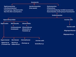
1. Ascomycota
Taphrinomycotina
(=Archiascomycetes)
( basal group- orignated earlier
than the other ascomycetes )
Saccharomycotina
(true yeasts)
Pezizomycotina
(Filamentous,
sporocarp-producing, Conidial ascomycetes
Euascomycetes, Crown group of ascomycetes
Sordariomycetes
Hypocreomycetidae Incertae sedis
Hypocreales Microascales Glomerellales
Hypocrea sp
(Trichoderma)
Hypocreaceae Nectriaceae Clavicipitaceae Incertae sedis
Nectria sp
Gibberella sp
Claviceps sp Verticillium sp
Ceratocystaceae
Ceratocystis
Glomerellaceae
Glomerella sp
Pestalotia sp
Incertae sedis
Magnaporthaceae
Magnaporthe sp
2. Diseases caused by Ceratocystis spp (anamorph: Chalara)
1. Sugar cane sett rot – C. paradoxa
2. Coconut stem bleeding – C. paradoxa
3. Oak wilt- C. Fagacearum
4. Black rot of sweet potato- C. fimbriata
Fungal characters
1. Perithicium- sexual fruiting body
2. Conidia asexual spores
3. Colletotrichum (anamorph) - Glomerella (Teleomorph)
Gleosporium (anamorph) - Glomerella (Teleomorph)
common name anthracnose fungi
Characteristic features
Foliar pathogens. Attacks more than 3000 species of monocots (maize,
sorghum) and dicots plants (pulses and fruits as post-harvest disease)
Life style : Hemi-biotrophs
Symptoms - anthracnose disease – Anthracnose disease is characterized
by sunken, necrotic and ulcer like lesions on leaves, stem, fruits/pods. This
symptom is produced by Colletotrichum spp. Production of acervulus is the
major sign on the lesions
Asexual stage – asexual fruiting body –acervulus and asexual spore –
conidia- are single celled, hyaline, mostly sickle shaped and cylindrical
shaped and contains oil globules
Sexual stage – sexual fruiting body – perithecia and sexual spores –
ascospores- are single celled, hyaline
6. Infection process (pathogenesis)
Life style : it is a hemi-biotroph
Conidia germinate to form dome-shaped appressorium which is darkly pigmented with melanin
a appressorial wall reinforcement chemical that prevent the bursting of appressorium due to
enormous turgor-pressure generated inside it upon accumulation of glycerol. This pressure
helps in puncturing of host cuticle and epidermal cells through the formation of infection peg.
A penetration hyphae / infection peg emerge from the base of the appressorium that break the
cuticle and cell wall of the epidermal cell
Biotrophic stage (symptomless infection stage): about 24 hours of post infection, the
penetration hyphae/infection peg then differentiates into bulbous primary hyphae (structurally
and functionally analogous to haustoria) which are surrounded by the plasmamembrane of the
plant epidermal cells. Through the primary hyphae, pathogen absorbs nutrients from the host
and releases the effector molecules from its thallus to the host cell. At this stage of infection, the
pathogen does not kill the host cell and host cell does not show any symptoms. Mostly this
biotrophic stage is confined to the first infected epidermal cells.
Necrotrophic stage : about 48 hours of post infection, thin, filamentous secondary hyphae
emerges from the tip of the bulbous primary hyphae and invades the adjacent epidermal and
parenchymatous cells. Later, secondary hyphae rapidly colonize the entire host tissue degrading
cell wall (tissue maceration) and killing cells (browing) ahead of its further spread and infection
producing characteristic anthracnose symptoms
7. Diseases caused by Glomerella spp (anamorph: Colletotrichum/Gleosporium)
Mango anthracnose Glomerella cingulata (Colletotrichum mangiferae)
Bean anthracnose Glomerella cingulata/ Collectotrichum lindemuthianum)
Chilli die back and fruit rot Glomerella cingulata (Colletotrichum capsici)
Banana anthracnose Glomerella musae (Gloeosporium musarum)
Red rot of sugarcane - Glomerella tucamanesis (Colletotrichum falcatum)
8. Disease caused by Botrydiplodia
(Dothidiomycetes)
• Stem end rot of mango, citrus
• Nut cracking in coconut
• Collar rot in Jatropha
9. Fungal characters
Young conidia matured conidia
Thallus – filamentous, Dark grey septate branched mycelium
Asexual reproduction - Pycnidia: Black, erumpent, conidiaare initially single celled, later
bicelled.
11. Life cycle of Magnoporthe oryzae and its pathogenesis
12. Blast - Pyricularia grisea (Magnaporthe grisea)
Symptoms:
All aerial parts of the plant (leaf blade, node neck of the panicle
rachis and also glumes) are affected both in the nursery and main
field.
Minute brown specks appear on leaves which become spindle –
shaped with dark brown margin and grayish center with yellow
halo (Leaf blast)
Drying of the leaves occur in severe cases.
Severely affected seedlings are completely killed.
The nodes turn black and rot (Node blast)
Neck region rots and the ear head becomes black and the panicle
break (neck rot /neck blast). Complete chaffiness of grains or
partial filling of grains occur.
13. Fungal characters:
Thallus :The mycelium is highly septate, intercellular at first and
later on intracellular. Young hyphae are colourless later become
olivaceous.
Parasitism - Hemibiotroph
Asexual - Conidiophores are unbranched and bear conidia at the
tips.The conidium has a slightly projection called hilum at its base
where it is attached to the conidiophores. Conidia are olive green
color and three celled, pyriform shaped.
Sexual reproduction – perithecia, ascospores