Fungi is most abundantly found organism in earth, almost all parts of earth we found earth, here we represent some characteristic with their uses and disadvantages .
Fungi are eukaryotic organisms that include microorganisms such as yeasts, moulds and mushrooms. These organisms are classified under kingdom fungi.
microbiology is a diverse and fascinating field that encompasses various branches this ppt includes only a few of them.
the collective efforts of researchers and scientists across these branches continue to expand our knowledge of the microbial world and derive innovations for human health, environmental preservation, agriculture, and industry. it's an exciting and essential field to study.
Biological Classification
This ppt shows the details of biological classification. it gives a brief idea about the five kingdom classification with a detailed description of kingdoms monera, protista and fungi. a detailed description of viruses, viroids, prions and lichens have also been given....
For more details visit my youtube channel: (VIHIRA ACADEMY)
https://www.youtube.com/channel/UCxo06Nj-QWo_7SNvMyDnJCQ?view_as=subscriber
Ultrastructure and characterstic features of bacteria.Archana Shaw
Ultrastructure and characterstic features of bacteria: BACTERIA AS A MODEL ORGANISM
THIS WAS MY PRESENTATION TOPIC IN CLASS. THOUGHT OF SHARING IT AND HOPE IT HELPS.
1) Strategies and structuresIn Protozoans the method of movement .pdfaptelecom16999
1) Strategies and structures:
In Protozoans the method of movement is determined by the type of organism and the
surrounding environment. Protozoans mainly move by cell extension, flagella or pseudopodia
and cilia, the movement as per the presence of structure can be classified as ciliary, flagellar and
amoeboid movement.
Ciliates : Ciliates form the largest group of protozoa. These organisms vary in size and often live
in watery environments, including oceans, marshes, bays and streams. Ciliates move using tiny
cilia, which are hair-like strands that act as sensors and tiny limbs.
Flagella are longer and less numerous that cilia, they use their long tail like flagella to move.
Amoebas : In these two cytoskeleton get polymerized . This creates a vacancy and cytoplasmice
material flow to cover the vacancy created. When amoeba moves cytoplasm moves to the arm
like extension called pseudopodium. This pseudopodium extends and enlarge and hence this
push the animal body towards that respective direction.
2) A) Flagellates can live as single cells, in colonies, or as parasites.
Commonly live in niche\'s of water.
They conduct photosynthesis and have a cell wall.
They contain flagella for propulsion or to create a current to bring in food.
They can inhabit the reproductive tract, alimentary canal, tissue sites and also the blood stream,
lymph vessels and cerebrospinal canal.
B) Pseudopods : Also called as false feet , are projections that can appear and disappear from the
organism\'s body. These are used for movement and to engulf prey and digest them using
enzymes.
C) Apicomplexa : Unicellular and spore forming, most of them possess a unique form of
organelle that comprises a type of plastid called an apicoplast, and an apical complex structure.
They have apicoplast(non photosynthetic plastid) , mitochondria and nuclear genomes.
Lack of cilia, sexual reproduction, use micropores for feeding, and the production of oocysts
containing sporozoites as the infective form.
They have unique gliding capability which enables them to cross through tissues and enter and
leave their host cells. This gliding ability is made possible by the use of adhesions and small
static myosin motors.
3) Key characteristics of fungi :
Fungi are unicellular or multicellular.
Most of the fungi grow as tubular filaments called hyphae
They are haploid.
Fungus are heterotrophs (they can obtain nutrients by absorption) . They absorb food and secrete
enzymes to digest complex molecules
Propogate by spores
Asexual or sexual reproduction
They can be multinucleated
Fungi are achlorophyllous (lack of cholorphyll pigment)
Both Fungi and protists belong to same kingdom but fungi is different from protist, protists are
able to live in an anaerobic environment without oxygen but fungi need aerobic respiration to
survive.
Protists are unicellular but fungi are multicellular. Protists are autotrophic (make their own
energy) and heterotrophic (rely on outside source to get energy), but fungi a.
Microbiology - Algae
Algae is an informal term for a large and diverse group of photosynthetic eukaryotic organisms. It is a polyphyletic grouping that includes species from multiple distinct clades.
Algae are sometimes considered plants and sometimes considered "protists" (a grab-bag category of generally distantly related organisms that are grouped on the basis of not being animals, plants, fungi, bacteria, or archaeans).
This ppt features the different kinds of protozoa, an animal-like protist . They are animal-like because they are heterotrophs, and are capable of moving.
Multi-source connectivity as the driver of solar wind variability in the heli...Sérgio Sacani
The ambient solar wind that flls the heliosphere originates from multiple
sources in the solar corona and is highly structured. It is often described
as high-speed, relatively homogeneous, plasma streams from coronal
holes and slow-speed, highly variable, streams whose source regions are
under debate. A key goal of ESA/NASA’s Solar Orbiter mission is to identify
solar wind sources and understand what drives the complexity seen in the
heliosphere. By combining magnetic feld modelling and spectroscopic
techniques with high-resolution observations and measurements, we show
that the solar wind variability detected in situ by Solar Orbiter in March
2022 is driven by spatio-temporal changes in the magnetic connectivity to
multiple sources in the solar atmosphere. The magnetic feld footpoints
connected to the spacecraft moved from the boundaries of a coronal hole
to one active region (12961) and then across to another region (12957). This
is refected in the in situ measurements, which show the transition from fast
to highly Alfvénic then to slow solar wind that is disrupted by the arrival of
a coronal mass ejection. Our results describe solar wind variability at 0.5 au
but are applicable to near-Earth observatories.
A brief information about the SCOP protein database used in bioinformatics.
The Structural Classification of Proteins (SCOP) database is a comprehensive and authoritative resource for the structural and evolutionary relationships of proteins. It provides a detailed and curated classification of protein structures, grouping them into families, superfamilies, and folds based on their structural and sequence similarities.
Observation of Io’s Resurfacing via Plume Deposition Using Ground-based Adapt...Sérgio Sacani
Since volcanic activity was first discovered on Io from Voyager images in 1979, changes
on Io’s surface have been monitored from both spacecraft and ground-based telescopes.
Here, we present the highest spatial resolution images of Io ever obtained from a groundbased telescope. These images, acquired by the SHARK-VIS instrument on the Large
Binocular Telescope, show evidence of a major resurfacing event on Io’s trailing hemisphere. When compared to the most recent spacecraft images, the SHARK-VIS images
show that a plume deposit from a powerful eruption at Pillan Patera has covered part
of the long-lived Pele plume deposit. Although this type of resurfacing event may be common on Io, few have been detected due to the rarity of spacecraft visits and the previously low spatial resolution available from Earth-based telescopes. The SHARK-VIS instrument ushers in a new era of high resolution imaging of Io’s surface using adaptive
optics at visible wavelengths.
Professional air quality monitoring systems provide immediate, on-site data for analysis, compliance, and decision-making.
Monitor common gases, weather parameters, particulates.
Slide 1: Title Slide
Extrachromosomal Inheritance
Slide 2: Introduction to Extrachromosomal Inheritance
Definition: Extrachromosomal inheritance refers to the transmission of genetic material that is not found within the nucleus.
Key Components: Involves genes located in mitochondria, chloroplasts, and plasmids.
Slide 3: Mitochondrial Inheritance
Mitochondria: Organelles responsible for energy production.
Mitochondrial DNA (mtDNA): Circular DNA molecule found in mitochondria.
Inheritance Pattern: Maternally inherited, meaning it is passed from mothers to all their offspring.
Diseases: Examples include Leber’s hereditary optic neuropathy (LHON) and mitochondrial myopathy.
Slide 4: Chloroplast Inheritance
Chloroplasts: Organelles responsible for photosynthesis in plants.
Chloroplast DNA (cpDNA): Circular DNA molecule found in chloroplasts.
Inheritance Pattern: Often maternally inherited in most plants, but can vary in some species.
Examples: Variegation in plants, where leaf color patterns are determined by chloroplast DNA.
Slide 5: Plasmid Inheritance
Plasmids: Small, circular DNA molecules found in bacteria and some eukaryotes.
Features: Can carry antibiotic resistance genes and can be transferred between cells through processes like conjugation.
Significance: Important in biotechnology for gene cloning and genetic engineering.
Slide 6: Mechanisms of Extrachromosomal Inheritance
Non-Mendelian Patterns: Do not follow Mendel’s laws of inheritance.
Cytoplasmic Segregation: During cell division, organelles like mitochondria and chloroplasts are randomly distributed to daughter cells.
Heteroplasmy: Presence of more than one type of organellar genome within a cell, leading to variation in expression.
Slide 7: Examples of Extrachromosomal Inheritance
Four O’clock Plant (Mirabilis jalapa): Shows variegated leaves due to different cpDNA in leaf cells.
Petite Mutants in Yeast: Result from mutations in mitochondrial DNA affecting respiration.
Slide 8: Importance of Extrachromosomal Inheritance
Evolution: Provides insight into the evolution of eukaryotic cells.
Medicine: Understanding mitochondrial inheritance helps in diagnosing and treating mitochondrial diseases.
Agriculture: Chloroplast inheritance can be used in plant breeding and genetic modification.
Slide 9: Recent Research and Advances
Gene Editing: Techniques like CRISPR-Cas9 are being used to edit mitochondrial and chloroplast DNA.
Therapies: Development of mitochondrial replacement therapy (MRT) for preventing mitochondrial diseases.
Slide 10: Conclusion
Summary: Extrachromosomal inheritance involves the transmission of genetic material outside the nucleus and plays a crucial role in genetics, medicine, and biotechnology.
Future Directions: Continued research and technological advancements hold promise for new treatments and applications.
Slide 11: Questions and Discussion
Invite Audience: Open the floor for any questions or further discussion on the topic.
Earliest Galaxies in the JADES Origins Field: Luminosity Function and Cosmic ...Sérgio Sacani
We characterize the earliest galaxy population in the JADES Origins Field (JOF), the deepest
imaging field observed with JWST. We make use of the ancillary Hubble optical images (5 filters
spanning 0.4−0.9µm) and novel JWST images with 14 filters spanning 0.8−5µm, including 7 mediumband filters, and reaching total exposure times of up to 46 hours per filter. We combine all our data
at > 2.3µm to construct an ultradeep image, reaching as deep as ≈ 31.4 AB mag in the stack and
30.3-31.0 AB mag (5σ, r = 0.1” circular aperture) in individual filters. We measure photometric
redshifts and use robust selection criteria to identify a sample of eight galaxy candidates at redshifts
z = 11.5 − 15. These objects show compact half-light radii of R1/2 ∼ 50 − 200pc, stellar masses of
M⋆ ∼ 107−108M⊙, and star-formation rates of SFR ∼ 0.1−1 M⊙ yr−1
. Our search finds no candidates
at 15 < z < 20, placing upper limits at these redshifts. We develop a forward modeling approach to
infer the properties of the evolving luminosity function without binning in redshift or luminosity that
marginalizes over the photometric redshift uncertainty of our candidate galaxies and incorporates the
impact of non-detections. We find a z = 12 luminosity function in good agreement with prior results,
and that the luminosity function normalization and UV luminosity density decline by a factor of ∼ 2.5
from z = 12 to z = 14. We discuss the possible implications of our results in the context of theoretical
models for evolution of the dark matter halo mass function.
Introduction:
RNA interference (RNAi) or Post-Transcriptional Gene Silencing (PTGS) is an important biological process for modulating eukaryotic gene expression.
It is highly conserved process of posttranscriptional gene silencing by which double stranded RNA (dsRNA) causes sequence-specific degradation of mRNA sequences.
dsRNA-induced gene silencing (RNAi) is reported in a wide range of eukaryotes ranging from worms, insects, mammals and plants.
This process mediates resistance to both endogenous parasitic and exogenous pathogenic nucleic acids, and regulates the expression of protein-coding genes.
What are small ncRNAs?
micro RNA (miRNA)
short interfering RNA (siRNA)
Properties of small non-coding RNA:
Involved in silencing mRNA transcripts.
Called “small” because they are usually only about 21-24 nucleotides long.
Synthesized by first cutting up longer precursor sequences (like the 61nt one that Lee discovered).
Silence an mRNA by base pairing with some sequence on the mRNA.
Discovery of siRNA?
The first small RNA:
In 1993 Rosalind Lee (Victor Ambros lab) was studying a non- coding gene in C. elegans, lin-4, that was involved in silencing of another gene, lin-14, at the appropriate time in the
development of the worm C. elegans.
Two small transcripts of lin-4 (22nt and 61nt) were found to be complementary to a sequence in the 3' UTR of lin-14.
Because lin-4 encoded no protein, she deduced that it must be these transcripts that are causing the silencing by RNA-RNA interactions.
Types of RNAi ( non coding RNA)
MiRNA
Length (23-25 nt)
Trans acting
Binds with target MRNA in mismatch
Translation inhibition
Si RNA
Length 21 nt.
Cis acting
Bind with target Mrna in perfect complementary sequence
Piwi-RNA
Length ; 25 to 36 nt.
Expressed in Germ Cells
Regulates trnasposomes activity
MECHANISM OF RNAI:
First the double-stranded RNA teams up with a protein complex named Dicer, which cuts the long RNA into short pieces.
Then another protein complex called RISC (RNA-induced silencing complex) discards one of the two RNA strands.
The RISC-docked, single-stranded RNA then pairs with the homologous mRNA and destroys it.
THE RISC COMPLEX:
RISC is large(>500kD) RNA multi- protein Binding complex which triggers MRNA degradation in response to MRNA
Unwinding of double stranded Si RNA by ATP independent Helicase
Active component of RISC is Ago proteins( ENDONUCLEASE) which cleave target MRNA.
DICER: endonuclease (RNase Family III)
Argonaute: Central Component of the RNA-Induced Silencing Complex (RISC)
One strand of the dsRNA produced by Dicer is retained in the RISC complex in association with Argonaute
ARGONAUTE PROTEIN :
1.PAZ(PIWI/Argonaute/ Zwille)- Recognition of target MRNA
2.PIWI (p-element induced wimpy Testis)- breaks Phosphodiester bond of mRNA.)RNAse H activity.
MiRNA:
The Double-stranded RNAs are naturally produced in eukaryotic cells during development, and they have a key role in regulating gene expression .
THE IMPORTANCE OF MARTIAN ATMOSPHERE SAMPLE RETURN.Sérgio Sacani
The return of a sample of near-surface atmosphere from Mars would facilitate answers to several first-order science questions surrounding the formation and evolution of the planet. One of the important aspects of terrestrial planet formation in general is the role that primary atmospheres played in influencing the chemistry and structure of the planets and their antecedents. Studies of the martian atmosphere can be used to investigate the role of a primary atmosphere in its history. Atmosphere samples would also inform our understanding of the near-surface chemistry of the planet, and ultimately the prospects for life. High-precision isotopic analyses of constituent gases are needed to address these questions, requiring that the analyses are made on returned samples rather than in situ.
THE IMPORTANCE OF MARTIAN ATMOSPHERE SAMPLE RETURN.
L 3-4. Causes of PD.ppt
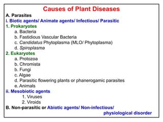
1. Causes of Plant Diseases
A. Parasites
i. Biotic agents/ Animate agents/ Infectious/ Parasitic
1. Prokaryotes
a. Bacteria
b. Fastidious Vascular Bacteria
c. Candidatus Phytoplasma (MLO/ Phytoplasma)
d. Spiroplasma
2. Eukaryotes
a. Protozoa
b. Chromista
b. Fungi
c. Algae
d. Parasitic flowering plants or phanerogamic parasites
e. Animals
ii. Mesobiotic agents
1. Viruses
2. Viroids
B. Non-parasitic or Abiotic agents/ Non-infectious/
physiological disorder
2. Bacteria
Bacteria are microscopic, unicellular prokaryotes, which lack
definitely organized nucleus and chlorophyll.
These microorganisms are with a primitive nucleus lacking a
clearly defined membrane. Majority of them are saprophytes
The bacteria are smaller than fungi and measure about
0.5 to 1.0 x 2.0 to 5.0 µm in size. In general, they vary from
1.0 to 5.0 microns in different forms.
Each bacterial cell is surrounded by a definite cell wall or
membrane, which is nitrogenous in chemical nature rather than
carbon compound (cellulose in fungi and higher plants).
3. Bacteria
Bacteria have been defined by Clifton as "extremely minute,
rigid, essentially unicellular organisms, free of true chlorophyll and
generally devoid of any photosynthetic pigments; most commonly
multiplying asexually by simple transverse fission, the resulting cells
being of equal or nearly equal size".
4. • More than 1,600 bacterial species are known. Of which about
200 spp. cause diseases in plants and are mostly rod-shaped
• Either non-motile or motile by means of one or more flagella on
their body arranged in a specific manner.
• They multiply mostly by binary fission.
• Most bacteria attacking plants are Gram-negative except the
genera, Streptomyces and Clavibacter (Corynebacterium)
• These are passive pathogens (cannot directly penetrate into the
host but need natural openings or wounds for their entry).
• First report of plant bacterial disease was made by T. J. Burrill
(Fire blight of apple and pear, Erwinia amylovora).
• Bacteria cause extensive and destructive plant diseases.
e.g. soft rot, leaf spot, blight, cankers, galls, tumours, wilt, etc.
5. Characteristics of bacteria
• Microscopic, unicellular, prokaryotic organism occur in single or
in colonies
• Possess rigid cell wall which is made up of peptidoglycan and
lipopolysaccharides
• Absence of well defined nucleus
• Mostly heterotrophic and some autotrophic
• Reproduce mostly by asexual method (binary fission).
• Sexual reproduction is lacking.
• Motile bacteria possess one or more flagella.
• Ribosomes are scattered in the cytoplasm
• Bacteria are named by shaped viz., cocci (ball-shaped), bacillus
(rod-shaped) and spirilli (spiral-shaped).
6. Fastidious vascular bacteria
[Rickettsia-like bacteria (RLB) /Rickettsia-like organism (RLO)]
• They are smaller bacteria with a cellular ultrastructure of typical
Gram-negative bacteria
• These are fastidious in their nutritional requirements, refusing to
grow on routine bacteriological media.
• They are rod shaped bacteria without flagella and are bounded
by a cell membrane and cell wall unlike MLO and
spiroplasma. The size of rod shaped cells varies from
0.2-0.5 µm dia with 1-4 µm long.
• They reproduce by binary fission.
7. • Mostly insect vectors transmit them.
Nematode (Xiphinema index) also helps in
transmission (yellow disease of grapevine).
• Mechanical inoculations (as in Pierce's disease of
grapevine) or vegetative propagation also reproduce
disease symptoms.
• Penicillin is effective against FVB.
• They may be confined to the phloem tissues or xylem
tissues of the host plants.
8. The FVB can be divided into three groups.
1. Xylem-limited FVB
The FVB causing these diseases is named as Xylella fastidiosa.
In general xylem-limited Gram-negative bacteria have elongated
cells of 0.2 to 0.5 into 1.4 m size. They are susceptible to
tetracycline but not to penicillin. Transmission mostly through
xylem feeding insects. E.g., Pierce's disease of grapevine.
2. Phloem-limited FVB
They are mostly rigid rods and Gram-negative and sensitive to
penicillin. Transmission is by leafhoppers, dodder and grafting.
e.g., Citrus greening is transmitted by citrus psyllid.
3. Non-tissue restricted FVB (e.g., Yellows of grapevine and
necrosis of grapevine)
9. Candidatus Phytoplasma
• They are very small, filterable, unicellular, normally non-motile,
wall-less prokaryote (mollicutes – mollis means soft or pliable
and cutis means skin) distinct from true bacteria in its
morphology, mode of reproduction, biochemistry, physiology,
phylogeny and taxonomy
•They are pleomorphic (varied shapes).
•They lack rigid cell wall, are bounded by a triple layered membrane
and contain cytoplasm, ribosomes and strands of nuclear
material.
•They are generally confined to sieve elements of the phloem and in
some cases in the cortical parenchymatous tissues
10. •They have fried egg appearance of colony (non-pathogenic type)
•They have both DNA and RNA.
•They do not have flagella, no spores and are Gram-negative.
•They produce symptoms like little leaf, phyllody, spike, yellows,
stunting, witches’ broom etc.
•They are mostly transmitted by leafhoppers.
•Multiplication of phytoplasma occur both in vectors and in
host plants.
•They are insensitive to penicillin and sensitive to tetracycline.
11. Spiroplasma
Spiroplasma are helical/ filamentous, wall-less prokaryotes
lacking rigid cell wall but bounded by unit membrane with
RNA granules (ribosomes) and DNA strand in the nuclear
region.
They are restricted to phloem region, requiring cholesterol for
growth and cause diseases in plants, insects and rats.
They are Gram positive, facultative anaerobic.
They are insensitive to penicillin and sensitive to erythrocin
and tetracycline.
12. Their motility is in a corkscrew pattern.
It can be cultured on artificial media.
Their major site of existence is either
phloem in plants or hemolymph of insect
vectors.
The average size of cells ranges from 2-4
µm long with a width of 120 nm.
E.g. corn stunt, citrus stubborn.
13. Flagellate Protozoa
Protozoa derived from two Greek words
viz., protos and zoan, which means first animal.
They are the smallest among the animal kingdom.
They are mostly single celled and vary in shape.
They produce spores.
Flagellate protozoa attack several crop plants.
e.g., Phloem necrosis of coffee, Hart rot of coconut.
14. Chromista
• The name Chromista means "coloured", and although some
chromists, like mildews (downy mildews) are colourless,
most are photosynthetic.
• It comprises of single celled or multicelled, eukaryotic, walled
microorganisms that produce heterokont, wall-less cells in
their life cycles.
• The members are photosynthetic organelle (plastid) which
contain chlorophyll c and do not store their energy in the
form of starch, like plants or even other algae.
• Chromists have closer phylogenetic relationship with brown
algae and diatoms than true fungi belong to kingdom Fungi
E.g., downy mildews and damping off pathogens.
15. Fungi
Fungus (pl. fungi) is a Latin word which means “mushroom”.
Fungi are microscopic, eukaryotic, heterotrophic,
achlorophyllous, nucleated, uni or multicellular organisms that
may reproduce sexually and asexually and whose filamentous
branched somatic structures are typically surrounded by cell walls
containing chitin or cellulose.
The branch of biology that deals with fungi is called Mycology
(Gr. myke= mushroom + logos= study of)
16. Fungi
Fungi are eukaryotic, achlorophyllous, unicellular or multicellular
heterotrophic organisms which obtain nutrients by absorption and
reproduce by sexual or asexual spores.
17. Characteristics of fungi
1. All are eukaryotic
(Possess membrane-bound nuclei (containing
chromosomes) and a range of membrane-bound
cytoplasmic organelles (e.g. mitochondria, vacuoles,
endoplasmic reticulum, golgi bodies, etc. ).
2. Most of them are filamentous
3. Some are unicellular E.g. Yeasts
4. Cell surrounded by rigid cell wall, made up of chitin and glucans
(some contain cellulose).
5. Many reproduce both sexually and asexually
18. 6. Their nuclei are typically haploid and hyphal compartments are
often multinucleate
7. All are achlorophyllous
8. All are chemoheterotrophic (chemo-organotrophic)
(They utilise pre-existing organic sources of carbon in their
environment and the energy from chemical reactions to
sythesize the organic compounds they require for growth
and energy)
9. Possess characteristic range of storage compounds
E.g., trehalose, glycogen, sugar alcohols and lipids.
10. May be free-living or may form intimate relationships with
other organisms i.e. free-living, parasitic or mutualistic.
19. Algae
Algae are eukaryotic, unicellular or multicellular organisms and
mostly occur in aquatic environments. Many algae thrive
as terrestrial or subterranean algae.
The size of algae ranges from 1.0 mm to many cm in length.
They are pigmented and vary in colour. They possess chlorophyll
and are photosynthetic.
They reproduce by asexual and sexual processes.
The study of algae is called phycology or algology.
E.g., Red rust of tea, mango, guava, citrus and Sapota –
Cephaleuros parasiticus
20. Phanerogamic parasites
Phanerogamic parasites are flowering plants or seed plants, which
lead a parasitic life on other living plants.
The phanerogamic parasites invade stem or root of the host plants.
Some of these parasites possess chlorophyll, which manufacture
carbohydrates to a limited extent and depend on the host for
mineral, salts and water. These are generally called as semi or
partial parasites.
Some of the parasites, which do not have chlorophyll, depend
entirely on the host plants for their food materials. They are called
holo or total parasites.
22. Character Prokaryotes Eukaryotes
Cellular
organization
Primitive Advanced
Size Smaller sized cells 1-2 x 1-5µm
or less
Larger than prokaryotes (More
than 5 µm in width or dia
Cell wall Peptidoglycans Cellulose
Nucleus and nuclear
membrane
No organized nucleus. Nuclear
membrane and nucleolus are
absent
Possess organized nucleus.
Nuclear membrane and nucleolus
are present
Membrane bound
cell organelles
Membrane bound cell organelles
such as endoplasmic reticulum,
golgi complex, mitochondria,
chloroplasts amd vacuoles are
absent.
Membrane bound cell organelles
such as endoplasmic reticulum,
golgi complex, mitochondria,
chloroplasts amd vacuoles are
present.
Ribosomes Smaller and made of 70S units Larger and made of 80S units
Number of
chromosomes per
nucleus
One More than one
Chromosome
replication by
mitosis
No Yes
Genetic material
(DNA)
DNA is not found in well
organized chromosome. Found in
nucleoids and in extra nuclear
plasmids and neither of which are
membrane bound. DNA is
shorter, circular and not histone
(protein) bound.
DNA is found in well organized
chromosome. DNA is long and
linear, histone bound.
Genetic
recombination
Unidirectional transfer of DNA
forms partial diploids
Fusion of gametes forms diploids
that segregate by meiosis
Cell division Only by fission (amitosis) By mitosis and meiosis
Flagella Single fibrillar type Bundle of nine fused pairs of
microtubule surrounding two
central single microtubules (9+2)
23. Virus
The word ‘Virus’ means slimy liquid, poison, venom or
infectious matter.
Viruses are ultramicroscopic, nucleoprotein entities, which are
infectious agents and obligate parasitic pathogens.
They are less than 200 m in size (pass through bacterial filters)
24. They are nucleoproteins (nucleic acid and protein) and devoid of
enzymes and depend on the hosts protein synthesizing machinery
(ribosomes) for multiplication.
They neither divide nor produce any structures like spores, but
multiply by stimulating host cells to replicate more viruses.
They have either DNA or RNA . Most of the plant virus is having
RNA e. g., TMV. Few viruses contain DNA. E.g., Cauliflower
mosaic virus, banana bunchy top virus.
Viruses are considered as mesobiotic agents (in between living
non-living) as their biological status is not clear.
25. Viruses are living because
• They have ability to assimilate (metabolism) with the release of
energy
• They are able to multiply
• They exhibit response to environment like temperature,
chemicals, etc.,
• They have genetic materials like RNA or DNA
• They have the ability to infect
26. Viruses are non-living because
• Viruses can be crystallized.
• Stanley (1935) considered virus as a molecule and a molecule is
not capable of self-replication
• Viruses are inert outside the living host
• Viruses do not have cell wall or cell membrane of any type
• They do not show functional autonomy
• They do not respire or excrete
• They lack any energy producing system.
27. Viroids
They are ultramicroscopic, small low molecular weight
ribonucleic acids without protein sheath that can infect plant
cells replicate them and cause disease.
They are also called as mini viruses (without protein coat).
E. g., Potato spindle tuber
Chrysanthemum stunt
Coconut cadang cadang
Citrus exocortis
28. Abiotic disorders
a. Unfavourable temperature (Low temperature, high temperature)
b. Unfavourable light
c. Unfavourable soil moisture
d. Relative humidity
e. Unfavourable oxygen relations
f. Atmospheric impurities (Sulphur dioxide, nitrogen dioxide,
ozone, ethylene, etc.)
g. Toxic effects of decomposing organic matter
h. Nutritional disorders
Excessive minerals - internal bark necrosis in red delicious apple;
Deficiency of magnesium (Red leaf in cotton),
Zinc (Khaira disease in rice, Foliocellosis in citrus),
Molybdenum (whip tail of cauliflower),
Calcium (blossom end rot in tomato),
Micronutrients (coconut pencil point disease).