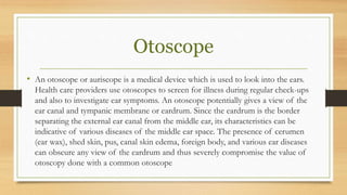
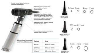
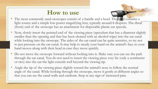
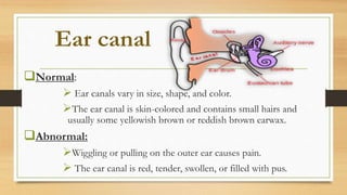
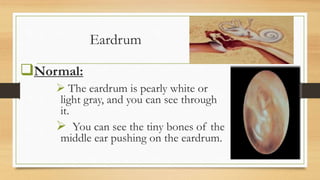
Otoscopy
Submitted By: Muhammad Awais.
Roll No. 1323 B group
Session 2012-16
Otoscope
• An otoscope or auriscope is a medical device which is used to look into the ears.
Health care providers use otoscopes to screen for illness during regular check-ups
and also to investigate ear symptoms. An otoscope potentially gives a view of the
ear canal and tympanic membrane or eardrum. Since the eardrum is the border
separating the external ear canal from the middle ear, its characteristics can be
indicative of various diseases of the middle ear space. The presence of cerumen
(ear wax), shed skin, pus, canal skin edema, foreign body, and various ear diseases
can obscure any view of the eardrum and thus severely compromise the value of
otoscopy done with a common otoscope
How to use
• The most commonly used otoscopes consist of a handle and a head. The head contains a
light source and a simple low-power magnifying lens, typically around 8 diopters. The distal
(front) end of the otoscope has an attachment for disposable plastic ear specula.
• Now, slowly insert the pointed end of the viewing piece (speculum that has a diameter slightly
smaller than the opening and that has been cleaned with an alcohol wipe) into the ear canal
while looking into the otoscope. The sides of the ear canal can be quite sensitive, so try not
to put pressure on the ear canal. It may help to steady your hand on the animal's face so your
hand moves along with their head in case they move quickly.
• Do not move the otoscope forward without looking into it. Make sure you can see the path
through the ear canal. You do not need to insert the viewing piece very far (only a centimeter
or two) into the ear-the light extends well beyond the viewing tip.
• Angle the tip of the viewing piece slightly toward the animal's nose to follow the normal
angle of the canal. While looking through the otoscope, move it gently at different angles so
that you can see the canal walls and eardrum. Stop at any sign of increased pain.
Ear canal
Normal:
 Ear canals vary in size, shape, and color.
The ear canal is skin-colored and contains small hairs and
usually some yellowish brown or reddish brown earwax.
Abnormal:
Wiggling or pulling on the outer ear causes pain.
 The ear canal is red, tender, swollen, or filled with pus.
Eardrum
Normal:
 The eardrum is pearly white or
light gray, and you can see through
it.
 You can see the tiny bones of the
middle ear pushing on the eardrum.
Abnormal:
 The light reflex on the eardrum is dull or absent The eardrum
is red and bulging.
You can often see amber liquid or bubbles behind the eardrum.
 You can see a hole in the eardrum (perforation).
 You can see whitish scars on the surface of the eardrum.
The eardrum is blocked by earwax or an object, such as a bean
or a bead.
Otoscopy

Otoscopy

  • 2.
    Otoscopy Submitted By: MuhammadAwais. Roll No. 1323 B group Session 2012-16
  • 4.
    Otoscope • An otoscopeor auriscope is a medical device which is used to look into the ears. Health care providers use otoscopes to screen for illness during regular check-ups and also to investigate ear symptoms. An otoscope potentially gives a view of the ear canal and tympanic membrane or eardrum. Since the eardrum is the border separating the external ear canal from the middle ear, its characteristics can be indicative of various diseases of the middle ear space. The presence of cerumen (ear wax), shed skin, pus, canal skin edema, foreign body, and various ear diseases can obscure any view of the eardrum and thus severely compromise the value of otoscopy done with a common otoscope
  • 6.
    How to use •The most commonly used otoscopes consist of a handle and a head. The head contains a light source and a simple low-power magnifying lens, typically around 8 diopters. The distal (front) end of the otoscope has an attachment for disposable plastic ear specula. • Now, slowly insert the pointed end of the viewing piece (speculum that has a diameter slightly smaller than the opening and that has been cleaned with an alcohol wipe) into the ear canal while looking into the otoscope. The sides of the ear canal can be quite sensitive, so try not to put pressure on the ear canal. It may help to steady your hand on the animal's face so your hand moves along with their head in case they move quickly. • Do not move the otoscope forward without looking into it. Make sure you can see the path through the ear canal. You do not need to insert the viewing piece very far (only a centimeter or two) into the ear-the light extends well beyond the viewing tip. • Angle the tip of the viewing piece slightly toward the animal's nose to follow the normal angle of the canal. While looking through the otoscope, move it gently at different angles so that you can see the canal walls and eardrum. Stop at any sign of increased pain.
  • 7.
    Ear canal Normal:  Earcanals vary in size, shape, and color. The ear canal is skin-colored and contains small hairs and usually some yellowish brown or reddish brown earwax. Abnormal: Wiggling or pulling on the outer ear causes pain.  The ear canal is red, tender, swollen, or filled with pus.
  • 8.
    Eardrum Normal:  The eardrumis pearly white or light gray, and you can see through it.  You can see the tiny bones of the middle ear pushing on the eardrum.
  • 9.
    Abnormal:  The lightreflex on the eardrum is dull or absent The eardrum is red and bulging. You can often see amber liquid or bubbles behind the eardrum.  You can see a hole in the eardrum (perforation).  You can see whitish scars on the surface of the eardrum. The eardrum is blocked by earwax or an object, such as a bean or a bead.