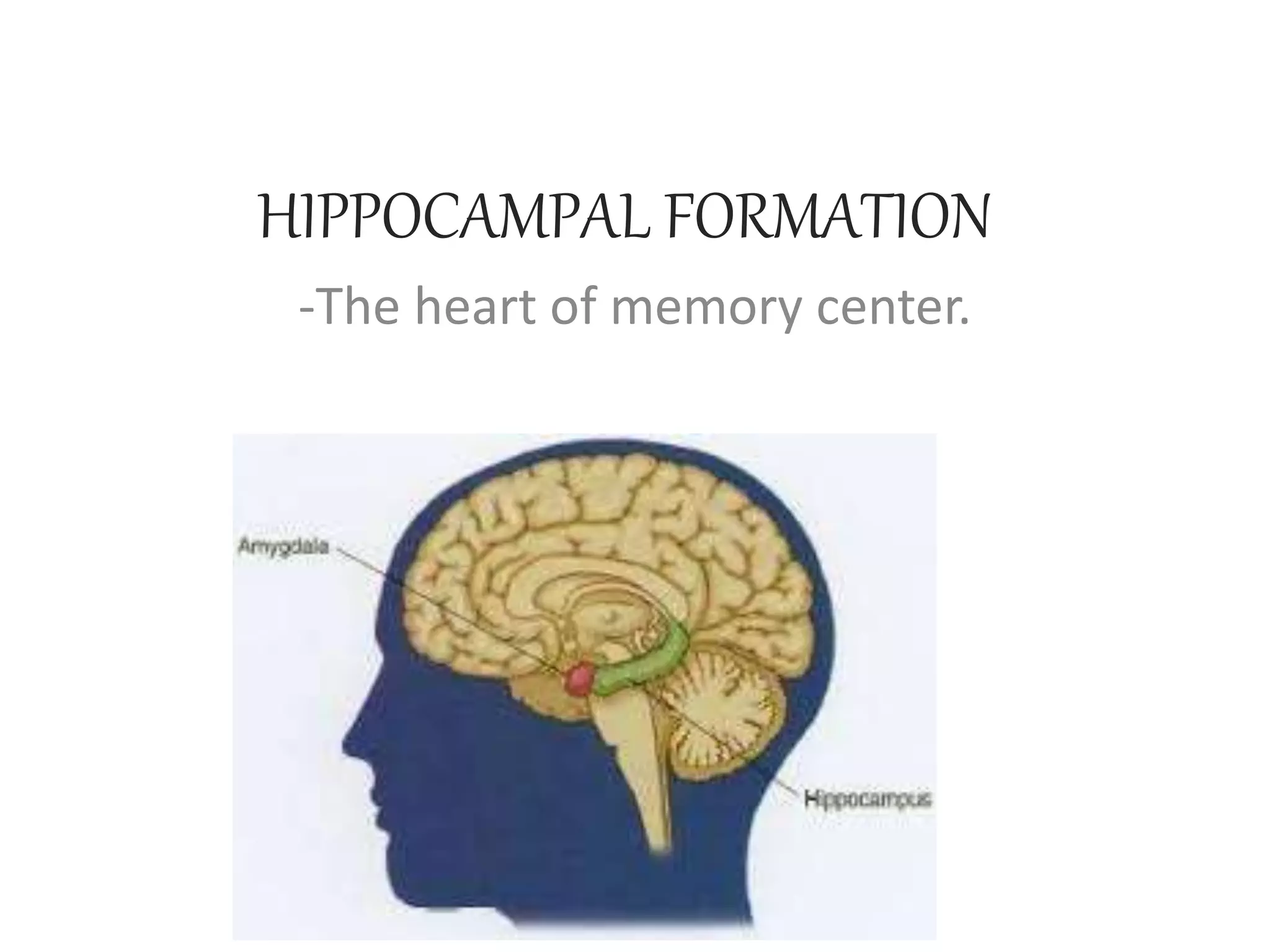
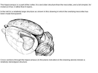
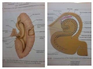
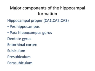
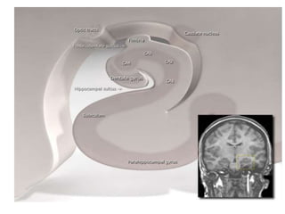
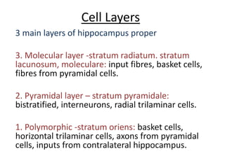
HIPPOCAMPAL FORMATION
-The heart of memory center.
Contents
1. Gross Structure
2. Cell layers
3. Connections
4. Evolution of hippocampus
5. Function
STRUCTURE
• The hippocampus, as its name suggests resembles a
sea horse shaped cortex in the cross section of the
middle temporal lobe of the brain.
• The nomenclature of its structure is also derived
from its resemblance with the Ram’s horn-Cornu
Ammons.
• it curves the entire length of the floor of inferior
horn of lateral ventricles.
• Primates have two Hippocampus on each side of the
hemispheres.
Major components of the hippocampal
formation
Hippocampal proper (CA1,CA2,CA3)
• Pes hippocampus
• Para hippocampus gurus
Dentate gyrus
Entorhinal cortex
Subiculum
Presubiculum
Parasubiculum
Cell Layers
3 main layers of hippocampus proper
3. Molecular layer -stratum radiatum. stratum
lacunosum, moleculare: input fibres, basket cells,
fibres from pyramidal cells.
2. Pyramidal layer – stratum pyramidale:
bistratified, interneurons, radial trilaminar cells.
1. Polymorphic -stratum oriens: basket cells,
horizontal trilaminar cells, axons from pyramidal
cells, inputs from contralateral hippocampus.
Beneath the ependyma of the floor
of the ventricle, a thin layer of white
mater called Alveus is present.
Nerve fibres of Alveus originated from
hippocampus – converge medially-
forming fimbria- becomes continious
with crus of fornix
Dentate gyral(DG) layers
• molecular layer
• granule cell layer (striatum granulosum)
• Polymorphic layer (or hilus)
DG cell types
diverse variety of interneurons
mossy fibers – axons from DG
granule cells – unmyelinated
axons – synapse on CA3
only excitatory output from DG
Cell layers of Dentate Gyrus
1- granule cell; unmyelinated axons (mossy fiber) – collaterals in PL
and then to CA3
2- dentate pyramidal basket cell 3 – stellate cells (give rise to
basket plexus)
4- mossy cell 5- inhibitory cells
Entorhinal cortex
•Located in medial temporal lobe.
• functions in assisting hippocampus in memory
and navigation.
• divided into 3 bands: medial & lateral with
distinct properties and connectivity.
• It lacks a layer of cells that should be the IVth
layer. This layer is called lamina dessicans.
CONNECTIONS
• Afferent connections: fibres
> from cingulate gyrus
> from septal nucleus – fornix – hippocampus.
> from one hippocampus to another via
commissure of fornix
Schaffer collaterals- projections from
CA3 to CA1 (ipsilateral & contralateral)
> from indusium griseum to hippocampus via
londitudinal striae.
> Preforant pathway- from olfactory cortex,
association areas, subcortical structures-
entorhinal cortex(from layer II & III)-
hippocampal areas.
> mossy fibers – axons from DG granule
cells – unmyelinated axons – synapse on
CA3
-sole excitatory output from DG
-multiple granule cells can synapse on
a single pyramidal cell
> From Brain stem:
Raphe nucleus- in the reticular formation- projection
fibres to hippocampus. Serotonergic in function.
Locus Ceruleus- in pons – sends projections. Involved
in secreting nE in physiological response to stress and
panic.
Ventral tegmental area- sends projections of
dopaminergic neurons. Involves in drug and reward
circuitry of brain.
• Efferents of Hippocampus
> the major efferents is Fornix.
*Fibres from the large pyramidal cells
forms the alveus- fibres converge at the middle
border of hippocampus forming a bundle of
fibers called Fimbria. It leaves the hippocampus
as crus of fornix.
*It forms the body of fornix attached to
the inferior part of septum pellucidum.
*body of fornix splits into two columns of
fornix near the anterior commissure at the
preoptic nucleus of hypothalamus.
*Columns of fornix gives anterior and
posterior divergence.
:: The anterior divergence (precommissural
fornix) ends at septal nuclei, lateral preoptic and
anterior part of hypothalamus, and nucleus
accumbens, which is present rostral to preoptic area,
forms the ventral striatum, a part of basal ganglia, with
the olfactory tubercle.
:: The posterior diverging fibers(post
commissural fornix) on each side continue to the
mammilary bodies to its medial nucleus.
:: to the tegmentum of midbrain.
:: fibres join the stria medularis thalami to
reach the habenular nuclei.
:: fibres join the stria terminalis to reach
amygdala.
Evolution of Hippocampus?
*The appreance of hippocampus has been same
across all the range of mammals starting from
monotrems like Echidna to primates like Human.
* in lower vertebrates and non mammalian
species there is no hippocampus, but an analogus part
called Pallium is present (the middle pallium creates
memory)
*The hippocampal size to body size ratio
increases as we go up the phylogenic tree of mammals.
But the neocortex size to body size increases
twice the hippocampal size in primates.
* Thus the hippocampus to cortex ratio is
larger in lower vertebrates than in primates
* Species like bats has larger hippocampal
volume due to their higher capacity in spatial
memory
* the hippocampal formation in lower
mammals help them for predation and hide,
Search of mate, socialization etc.
Functions of hippocampus
* consolidating short term memory to long term.
* the subiculum as well as hypothalamus
process, establishes, & retrieve short term memory.
* the Entorhinal cortex involvs in declarative,
spatial navigation memory as well as memory
formation
* the hippocampus converts STM to LTM via long
term potentiation, which allows altering strength of
connections in neurons which are simultaneously
active.
PATHOLOGY
* Age related – Alzheimer’s disease : loss of substantial
cells, demyelination.
Due to chronic cerebral hypoperfusion.
Effect in cognition, working and episodic memory.
* Stress- chronic depression- reducing excitability , long
term potentiation, atrophy of dendrites in CA3 pyramidal
cells.
* Epilepsy- hippocampal sclerosis in medial temporal
epilepsy.
* anterograde amnesia- lesion of hypothalamus-
memories of remote past remains episodic but unable to form
new memories for a long time.
* Triensent global amnesia- venous congestion of brain
leading to ischemia- dramatic, temporary sudden loss of short
term memory.
THANK YOU

hippocampal formation

  • 1.
  • 2.
    Contents 1. Gross Structure 2.Cell layers 3. Connections 4. Evolution of hippocampus 5. Function
  • 3.
    STRUCTURE • The hippocampus,as its name suggests resembles a sea horse shaped cortex in the cross section of the middle temporal lobe of the brain. • The nomenclature of its structure is also derived from its resemblance with the Ram’s horn-Cornu Ammons. • it curves the entire length of the floor of inferior horn of lateral ventricles. • Primates have two Hippocampus on each side of the hemispheres.
  • 6.
    Major components ofthe hippocampal formation Hippocampal proper (CA1,CA2,CA3) • Pes hippocampus • Para hippocampus gurus Dentate gyrus Entorhinal cortex Subiculum Presubiculum Parasubiculum
  • 8.
    Cell Layers 3 mainlayers of hippocampus proper 3. Molecular layer -stratum radiatum. stratum lacunosum, moleculare: input fibres, basket cells, fibres from pyramidal cells. 2. Pyramidal layer – stratum pyramidale: bistratified, interneurons, radial trilaminar cells. 1. Polymorphic -stratum oriens: basket cells, horizontal trilaminar cells, axons from pyramidal cells, inputs from contralateral hippocampus.
  • 9.
    Beneath the ependymaof the floor of the ventricle, a thin layer of white mater called Alveus is present. Nerve fibres of Alveus originated from hippocampus – converge medially- forming fimbria- becomes continious with crus of fornix
  • 11.
    Dentate gyral(DG) layers •molecular layer • granule cell layer (striatum granulosum) • Polymorphic layer (or hilus)
  • 12.
    DG cell types diversevariety of interneurons mossy fibers – axons from DG granule cells – unmyelinated axons – synapse on CA3 only excitatory output from DG
  • 13.
    Cell layers ofDentate Gyrus 1- granule cell; unmyelinated axons (mossy fiber) – collaterals in PL and then to CA3 2- dentate pyramidal basket cell 3 – stellate cells (give rise to basket plexus) 4- mossy cell 5- inhibitory cells
  • 14.
    Entorhinal cortex •Located inmedial temporal lobe. • functions in assisting hippocampus in memory and navigation. • divided into 3 bands: medial & lateral with distinct properties and connectivity. • It lacks a layer of cells that should be the IVth layer. This layer is called lamina dessicans.
  • 15.
    CONNECTIONS • Afferent connections:fibres > from cingulate gyrus > from septal nucleus – fornix – hippocampus. > from one hippocampus to another via commissure of fornix Schaffer collaterals- projections from CA3 to CA1 (ipsilateral & contralateral) > from indusium griseum to hippocampus via londitudinal striae.
  • 16.
    > Preforant pathway-from olfactory cortex, association areas, subcortical structures- entorhinal cortex(from layer II & III)- hippocampal areas. > mossy fibers – axons from DG granule cells – unmyelinated axons – synapse on CA3 -sole excitatory output from DG -multiple granule cells can synapse on a single pyramidal cell
  • 17.
    > From Brainstem: Raphe nucleus- in the reticular formation- projection fibres to hippocampus. Serotonergic in function. Locus Ceruleus- in pons – sends projections. Involved in secreting nE in physiological response to stress and panic. Ventral tegmental area- sends projections of dopaminergic neurons. Involves in drug and reward circuitry of brain.
  • 18.
    • Efferents ofHippocampus > the major efferents is Fornix. *Fibres from the large pyramidal cells forms the alveus- fibres converge at the middle border of hippocampus forming a bundle of fibers called Fimbria. It leaves the hippocampus as crus of fornix. *It forms the body of fornix attached to the inferior part of septum pellucidum.
  • 20.
    *body of fornixsplits into two columns of fornix near the anterior commissure at the preoptic nucleus of hypothalamus. *Columns of fornix gives anterior and posterior divergence. :: The anterior divergence (precommissural fornix) ends at septal nuclei, lateral preoptic and anterior part of hypothalamus, and nucleus accumbens, which is present rostral to preoptic area, forms the ventral striatum, a part of basal ganglia, with the olfactory tubercle.
  • 21.
    :: The posteriordiverging fibers(post commissural fornix) on each side continue to the mammilary bodies to its medial nucleus. :: to the tegmentum of midbrain. :: fibres join the stria medularis thalami to reach the habenular nuclei. :: fibres join the stria terminalis to reach amygdala.
  • 22.
    Evolution of Hippocampus? *Theappreance of hippocampus has been same across all the range of mammals starting from monotrems like Echidna to primates like Human. * in lower vertebrates and non mammalian species there is no hippocampus, but an analogus part called Pallium is present (the middle pallium creates memory) *The hippocampal size to body size ratio increases as we go up the phylogenic tree of mammals. But the neocortex size to body size increases twice the hippocampal size in primates.
  • 23.
    * Thus thehippocampus to cortex ratio is larger in lower vertebrates than in primates * Species like bats has larger hippocampal volume due to their higher capacity in spatial memory * the hippocampal formation in lower mammals help them for predation and hide, Search of mate, socialization etc.
  • 25.
    Functions of hippocampus *consolidating short term memory to long term. * the subiculum as well as hypothalamus process, establishes, & retrieve short term memory. * the Entorhinal cortex involvs in declarative, spatial navigation memory as well as memory formation * the hippocampus converts STM to LTM via long term potentiation, which allows altering strength of connections in neurons which are simultaneously active.
  • 26.
    PATHOLOGY * Age related– Alzheimer’s disease : loss of substantial cells, demyelination. Due to chronic cerebral hypoperfusion. Effect in cognition, working and episodic memory. * Stress- chronic depression- reducing excitability , long term potentiation, atrophy of dendrites in CA3 pyramidal cells. * Epilepsy- hippocampal sclerosis in medial temporal epilepsy. * anterograde amnesia- lesion of hypothalamus- memories of remote past remains episodic but unable to form new memories for a long time. * Triensent global amnesia- venous congestion of brain leading to ischemia- dramatic, temporary sudden loss of short term memory.
  • 27.