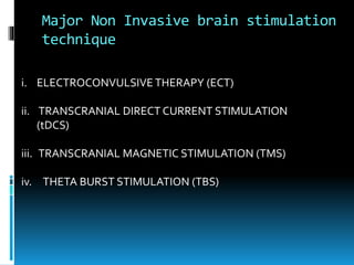
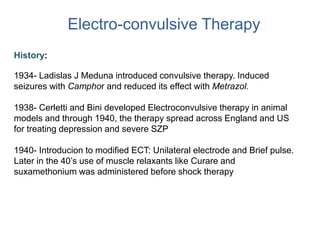
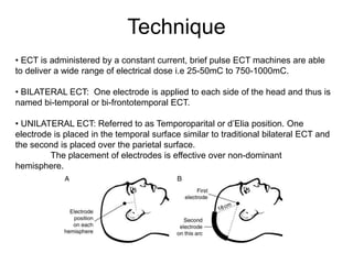
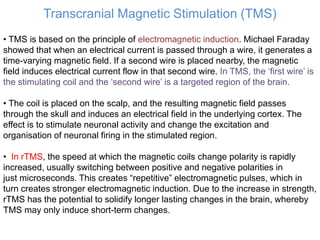
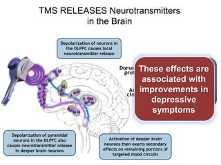
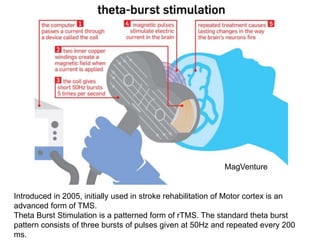
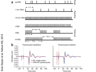
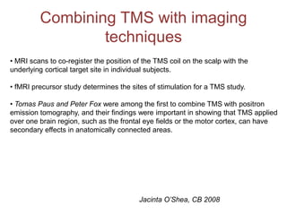
DIFFERENT ‘NON INVASIVE’
BRAIN STIMULATION
PROCEDURES
Presented by-
Moitrayee Majumder
M.Phil Neuroscience 1st year
Major Non Invasive brain stimulation
technique
i. ELECTROCONVULSIVE THERAPY (ECT)
ii. TRANSCRANIAL DIRECT CURRENT STIMULATION
(tDCS)
iii. TRANSCRANIAL MAGNETIC STIMULATION (TMS)
iv. THETA BURST STIMULATION (TBS)
Electro-convulsive Therapy
History:
1934- Ladislas J Meduna introduced convulsive therapy. Induced
seizures with Camphor and reduced its effect with Metrazol.
1938- Cerletti and Bini developed Electroconvulsive therapy in animal
models and through 1940, the therapy spread across England and US
for treating depression and severe SZP
1940- Introducion to modified ECT: Unilateral electrode and Brief pulse.
Later in the 40’s use of muscle relaxants like Curare and
suxamethonium was administered before shock therapy
Technique
• ECT is administered by a constant current, brief pulse ECT machines are able
to deliver a wide range of electrical dose i.e 25-50mC to 750-1000mC.
• BILATERAL ECT: One electrode is applied to each side of the head and thus is
named bi-temporal or bi-frontotemporal ECT.
• UNILATERAL ECT: Referred to as Temporoparital or d’Elia position. One
electrode is placed in the temporal surface similar to traditional bilateral ECT and
the second is placed over the parietal surface.
The placement of electrodes is effective over non-dominant
hemisphere.
Dosing Strategy
• Stimulus dosing: The selection of electrical dose for an individual
patient is contingent upon the patient’s seizure threshold.
• Empirical measurement is the best available means of establishing, the
initial seizure threshold. Developing experience for the initial dose by
assessing the severity of the patient’s condition.
• During management of life threatening events, available schedules like
the Clinic’s dosing strategy is to be followed.
• In Unilateral ECT: The empirical techniques may not be crucial for
electrical doses intended to be only two or three times the seizure
threshold whereas it is practiced when yet higher dose is to be
implemented.
NICE guidelines
Mechanism of action
• ECT might have its effects on many systems in the brain but focused on
neurotransmitter release and regulation system.
• Modulating monoamine systems viz Serotonergic and nor-adrenergic pathways in
the CNS.
• Enhancing activity of dopaminergic systems which explains its effectiveness in
Depressive as well as Parkinsonism. Studies in animal model showed
expression of neuroprotective protein BDNF which antagonises neurotoxic effects
of stress on brain, on stimulations with ECT.
• Anticonvulsant in treatment and prophylaxis of Bipolar Disorder.
• Effectiveness on excitatory amino acid systems in treating psychosis in Mania
and Schizophrenia.
• Animal models treated with ECT showed potent effects in strengthening neuronal
survival and promotes the production of new neural processes.
• Chronic depression is associated with atrophy of brain structures like fronto-
temporal lobes and ECT was found to reverse it effectively
ECT in Depressive Illness
• Emergency treatment of Depression: Psychomotor retardation with eating and
drinking or physical deterioration. Suicidal tendencies.
• Treatment resistant Depression: Substantial response to ECT compared to
non-treatment resistants.
• Depressive Stupor and Catatonic depression: Increased motor immobility and
withdrawl or purposeless excessive motor activity in absence of external stimuli
is treated with short acting BDZ primarilly but in treatment unaffected or non-
severe cases ECT shows therapeutic effect
• Treating Elderly patients with depression: Older people have higher seizure
threshold but they show significant improvement with remission in most of them.
ECT techniques need to modified to reduce adverse cognitive effect.
• Depression in Pregnancy: 23% Postnatal depressive states initiates during
pregnancy. Miller(1994) reviewed cases treated with ECT and found out
statistically reduced postnatal depressive symptoms with less unaccountable
genetic malformations of the neonate.
ECT Handbook: Allan Scott
Adverse Effects of ECT
Cognitive adverse effects: Weiner summarised about retrograde amnesia that
can occur with ECT and noted that:
• It produces deficits in both autobiographical and impersonal memory domains.
• The severity and persistance of retrograde amnesia is greater with bilateral
electrode placement and with sine-wave stimuli than brief-pulse.
• Objective memory impairment may be persisting pre and post therapy session.
•Post therapy, some patients experience variable periods of disorientation
associated with impaired attention, memory and praxis. These effects are short
lived.
• Acute confusional states between treatment in patients with history of taking
psychotropic drugs or those with pre existing neurological conditions. In case of
which treatment modifications are recommended.
• Rarely patients develop postictal delerium manifesting as restlessness,
aggression or agitation which responds to treatment with BDZ.
• Headaches, muscular aches, drowsiness, weakness, nausea and anorexia.
Transcranial Direct Current Stimulations
HISTORY:
19th Century- rudimentary experiments of current on animals and human by Luigi
Galvani and Alessandro Volta.
1801- Giovanni Aldini used and studied application of DC to stabilize mood of
melancholy patients
1960- D J Albert showed DC induced cortical excitability.
Bindman et al performed experiments that resulted in long-lasting
polarization effects following electric stimulation of the exposed motor cortex of
animals.
Current View- With illustrated understanding
of basic brain functioning and therapeutic
applications, reliable tDC stimulation
technique supported with research using
brain imaging techniques such as fMRI,
tDCS is being more frequently used as a
method of non invasive psychiatric
treatment with proper safety protocols.
PC: JAMA Neurology
Mechanism of tDCS
• A constant current stimulator and surface electrodes soaked in isotonic sodium chloride
solution are applied and secured onto the scalp over desired areas such as the left or
right precentral gyrus region (corresponding to C3 or C4 of the international 10-20
electroencephalographic system), form terminals relaying currents across the scalp and
through the underlying brain tissue.
• McCreery et al, found that current densities below 25 mA/cm2 did not cause brain
tissue damage, and the protocols that apply 1 to 2 mA as in present-day studies fall well
within these limits.
• Despite a fraction of the direct current being shunted through the scalp, tDCS carries
adequate currents to the underlying cortex that are sufficient for neuronal excitability
shifts.
• The advantages of tDCS over other non-invasive brain stimulation methods include its
ease of use, large electrode size allowing effect over a larger neural network, sham
mode allowing controlled experiments and randomized controlled clinical trials, and
portability that makes it possible to apply stimulation while the patient receives
occupational or physical therapy.
•LIMITATIONS: TDCS is limited by its poor temporal resolution and anatomical
localization. Furthermore, interindividual variation in conductivity due to differences in
Brain Polarization in tDCS
• Nitsche and Paulus demonstrated modulating effects of anodal (increases cortical
excitability) and cathodal (decreases cortical excitability) TDCS on brain tissue in which
the effects surprisingly outlasted the duration of stimulation.
• Adequate tDCS shows neuronal excitability shifts and change in measured of cerebral
blood flow, which acts as neurotrophic to depressed brain regions.
• The prolonged sensory, motor, and cognitive effects of TDCS have been attributed to
persistent bidirectional modification of postsynaptic connections similar to long-term
potentiation and
long-term depression effects.
TDCS stimulation involves the alteration of synaptic transmission ability through
modifications of intracellular cAMP and calcium levels, as well as glial activation
• Dextromethorphan, an NMDA antagonist, suppressed anodal and cathodal TDCS
effects, strongly suggesting the involvement of receptors of the antagonist in both types
of direct
current–induced neuroplasticity. In contrast Carbamazepine selectively eliminated
anodal current by stabilizing the inactive state of the voltage gated Na+ channels.
SIDE EFFECTS: Skin irritation, Phosphene at onset of stimulation, nausea, headache
Pryamidal tract
of Patient 2
Pyramidal tract
of Patient 1
Bilateral
TDCS
stimulation
Gottfried
Schlaug et
al, Arch
Neurol, 2008
Indications of using tDCS
• Major Depressive disorder and Mania: The dorsal system (cognitive
control system) and the Ventral system (emotional evaluation system)
counterbalances cognitive processing of emotional output. Impairment of
one pathway results in depression or mania.
Neuromodulatory effects of tDCS, reverses the imbalance
between hypoactive cortical areas and hyperactive subcortical areas.
• Schizophrenia: Auditory verbal hallucinations have been linked to
fronto-temporal abnormalities with an hyperactivity in the left temporo-
parietal junction and negative symptoms have been linked to hypoactivity
in the right and left prefrontal cortex.
• Sleep disoeders
• Anxiety
• Stroke
• Alzheimer’s disease: Improving Visual recognition memory
Gabriel Tortella et al, World J
Psychiatry, 2015
Transcranial Magnetic Stimulation (TMS)
• TMS is based on the principle of electromagnetic induction. Michael Faraday
showed that when an electrical current is passed through a wire, it generates a
time-varying magnetic field. If a second wire is placed nearby, the magnetic
field induces electrical current flow in that second wire. In TMS, the ‘first wire’ is
the stimulating coil and the ‘second wire’ is a targeted region of the brain.
• The coil is placed on the scalp, and the resulting magnetic field passes
through the skull and induces an electrical field in the underlying cortex. The
effect is to stimulate neuronal activity and change the excitation and
organisation of neuronal firing in the stimulated region.
• In rTMS, the speed at which the magnetic coils change polarity is rapidly
increased, usually switching between positive and negative polarities in
just microseconds. This creates “repetitive” electromagnetic pulses, which in
turn creates stronger electromagnetic induction. Due to the increase in strength,
rTMS has the potential to solidify longer lasting changes in the brain, whereby
TMS may only induce short-term changes.
TMS RELEASES Neurotransmitters
in the Brain
Depolarization of neurons in
the DLPFC causes local
neurotransmitter release
Depolarization of pyramidal
neurons in the DLPFC also
causes neurotransmitter release
in deeper brain neurons
Activation of deeper brain
neurons then exerts secondary
effects on remaining portions of
targeted mood circuits
Dorsolateral
prefrontal
cortex
Anterior
cingulate
cortex
Kito (2008) J Neuropsychiatry Clin Neurosci
These effects are
associated with
improvements in
depressive
symptoms
TMS Protocols
• Single pulse TMS: Initial appliation of TMS on motor system enabled functional
representation in Primary Motor Cortex (M1) with spTMS and response in particular
muscle with Motor Evoked Potential (MEP). Suprathreshold spTMS over Primary visual
cortex (V1) induced Phosphenes and Transient Scotomas whereas over V5 induced
motion perception.
• Paired Pulse TMS: ppTMS was used to study two anatomically
or functionally connected brain areas and providing a
Subthreshold Conditioning stimuli prceeding a Suprathreshold
test stimuli, the inter-stimuli latency determines facilitatory or
inhibitory effects on cortico-spinal output as MEPs.
• Repetative TMS: rTMS is applied over a site of interest for
several minutes. The induced effects outlast the period of
stimulation, giving insight into the role of the specific
stimulated brain regions in plasticity and behavior.
Eg: Low frequency (1 Hz) inhibits cortical stimulation which can be
pharmacologically reversed and High Frequency (5-20 Hz) facilitates cortical excitability.
This model has been used to understand sequence motor learning at different time
points involving different cortical areas.
Eran Dayan et al, Nature NS, 2013
Introduced in 2005, initially used in stroke rehabilitation of Motor cortex is an
advanced form of TMS.
Theta Burst Stimulation is a patterned form of rTMS. The standard theta burst
pattern consists of three bursts of pulses given at 50Hz and repeated every 200
ms.
MagVenture
EranDayanetal,NatureNS,2013
Combining TMS with imaging
techniques
• MRI scans to co-register the position of the TMS coil on the scalp with the
underlying cortical target site in individual subjects.
• fMRI precursor study determines the sites of stimulation for a TMS study.
• Tomas Paus and Peter Fox were among the first to combine TMS with positron
emission tomography, and their findings were important in showing that TMS applied
over one brain region, such as the frontal eye fields or the motor cortex, can have
secondary effects in anatomically connected areas.
Jacinta O’Shea, CB 2008
Indications for TMS
 Auditory hallucinations in schizophrenia – 1 Hz TMS over superior temporal gyrus
 PTSD – 10 Hz over right prefrontal cortex
 ADHD – to target the right medial frontal gyrus.
 Post stroke rehabilitation
Adverse Effects
Seizure induction
Headache
Scalp facial muscle twitching
Mild tinnitus, hearing loss
THANKYOU
The presentation had been very peripheral as it requires decades to go
through 2051 documented material.

NIBS

  • 1.
    DIFFERENT ‘NON INVASIVE’ BRAINSTIMULATION PROCEDURES Presented by- Moitrayee Majumder M.Phil Neuroscience 1st year
  • 2.
    Major Non Invasivebrain stimulation technique i. ELECTROCONVULSIVE THERAPY (ECT) ii. TRANSCRANIAL DIRECT CURRENT STIMULATION (tDCS) iii. TRANSCRANIAL MAGNETIC STIMULATION (TMS) iv. THETA BURST STIMULATION (TBS)
  • 3.
    Electro-convulsive Therapy History: 1934- LadislasJ Meduna introduced convulsive therapy. Induced seizures with Camphor and reduced its effect with Metrazol. 1938- Cerletti and Bini developed Electroconvulsive therapy in animal models and through 1940, the therapy spread across England and US for treating depression and severe SZP 1940- Introducion to modified ECT: Unilateral electrode and Brief pulse. Later in the 40’s use of muscle relaxants like Curare and suxamethonium was administered before shock therapy
  • 4.
    Technique • ECT isadministered by a constant current, brief pulse ECT machines are able to deliver a wide range of electrical dose i.e 25-50mC to 750-1000mC. • BILATERAL ECT: One electrode is applied to each side of the head and thus is named bi-temporal or bi-frontotemporal ECT. • UNILATERAL ECT: Referred to as Temporoparital or d’Elia position. One electrode is placed in the temporal surface similar to traditional bilateral ECT and the second is placed over the parietal surface. The placement of electrodes is effective over non-dominant hemisphere.
  • 5.
    Dosing Strategy • Stimulusdosing: The selection of electrical dose for an individual patient is contingent upon the patient’s seizure threshold. • Empirical measurement is the best available means of establishing, the initial seizure threshold. Developing experience for the initial dose by assessing the severity of the patient’s condition. • During management of life threatening events, available schedules like the Clinic’s dosing strategy is to be followed. • In Unilateral ECT: The empirical techniques may not be crucial for electrical doses intended to be only two or three times the seizure threshold whereas it is practiced when yet higher dose is to be implemented. NICE guidelines
  • 6.
    Mechanism of action •ECT might have its effects on many systems in the brain but focused on neurotransmitter release and regulation system. • Modulating monoamine systems viz Serotonergic and nor-adrenergic pathways in the CNS. • Enhancing activity of dopaminergic systems which explains its effectiveness in Depressive as well as Parkinsonism. Studies in animal model showed expression of neuroprotective protein BDNF which antagonises neurotoxic effects of stress on brain, on stimulations with ECT. • Anticonvulsant in treatment and prophylaxis of Bipolar Disorder. • Effectiveness on excitatory amino acid systems in treating psychosis in Mania and Schizophrenia. • Animal models treated with ECT showed potent effects in strengthening neuronal survival and promotes the production of new neural processes. • Chronic depression is associated with atrophy of brain structures like fronto- temporal lobes and ECT was found to reverse it effectively
  • 7.
    ECT in DepressiveIllness • Emergency treatment of Depression: Psychomotor retardation with eating and drinking or physical deterioration. Suicidal tendencies. • Treatment resistant Depression: Substantial response to ECT compared to non-treatment resistants. • Depressive Stupor and Catatonic depression: Increased motor immobility and withdrawl or purposeless excessive motor activity in absence of external stimuli is treated with short acting BDZ primarilly but in treatment unaffected or non- severe cases ECT shows therapeutic effect • Treating Elderly patients with depression: Older people have higher seizure threshold but they show significant improvement with remission in most of them. ECT techniques need to modified to reduce adverse cognitive effect. • Depression in Pregnancy: 23% Postnatal depressive states initiates during pregnancy. Miller(1994) reviewed cases treated with ECT and found out statistically reduced postnatal depressive symptoms with less unaccountable genetic malformations of the neonate. ECT Handbook: Allan Scott
  • 8.
    Adverse Effects ofECT Cognitive adverse effects: Weiner summarised about retrograde amnesia that can occur with ECT and noted that: • It produces deficits in both autobiographical and impersonal memory domains. • The severity and persistance of retrograde amnesia is greater with bilateral electrode placement and with sine-wave stimuli than brief-pulse. • Objective memory impairment may be persisting pre and post therapy session. •Post therapy, some patients experience variable periods of disorientation associated with impaired attention, memory and praxis. These effects are short lived. • Acute confusional states between treatment in patients with history of taking psychotropic drugs or those with pre existing neurological conditions. In case of which treatment modifications are recommended. • Rarely patients develop postictal delerium manifesting as restlessness, aggression or agitation which responds to treatment with BDZ. • Headaches, muscular aches, drowsiness, weakness, nausea and anorexia.
  • 9.
    Transcranial Direct CurrentStimulations HISTORY: 19th Century- rudimentary experiments of current on animals and human by Luigi Galvani and Alessandro Volta. 1801- Giovanni Aldini used and studied application of DC to stabilize mood of melancholy patients 1960- D J Albert showed DC induced cortical excitability. Bindman et al performed experiments that resulted in long-lasting polarization effects following electric stimulation of the exposed motor cortex of animals. Current View- With illustrated understanding of basic brain functioning and therapeutic applications, reliable tDC stimulation technique supported with research using brain imaging techniques such as fMRI, tDCS is being more frequently used as a method of non invasive psychiatric treatment with proper safety protocols. PC: JAMA Neurology
  • 10.
    Mechanism of tDCS •A constant current stimulator and surface electrodes soaked in isotonic sodium chloride solution are applied and secured onto the scalp over desired areas such as the left or right precentral gyrus region (corresponding to C3 or C4 of the international 10-20 electroencephalographic system), form terminals relaying currents across the scalp and through the underlying brain tissue. • McCreery et al, found that current densities below 25 mA/cm2 did not cause brain tissue damage, and the protocols that apply 1 to 2 mA as in present-day studies fall well within these limits. • Despite a fraction of the direct current being shunted through the scalp, tDCS carries adequate currents to the underlying cortex that are sufficient for neuronal excitability shifts. • The advantages of tDCS over other non-invasive brain stimulation methods include its ease of use, large electrode size allowing effect over a larger neural network, sham mode allowing controlled experiments and randomized controlled clinical trials, and portability that makes it possible to apply stimulation while the patient receives occupational or physical therapy. •LIMITATIONS: TDCS is limited by its poor temporal resolution and anatomical localization. Furthermore, interindividual variation in conductivity due to differences in
  • 11.
    Brain Polarization intDCS • Nitsche and Paulus demonstrated modulating effects of anodal (increases cortical excitability) and cathodal (decreases cortical excitability) TDCS on brain tissue in which the effects surprisingly outlasted the duration of stimulation. • Adequate tDCS shows neuronal excitability shifts and change in measured of cerebral blood flow, which acts as neurotrophic to depressed brain regions. • The prolonged sensory, motor, and cognitive effects of TDCS have been attributed to persistent bidirectional modification of postsynaptic connections similar to long-term potentiation and long-term depression effects. TDCS stimulation involves the alteration of synaptic transmission ability through modifications of intracellular cAMP and calcium levels, as well as glial activation • Dextromethorphan, an NMDA antagonist, suppressed anodal and cathodal TDCS effects, strongly suggesting the involvement of receptors of the antagonist in both types of direct current–induced neuroplasticity. In contrast Carbamazepine selectively eliminated anodal current by stabilizing the inactive state of the voltage gated Na+ channels. SIDE EFFECTS: Skin irritation, Phosphene at onset of stimulation, nausea, headache
  • 12.
    Pryamidal tract of Patient2 Pyramidal tract of Patient 1 Bilateral TDCS stimulation Gottfried Schlaug et al, Arch Neurol, 2008
  • 13.
    Indications of usingtDCS • Major Depressive disorder and Mania: The dorsal system (cognitive control system) and the Ventral system (emotional evaluation system) counterbalances cognitive processing of emotional output. Impairment of one pathway results in depression or mania. Neuromodulatory effects of tDCS, reverses the imbalance between hypoactive cortical areas and hyperactive subcortical areas. • Schizophrenia: Auditory verbal hallucinations have been linked to fronto-temporal abnormalities with an hyperactivity in the left temporo- parietal junction and negative symptoms have been linked to hypoactivity in the right and left prefrontal cortex. • Sleep disoeders • Anxiety • Stroke • Alzheimer’s disease: Improving Visual recognition memory Gabriel Tortella et al, World J Psychiatry, 2015
  • 14.
    Transcranial Magnetic Stimulation(TMS) • TMS is based on the principle of electromagnetic induction. Michael Faraday showed that when an electrical current is passed through a wire, it generates a time-varying magnetic field. If a second wire is placed nearby, the magnetic field induces electrical current flow in that second wire. In TMS, the ‘first wire’ is the stimulating coil and the ‘second wire’ is a targeted region of the brain. • The coil is placed on the scalp, and the resulting magnetic field passes through the skull and induces an electrical field in the underlying cortex. The effect is to stimulate neuronal activity and change the excitation and organisation of neuronal firing in the stimulated region. • In rTMS, the speed at which the magnetic coils change polarity is rapidly increased, usually switching between positive and negative polarities in just microseconds. This creates “repetitive” electromagnetic pulses, which in turn creates stronger electromagnetic induction. Due to the increase in strength, rTMS has the potential to solidify longer lasting changes in the brain, whereby TMS may only induce short-term changes.
  • 15.
    TMS RELEASES Neurotransmitters inthe Brain Depolarization of neurons in the DLPFC causes local neurotransmitter release Depolarization of pyramidal neurons in the DLPFC also causes neurotransmitter release in deeper brain neurons Activation of deeper brain neurons then exerts secondary effects on remaining portions of targeted mood circuits Dorsolateral prefrontal cortex Anterior cingulate cortex Kito (2008) J Neuropsychiatry Clin Neurosci These effects are associated with improvements in depressive symptoms
  • 16.
    TMS Protocols • Singlepulse TMS: Initial appliation of TMS on motor system enabled functional representation in Primary Motor Cortex (M1) with spTMS and response in particular muscle with Motor Evoked Potential (MEP). Suprathreshold spTMS over Primary visual cortex (V1) induced Phosphenes and Transient Scotomas whereas over V5 induced motion perception. • Paired Pulse TMS: ppTMS was used to study two anatomically or functionally connected brain areas and providing a Subthreshold Conditioning stimuli prceeding a Suprathreshold test stimuli, the inter-stimuli latency determines facilitatory or inhibitory effects on cortico-spinal output as MEPs. • Repetative TMS: rTMS is applied over a site of interest for several minutes. The induced effects outlast the period of stimulation, giving insight into the role of the specific stimulated brain regions in plasticity and behavior. Eg: Low frequency (1 Hz) inhibits cortical stimulation which can be pharmacologically reversed and High Frequency (5-20 Hz) facilitates cortical excitability. This model has been used to understand sequence motor learning at different time points involving different cortical areas. Eran Dayan et al, Nature NS, 2013
  • 17.
    Introduced in 2005,initially used in stroke rehabilitation of Motor cortex is an advanced form of TMS. Theta Burst Stimulation is a patterned form of rTMS. The standard theta burst pattern consists of three bursts of pulses given at 50Hz and repeated every 200 ms. MagVenture
  • 18.
  • 19.
    Combining TMS withimaging techniques • MRI scans to co-register the position of the TMS coil on the scalp with the underlying cortical target site in individual subjects. • fMRI precursor study determines the sites of stimulation for a TMS study. • Tomas Paus and Peter Fox were among the first to combine TMS with positron emission tomography, and their findings were important in showing that TMS applied over one brain region, such as the frontal eye fields or the motor cortex, can have secondary effects in anatomically connected areas. Jacinta O’Shea, CB 2008
  • 20.
    Indications for TMS Auditory hallucinations in schizophrenia – 1 Hz TMS over superior temporal gyrus  PTSD – 10 Hz over right prefrontal cortex  ADHD – to target the right medial frontal gyrus.  Post stroke rehabilitation
  • 21.
    Adverse Effects Seizure induction Headache Scalpfacial muscle twitching Mild tinnitus, hearing loss
  • 22.
    THANKYOU The presentation hadbeen very peripheral as it requires decades to go through 2051 documented material.

Editor's Notes

  • #4 Ladislas believed SZP and EL are antagonist. Inducing seizure can cure paranoia. Metrazol is a anticonvulsant Suxamethonium- used for pain and is a sedative
  • #5 According to National Institute for Clinical Excellence, the dose and position is standarized for clinical efficacy.
  • #7 Seizures called kindling occurs in BD due to sudden mood swings. Monoamine Nts regulate emotional and arousal behaviour.
  • #8 Effective across 3 days to 2 week therapy The Consensus group suggests, in absence of urgent need of shock therapy, failure to respond adequately to 2 different courses of antidepressant for a sufficient time is treatment resistance D
  • #12 Brief sessions of DC stimulation brings about the threshold for depolarization or hyperpolarization.
  • #13 Diffusion tensor imaging and stroke recovery potential
  • #14 The current neural models of depression propose that the emotional deregulation is due to abnormalities in the dorsal neural system (cognitive control system) and the ventral neural system (emotional evaluation system) CDS- DLPFC, dorsomedial PFC, the anterior dorsal cingulate gyrus and the hippocampus EES-  amygdala, insula, the ventral striate, dorsal cingulate gyrus and ventral PFC
  • #15  Anthony Barker and colleagues first demonstrated TMS in 1985 it has been used widely to stimulate both peripheral nerves and brain tissue in studies encompassing motor conduction in human development, motor control, movement disorders, swallowing, vision, attention, memory, speech and language, epilepsy, depression, stroke, pain and plasticity
  • #16 When the pulsed magnetic fields from the TMS coil are applied to the left dorsolateral prefrontal cortex, there are a series of events that are thought to underlie the therapeutic effects of TMS in the treatment of major depression: First, direct neuronal depolarization under the coil leads to local action potentials in neurons and the local release of neurotransmitters in the cortex. In addition to these local effects, neuronal depolarization of cortical pyramidal neurons is thought to occur (as represented by the blue neural pathway), reaching to deeper brain regions that lie outside the direct action of the pulsed magnetic fields. Activation of these deeper brain regions is then presumed to lead to secondary activation of brainstem neurotransmitter centers, which are then presumed to result in upward influences on the remaining brain regions involved in mood regulation (represented by the purple neural pathway). As a result, dopamine (Kanno 2004, pp. 75A, 76A, 77A) and serotonin (Juckel 1999, pp. 393A, 394A) activity are increased in areas of the brain whose low neurotransmitter activity have been linked to depression. The activity may be increased both in the short term by increasing release of neurotransmitters and in the long term by modulating expression of proteins involved in neurotransmitters signaling (Post 2001, p. 200A,B). Presumably, as a result of these changes, depression lifts (Slotema 2010, p. 876A). The net action of TMS is therefore targeted on the specific brain areas known to be involved in the regulation of mood, and is comprehensive in that its action has both direct effects on local neurons in the cerebral cortex, and then results in deeper actions on brain regions that are distant from the site of stimulation, but neurally connected to these cortical areas. These effects can be demonstrated in human neuroimaging studies of patients who have undergone treatment with TMS for their depression, as shown in the SPECT (single photon emission computed tomography) scan on the right (Kito, et al, 2008). In this image, the TMS coil has been positioned over the dorsolateral prefrontal cortex on the left side of the head. The area just underneath the coil is showing increased metabolic activity as a direct result of the magnetic stimulation. You can also see that the increase in metabolism reaches secondarily the deeper brain regions, in this case the regions of the cingulate cortex also show increased activation. References: Kanno M, Matsumoto M, et al. Effects of acute repetitive transcranial magnetic stimulation on dopamine release in rat dorsolateral striatum. J Neurological Sciences. 2004;217:73-81. Juckel G, Mendlin MA, et al. Electrical Stimulation of Rat Medial Prefrontal Cortex Enhances Forebrain Serotonin Output: Implications for Electroconvulsive Therapy and Transcranial Magnetic Stimulation in Depression. Neuropsychopharmacology. 1999;21(3):391-398. Slotema CW, Blom JD, et al. Should we expand the toolbox of psychiatric treatment methods to include repetitive transcranial magnetic stimulation (rTMS)? A meta-analysis of the efficacy of rTMS in psychiatric disorders. J Clin Psychiatry. 2010;71(7):873-884. Kito, S, Fujita, K, Koga, Y. Changes in Regional Cerebral Blood Flow After Repetitive Transcranial Magnetic Stimulation of the Left Dorsolateral Prefrontal Cortex in Treatment-Resistant Depression. J Neuropsychiatry Clin Neurosci. 2008; 20(1):74-80.
  • #17 Variation of the precise latency between the conditioning and test stimuli can result in intracortical inhibitory (if the conditioning stimulus precedes the test stimulus by <5 ms) or intracortical facilitory (if the conditioning stimulus precedes the test stimulus by latencies between 6 and 25 ms, or if the conditioning stimulus succeeds the test stimulus at 1.5-ms intervals between approximately 1 and 4.5 ms) effects on corticospinal output  rTMS has been extended to probe cognitive processes as well, including spatial attention, working memory, episodic memory and decision making
  • #18 The effects of synaptic plasticy lasts for 30 min post one session.
  • #19 Continuous TBS (cTBS) involves the application of burst trains for 20–40 s and has an inhibitory effect on corticospinal excitability. Conversely, for intermittent TBS (iTBS), burst trains with a duration of 2 s are applied over a total of 190 s, with the trains repeating every 10 s. Intermediate TBS, a third variant including 5-s burst trains repeated every 15 s for a total of 110 s, is typically used as a negative control for cTBS and iTBS as it shows no effects on corticospinal excitibility. These patterned stimulation protocols induce longer-lasting effects than conventional rTMS paradigms.