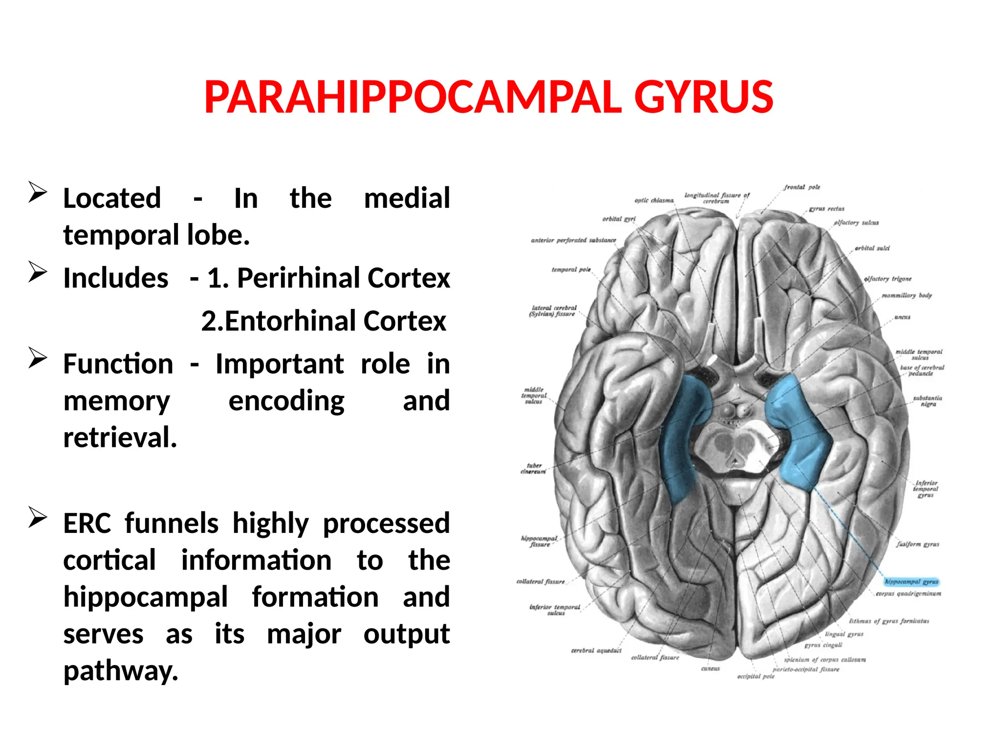
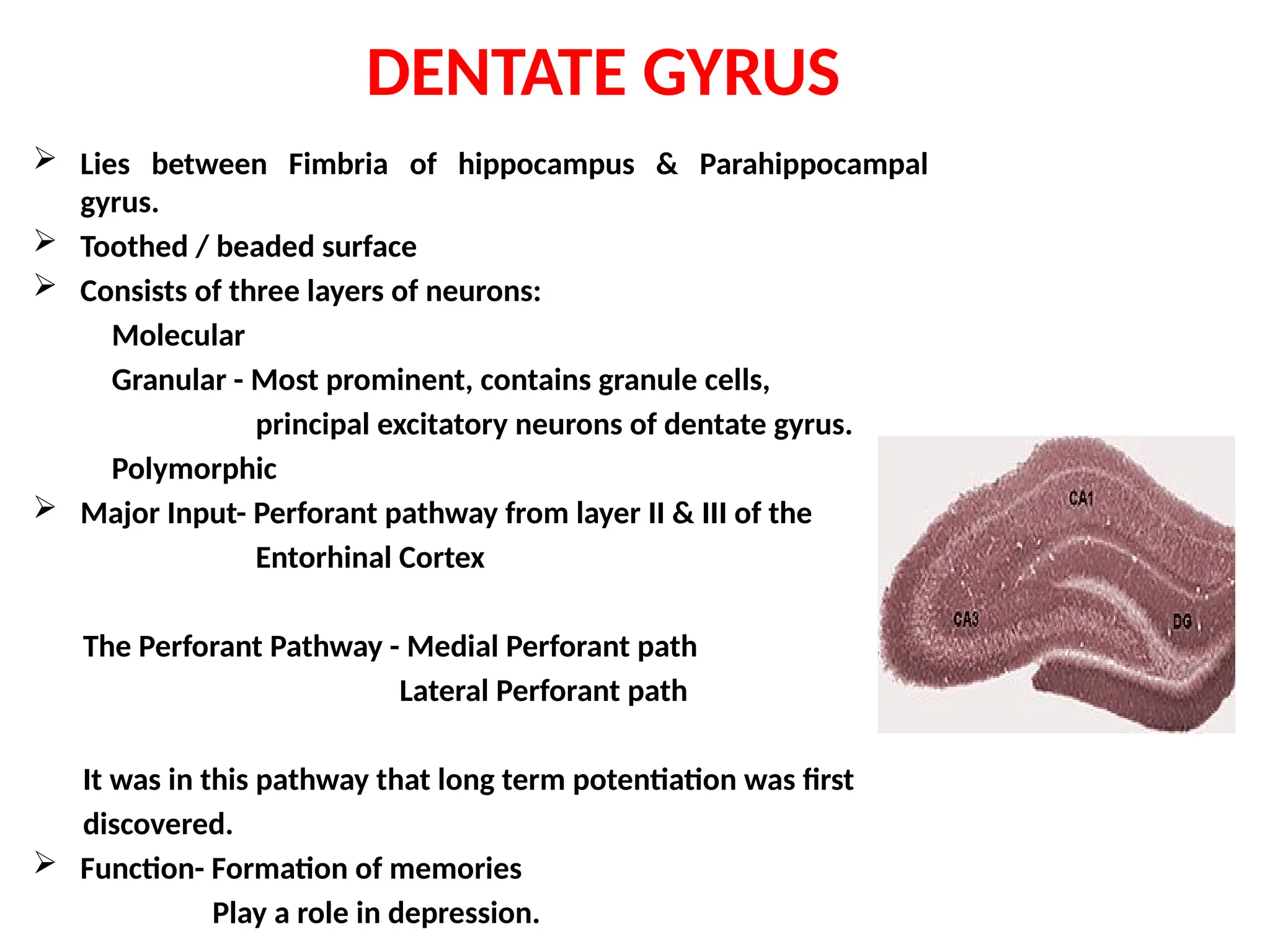
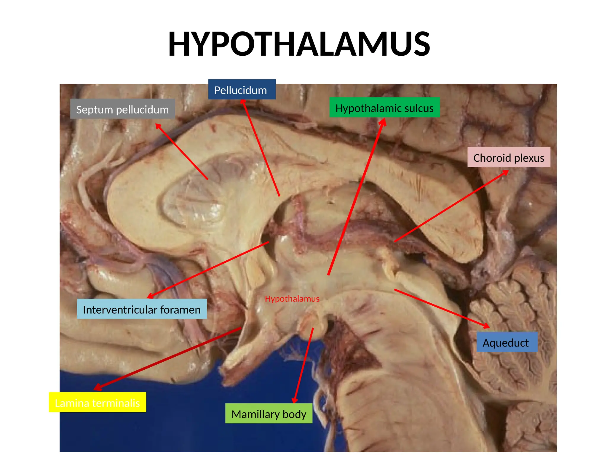
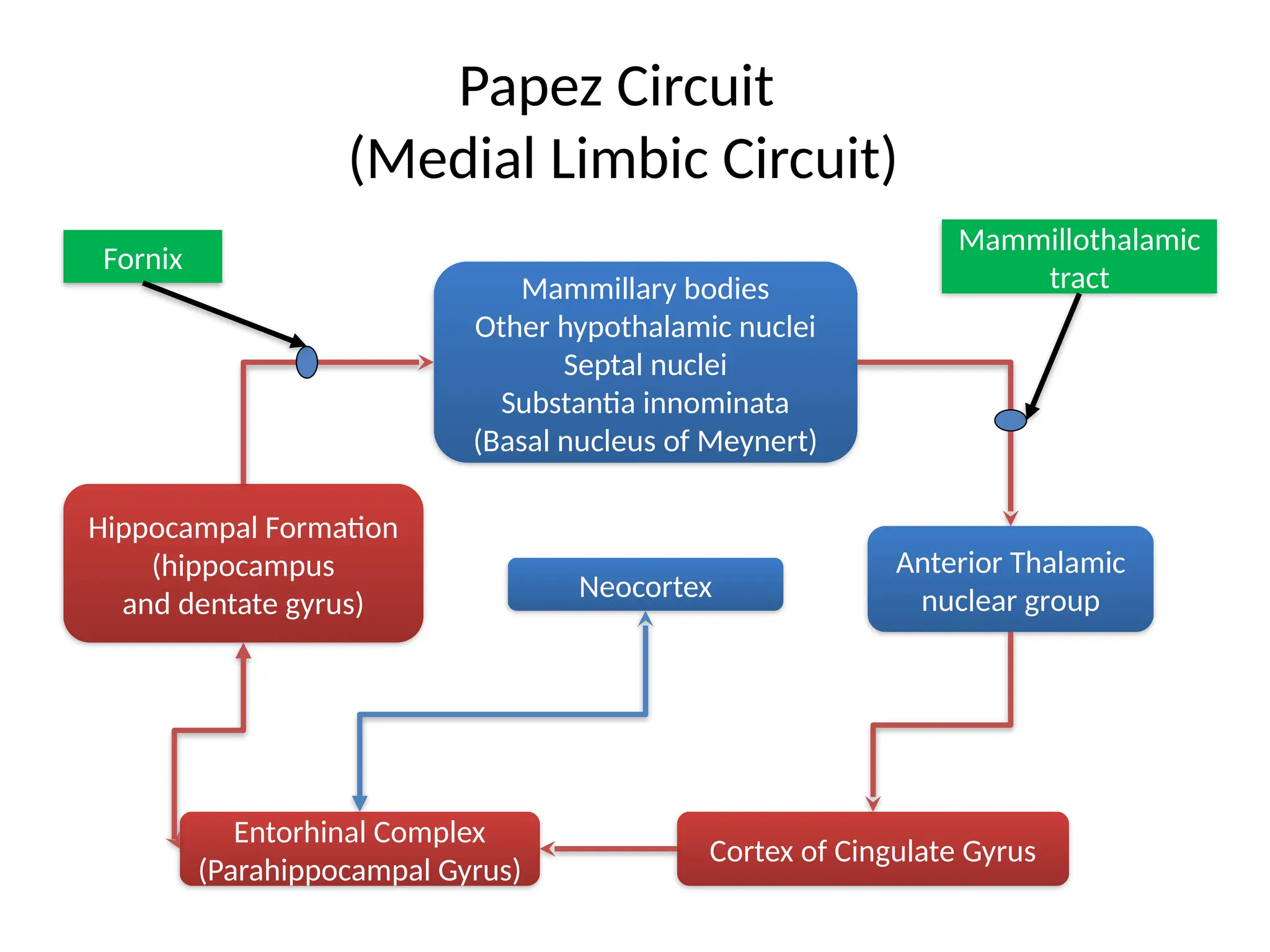
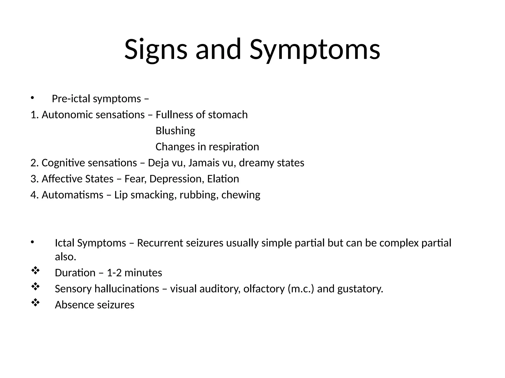
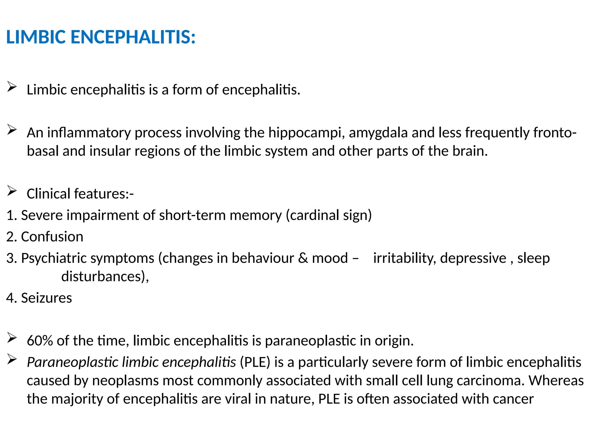
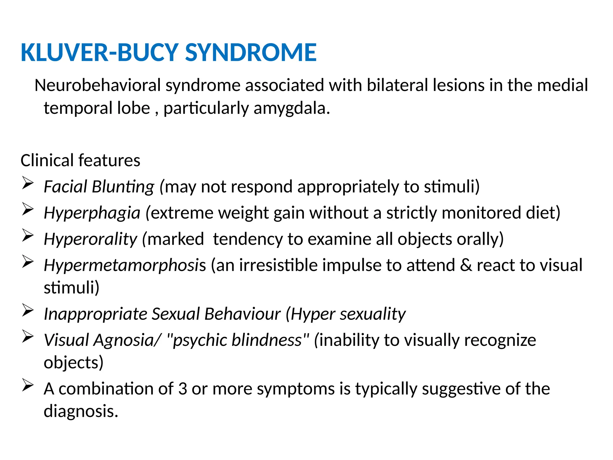
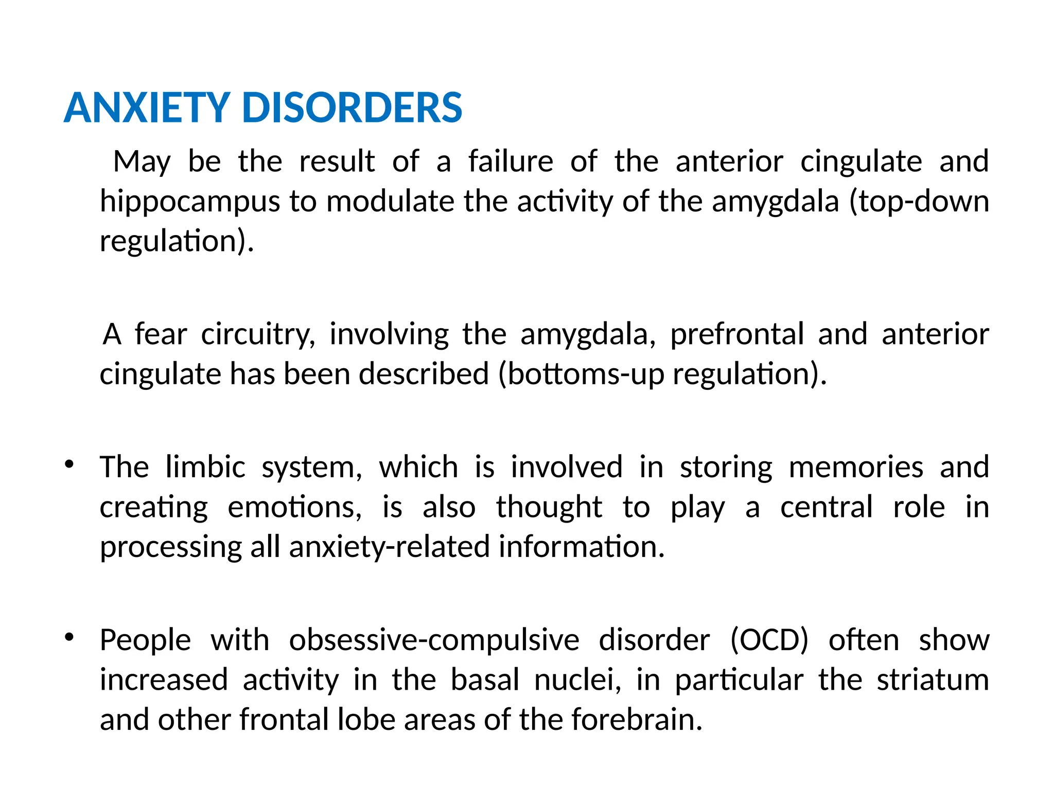
LIMBIC SYSTEM-IT’S CLINICAL IMPLICATION
PRESENTER – Dr. Shaitan Singh Jat
SR Neurology
MODERATORS – Dr. Trilochan Shrivastava Sir
Dr. Rahul Gupta Sir
Contents
• Historical Aspects
• Introduction
• Components of Limbic System
• Papez Circuit
• Clinical Implications
Historical Aspects
Paul Broca 1878 le grand lobe limbique
James Papez 1937 Papez Circuit
Paul Kluver & Heinrich Bucy First evidence of cortical
1939 representation
of emotions in limbic
system
Paul Maclean 1952 Limbic System
 Functional anatomic system of interconnected cortical and
sub cortical structures.
 Area of intimate processing between the hypothalamus and
cortical information.
 Plays a role in emotions, learning and autonomic regulation.
INTRODUCTION
THE BROAD FUNCTIONAL DIVISIONS
• Rostral limbic system: Amygdala, septum, orbitofrontal cortex,
anterior insula, and anterior cingulate.
– Important for emotion
• Caudal limbic system: Hippocampus, posterior parahippocampal
cortex and posterior cingulate.
– Important for memory and visual-spatial functions
1. Limbic lobe
a) Cingulate gyrus
b) Para hippocampal Gyrus
2. Hippocampal formation
a) Dentate Gyrus
b) Hippocampus proper
c) Subiculum
3. Amygdala
4. Septal area
5. Hypothalamus
Others - cortex and insula, uncus, stria terminalis
Components of limbic system
The amygdala
• Located in medial temporal lobe
• Just anterior to the hippocampal
formation
• It is fused with the tip of the tail of
the caudate nucleus
• Stria terminalis emerges from its post
aspect
Amygdala Inputs
AMYGDALA
Corticomedial Nuclear
Group
Basolateral Nuclear
Group
Central Nucleus
Olfactory
System
Temporal Lobe
(associated with visual,
auditory, tactile senses)
Brainstem (viscerosensory relay
Nuclei: solitary nucleus
and parbrachial nucleus)
Ventral
Amygdalofugal
Fibers
Mediates behaviors
triggered by olfactory
stimuli
Mediates emotional
responses
Attaching emotional
significance to a
stimulus
Amygdala Outputs
AMYGDALA
Corticomedial Nuclear
Group
Basolateral Nuclear
Group
Central Nucleus
Ventral
Amygdalofugal
Fibers
Septal Nuclei
Hypothalamus
Limbic Association Cortex
Prefrontal Cortex
Hippocampal Formation
Dorsal motor n. Of X
Reticular formation
Hypothalamus
Nuclei of ANS
Ventral
Amygdalofugal
Fibers
Stria Terminalis
Learning
emotional
significance
Autonomic
responses
Regulation of
“appetitive
behaviours”;
i.e., Eating in
response to
smells
•Emotional Learning - Formation and storage of memories
associated with emotional events.
•Modulation of Memory Consolidation - Following any learning
event, the long-term memory for the event is not formed
instantaneously. Rather, information regarding the event is slowly
assimilated into long-term storage over time, possibly via Long-term
Potentiation.
•Aggression - Animal studies have shown that stimulating the
amygdala appears to increase both sexual and aggressive behaviour.
•Alcoholism and binge drinking - Alcoholism is associated with
dampened activation in brain networks responsible for emotional
processing, including the amygdala.
Functions of the Amygdala
• Project into the limbic system one’s current status in relation to
both surroundings and thoughts.
• Make the person behavioral response appropriate for each
occasion.
• Relate environmental stimuli to coordinated behavioral
autonomic and endocrine responses seen in species-
preservation.
• Responses include:
Feeding and Drinking
Fighting behavior
Mating and maternal care
Responses to physical or emotional stresses
Continue..
Limbic Cortex
 Situated at the inferomedial aspect of thecerebral hemispheres.
 Consists of two concentric gyri surrounding the corpus callosum.
 Broca proposed - the larger outer gyrus -"
limbic gyrus"
smaller inner one "the intralimbic gyrus".
 The limbic gyrus (limbic lobe) consists
Isthmus of the cingulate gyrus,
Parahippocampal gyrus
Subcallosal area.
CINGULATE GYRUS
 Location - Dorsal to the corpus callosum
 Connections – 1. Heavily interconnected with the association areas of the cerebral cortex.
2. Receives inputs from the anterior nucleus of the thalamus and the
neocortex, somatosensory areas of the cerebral cortex.
3. It projects to the entorhinal cortex via the cingulum.
 Functions - Involved with emotion formation and processing, learning, and memory,
central role in attention, feelings of safety and security have also been attributed to this
part of the brain.
PARAHIPPOCAMPAL GYRUS
 Located - In the medial
temporal lobe.
 Includes - 1. Perirhinal Cortex
2.Entorhinal Cortex
 Function - Important role in
memory encoding and
retrieval.
 ERC funnels highly processed
cortical information to the
hippocampal formation and
serves as its major output
pathway.
A subcortical banana-shaped
structure composed of allocortex
structure.
Location - In the temporal lobe
as the floor of the inferior horn
of the lateral ventricle.
The 3 components
Hippocampal Formation
S shaped in coronal section.
 It has 3 distinct zones:
Dentate Gyrus
Hippocampus Proper
Subiculum
UPPER LIMB forms
hippocampus.
Two parts – Cornu Ammonis
Dentate Gyrus
MIDDLE LIMB connects cornu
ammonis with parahippcampal
gyrus – subiculum
LOWER LIMB – parahippocampal
gyrus
DENTATE GYRUS
 Lies between Fimbria of hippocampus & Parahippocampal
gyrus.
 Toothed / beaded surface
 Consists of three layers of neurons:
Molecular
Granular - Most prominent, contains granule cells,
principal excitatory neurons of dentate gyrus.
Polymorphic
 Major Input- Perforant pathway from layer II & III of the
Entorhinal Cortex
The Perforant Pathway - Medial Perforant path
Lateral Perforant path
It was in this pathway that long term potentiation was first
discovered.
 Function- Formation of memories
Play a role in depression.
 In cross section resembles “sea horse”.
 Is the inferomedial structure of the
parahippocampal formation.
 Stretches in a ‘C’ shape formation over the
corpus callosum.
 Divided into several zones of pyramidal
cells on the basis of the fiber connections:
CA1 – CA 4 fields
 Trilaminate structure
Molecular
Pyramidal
Polymorphic
 The most sensitive area in the brain for
Ischemic events in hippocampus is CA1
(Sommer’s Sector).
HIPPOCAMPUS
SUBICULAR COMPLEX
• Subiculum is the transitional zone between the six-layered
entorhinal cortex and the three-layered hippocampus.
• Components: Pre subiculum
Para subiculum
Subiculum
Hippocampal Circuits
Fornix
b
r
a
n
c
h
(P
o
s
tc
o
m
m
is
u
r
a
l
B
r
a
n
c
h
)
Fornix
(
P
r
e
c
o
m
m
i
s
s
u
r
a
l
)
(septal-hippocampal pathway) (Hippocampal commissure)
(Perforant
&
alvear path)
Afferents
Efferents
HIPPOCAMPUS
(Cortico-entorhinal projections)
ERC/Sub
(PHG)
Cortex
Septal nuclei
Mammillary Body
Contralateral
Hippocampus
1. & 2.
The Hippocampus Dentate Complex
(HC-DG)
Afferent Pathways
Pyramidal cell
(CA1,2)
PHG (ERC, Sub)
Dentate gyrus
(granule cells)
mossy fibers
Pyramidal cell
(CA3)
schaffer collaterals
perforant path
alvear path
Septal nuclei
septo-hippocampal
path - thru fornix
Functions of the Hippocampus
1. Declarative Memory – Facts and events.
2. Spatial Memory - The hippocampus contains
place cells that encode spatial memory
Recalling of place, and of the routes required
to navigate them requires hippocampal
activation.
Septal Area
• Location - Situated ventral to corpus callosum.
• Parts - Paraterminal Gyrus & Septum Pellucidum.
• Connections – 1. Receive olfactory fiber from medial olfactory
stria.
2. Septal nuclei receive reciprocal connections
from the Hippocampus via the fornix.
• Functions – 1. Also called as the Medial Olfactory Area has no
relation to the sense of smell, but it is considered a pleasure
zone in animals.
2. Septal nuclei play a role in reward and
reinforcement along with the Nucleus Accumbens.
Elements of Diencephalon
Habenular nucleus- Lies in habenular triangle at the root
of pineal body.
Mammillary Bodies
Anterior thalamic nucleus
Afferent from – Mammillary body
Efferent to – Cingulate gyrus
Thought to play a role in the modulation of alertness and
are involved in learning and episodic memory.
Hypothalamus
Mammillary Bodies
• Located at the ends of the
anterior arches of the fornix.
• Acts as a relay for impulses
coming from
the amygdala and hippocampi,
via the mamillo-thalamic
tract to the thalamus.
• This circuit, from amygdalae to
mammillary bodies, and then
on to the thalamus, is part of
the larger 'Papez circuit'.
• Fuctions – 1. Episodic Memory
2. Adding the
element of smell to the
memories.
Hypothalamus
Lamina terminalis
Mamillary body
Hypothalamic sulcus
Septum pellucidum
Pellucidum
Choroid plexus
Aqueduct
Interventricular foramen
HYPOTHALAMUS
The hypothalamus also divide anteroposteriorly into three regions
Supraoptic(chiasmatic)
-Lie above optic chiasma
Tuberal
(infundibulotuberal)
-includes infundibulum
tubercinereum
Mamillary
(posterior)
-consists of
mamillary
body and
area above it)
Functions
• Hormonal Release – Through its control of the anterior and posterior
pituitary.
• Hormonal and Behavioural Circadian Rhythms
• Control of food intake - Extreme lateral part of the ventromedial
nucleus of the hypothalamus is responsible for the control
of food intake.
• Fear processing - The medial zone of hypothalamus is part of a
circuitry that controls motivated behaviours, like defensive
behaviours.
Papez Circuit
(Medial Limbic Circuit)
Mammillary bodies
Other hypothalamic nuclei
Septal nuclei
Substantia innominata
(Basal nucleus of Meynert)
Hippocampal Formation
(hippocampus
and dentate gyrus)
Anterior Thalamic
nuclear group
Cortex of Cingulate Gyrus
Entorhinal Complex
(Parahippocampal Gyrus)
Neocortex
Fornix
Mammillothalamic
tract
Functions
1. Emotion – Initially believed that the Papez
circuit was involved in emotions. But no
further evidence of involvement in emotions.
2. Memory – Especially Episodic and Spatial
memory.
LIMBIC SYSTEM - CLINICAL IMPLICATIONS
TEMPORAL LOBE EPILEPSY
• Form of focal epilepsy, characterized by recurrent epileptic seizures arising from one
or both temporal lobes
• Two main types
1. Mesial temporal lobe epilepsy (MTLE) – Arises from Hippocampus,
Para hippocampal gyrus and Amygdala.
2. Lateral temporal lobe epilepsy (LTLE)
• Mesial temporal sclerosis –
Cause of 47-70% of all TLE
• Pathological abnormalities:-
1. Specific pattern of hippocampal neuron cell loss - Severe neuronal loss in CA1, May
spread to involve CA3 and CA4,
CA2 and dentate are only mildly involved
Associated with hippocampal atrophy and gliosis
2. Dispersion of granule cell layer in dentate gyrus
Signs and Symptoms
• Pre-ictal symptoms –
1. Autonomic sensations – Fullness of stomach
Blushing
Changes in respiration
2. Cognitive sensations – Deja vu, Jamais vu, dreamy states
3. Affective States – Fear, Depression, Elation
4. Automatisms – Lip smacking, rubbing, chewing
• Ictal Symptoms – Recurrent seizures usually simple partial but can be complex partial
also.
 Duration – 1-2 minutes
 Sensory hallucinations – visual auditory, olfactory (m.c.) and gustatory.
 Absence seizures
• Interictal Symptoms –
1. Personality changes –
 Hyposexuality
 Perseverative approach to interactions (viscosity)
 References to personal destiny and philosophical themes (left TLE)
 Excessive emotionality (right TLE)
2. Psychotic symptoms – Interictal are more common than intraictal psychosis.
 10% of all complex partial epilepsy patients develop psychotic symptoms.
 Classically they appear in those who have had epilepsy for a long time and
developed personality changes.
 Most characteristic symptoms – Hallucinations and paranoid delusions.
3. Violence
4. Affective symptoms – Seen less than psychotic symptoms.
 Episodic
 High incidence of suicide in patients with epilepsy.
LIMBIC ENCEPHALITIS:
 Limbic encephalitis is a form of encephalitis.
 An inflammatory process involving the hippocampi, amygdala and less frequently fronto-
basal and insular regions of the limbic system and other parts of the brain.
 Clinical features:-
1. Severe impairment of short-term memory (cardinal sign)
2. Confusion
3. Psychiatric symptoms (changes in behaviour & mood – irritability, depressive , sleep
disturbances),
4. Seizures
 60% of the time, limbic encephalitis is paraneoplastic in origin.
 Paraneoplastic limbic encephalitis (PLE) is a particularly severe form of limbic encephalitis
caused by neoplasms most commonly associated with small cell lung carcinoma. Whereas
the majority of encephalitis are viral in nature, PLE is often associated with cancer
ALZHEIMERS’ DISEASE
 Neurodegenerative changes in limbic
system.
 Amyloid proteins build up and form
amyloid plaques (outside cells).
 Neurofibrillary tangles (inside cells),
leads to neuronal death.
 Hippocampus is one of first areas to
degenerate, leads to anterograde
amnesia.
 Cortex also degenerates early, leads
to retrograde amnesia and dementia.
KLUVER-BUCY SYNDROME
Neurobehavioral syndrome associated with bilateral lesions in the medial
temporal lobe , particularly amygdala.
Clinical features
 Facial Blunting (may not respond appropriately to stimuli)
 Hyperphagia (extreme weight gain without a strictly monitored diet)
 Hyperorality (marked tendency to examine all objects orally)
 Hypermetamorphosis (an irresistible impulse to attend & react to visual
stimuli)
 Inappropriate Sexual Behaviour (Hyper sexuality
 Visual Agnosia/ "psychic blindness" (inability to visually recognize
objects)
 A combination of 3 or more symptoms is typically suggestive of the
diagnosis.
• The visual agnosia in KB syndrome presumably
results from damage to the amygdala, which
normally functions as a site of transfer of information
between sensory association cortex and the
Hypothalamus.
• After the damage to amygdala, visual stimuli can no
longer be paired with affective responses.
KORSAKOFF’S SYNDROME
 Amnestic syndrome, caused by thiamine deficiency.
 Associated with poor nutritional habits of people
with chronic alcohol abuse, gastric carcinoma,
haemodialysis etc.
 Leads to damage to mammillary bodies and
dorsomedial nucleus of thalamus.
 Symptoms
Amnesia, confabulation, attention deficit,
disorientation, and vision impairment, change in
personality like - lack of initiatives, lack of interest
or concern, executive function deficits.
 Recent memory more affected than remote,
Immediate recall is usually preserved.
LIMBIC SYSTEM IN SCHIZOPHRENIA
 Ventricular enlargement
 Reduced limbic volumes
 Decreased volume of hippocampus
 Decreased amygdala response during facial recognition tasks (fMRI)
 Decreased activity in dorsolateral prefrontal cortex (PET)
 Papez circuit is probably involved in schizophrenia.
 Distortion of cortical neuronal organization of layer II of the ERC
 Reduced number of GABAergic cells in the cingulate and anterior
thalamus with resultant glutamatergic excitotoxicity.
LIMBIC SYSTEM IN BIPOLAR DISORDER
REDUCTIONS IN VOLUME OF THE
 Frontal lobes
 Basal ganglia
 Amygdala
 Hippocampus
Functional studies have revealed decreased activity in the
prefrontal cortex and anterior cingulate gyrus, which is
the centre for integration of attentional and emotional
output and helps effortful control of emotional arousal.
ANXIETY DISORDERS
May be the result of a failure of the anterior cingulate and
hippocampus to modulate the activity of the amygdala (top-down
regulation).
A fear circuitry, involving the amygdala, prefrontal and anterior
cingulate has been described (bottoms-up regulation).
• The limbic system, which is involved in storing memories and
creating emotions, is also thought to play a central role in
processing all anxiety-related information.
• People with obsessive-compulsive disorder (OCD) often show
increased activity in the basal nuclei, in particular the striatum
and other frontal lobe areas of the forebrain.
THANK
YOU

limbic system and its clinical importance.pptx

  • 1.
    LIMBIC SYSTEM-IT’S CLINICALIMPLICATION PRESENTER – Dr. Shaitan Singh Jat SR Neurology MODERATORS – Dr. Trilochan Shrivastava Sir Dr. Rahul Gupta Sir
  • 2.
    Contents • Historical Aspects •Introduction • Components of Limbic System • Papez Circuit • Clinical Implications
  • 3.
    Historical Aspects Paul Broca1878 le grand lobe limbique James Papez 1937 Papez Circuit Paul Kluver & Heinrich Bucy First evidence of cortical 1939 representation of emotions in limbic system Paul Maclean 1952 Limbic System
  • 4.
     Functional anatomicsystem of interconnected cortical and sub cortical structures.  Area of intimate processing between the hypothalamus and cortical information.  Plays a role in emotions, learning and autonomic regulation. INTRODUCTION
  • 5.
    THE BROAD FUNCTIONALDIVISIONS • Rostral limbic system: Amygdala, septum, orbitofrontal cortex, anterior insula, and anterior cingulate. – Important for emotion • Caudal limbic system: Hippocampus, posterior parahippocampal cortex and posterior cingulate. – Important for memory and visual-spatial functions
  • 6.
    1. Limbic lobe a)Cingulate gyrus b) Para hippocampal Gyrus 2. Hippocampal formation a) Dentate Gyrus b) Hippocampus proper c) Subiculum 3. Amygdala 4. Septal area 5. Hypothalamus Others - cortex and insula, uncus, stria terminalis Components of limbic system
  • 7.
    The amygdala • Locatedin medial temporal lobe • Just anterior to the hippocampal formation • It is fused with the tip of the tail of the caudate nucleus • Stria terminalis emerges from its post aspect
  • 8.
    Amygdala Inputs AMYGDALA Corticomedial Nuclear Group BasolateralNuclear Group Central Nucleus Olfactory System Temporal Lobe (associated with visual, auditory, tactile senses) Brainstem (viscerosensory relay Nuclei: solitary nucleus and parbrachial nucleus) Ventral Amygdalofugal Fibers Mediates behaviors triggered by olfactory stimuli Mediates emotional responses Attaching emotional significance to a stimulus
  • 9.
    Amygdala Outputs AMYGDALA Corticomedial Nuclear Group BasolateralNuclear Group Central Nucleus Ventral Amygdalofugal Fibers Septal Nuclei Hypothalamus Limbic Association Cortex Prefrontal Cortex Hippocampal Formation Dorsal motor n. Of X Reticular formation Hypothalamus Nuclei of ANS Ventral Amygdalofugal Fibers Stria Terminalis Learning emotional significance Autonomic responses Regulation of “appetitive behaviours”; i.e., Eating in response to smells
  • 10.
    •Emotional Learning -Formation and storage of memories associated with emotional events. •Modulation of Memory Consolidation - Following any learning event, the long-term memory for the event is not formed instantaneously. Rather, information regarding the event is slowly assimilated into long-term storage over time, possibly via Long-term Potentiation. •Aggression - Animal studies have shown that stimulating the amygdala appears to increase both sexual and aggressive behaviour. •Alcoholism and binge drinking - Alcoholism is associated with dampened activation in brain networks responsible for emotional processing, including the amygdala. Functions of the Amygdala
  • 11.
    • Project intothe limbic system one’s current status in relation to both surroundings and thoughts. • Make the person behavioral response appropriate for each occasion. • Relate environmental stimuli to coordinated behavioral autonomic and endocrine responses seen in species- preservation. • Responses include: Feeding and Drinking Fighting behavior Mating and maternal care Responses to physical or emotional stresses Continue..
  • 12.
    Limbic Cortex  Situatedat the inferomedial aspect of thecerebral hemispheres.  Consists of two concentric gyri surrounding the corpus callosum.  Broca proposed - the larger outer gyrus -" limbic gyrus" smaller inner one "the intralimbic gyrus".  The limbic gyrus (limbic lobe) consists Isthmus of the cingulate gyrus, Parahippocampal gyrus Subcallosal area.
  • 13.
    CINGULATE GYRUS  Location- Dorsal to the corpus callosum  Connections – 1. Heavily interconnected with the association areas of the cerebral cortex. 2. Receives inputs from the anterior nucleus of the thalamus and the neocortex, somatosensory areas of the cerebral cortex. 3. It projects to the entorhinal cortex via the cingulum.  Functions - Involved with emotion formation and processing, learning, and memory, central role in attention, feelings of safety and security have also been attributed to this part of the brain.
  • 14.
    PARAHIPPOCAMPAL GYRUS  Located- In the medial temporal lobe.  Includes - 1. Perirhinal Cortex 2.Entorhinal Cortex  Function - Important role in memory encoding and retrieval.  ERC funnels highly processed cortical information to the hippocampal formation and serves as its major output pathway.
  • 15.
    A subcortical banana-shaped structurecomposed of allocortex structure. Location - In the temporal lobe as the floor of the inferior horn of the lateral ventricle. The 3 components Hippocampal Formation
  • 16.
    S shaped incoronal section.  It has 3 distinct zones: Dentate Gyrus Hippocampus Proper Subiculum UPPER LIMB forms hippocampus. Two parts – Cornu Ammonis Dentate Gyrus MIDDLE LIMB connects cornu ammonis with parahippcampal gyrus – subiculum LOWER LIMB – parahippocampal gyrus
  • 17.
    DENTATE GYRUS  Liesbetween Fimbria of hippocampus & Parahippocampal gyrus.  Toothed / beaded surface  Consists of three layers of neurons: Molecular Granular - Most prominent, contains granule cells, principal excitatory neurons of dentate gyrus. Polymorphic  Major Input- Perforant pathway from layer II & III of the Entorhinal Cortex The Perforant Pathway - Medial Perforant path Lateral Perforant path It was in this pathway that long term potentiation was first discovered.  Function- Formation of memories Play a role in depression.
  • 18.
     In crosssection resembles “sea horse”.  Is the inferomedial structure of the parahippocampal formation.  Stretches in a ‘C’ shape formation over the corpus callosum.  Divided into several zones of pyramidal cells on the basis of the fiber connections: CA1 – CA 4 fields  Trilaminate structure Molecular Pyramidal Polymorphic  The most sensitive area in the brain for Ischemic events in hippocampus is CA1 (Sommer’s Sector). HIPPOCAMPUS
  • 19.
    SUBICULAR COMPLEX • Subiculumis the transitional zone between the six-layered entorhinal cortex and the three-layered hippocampus. • Components: Pre subiculum Para subiculum Subiculum
  • 20.
    Hippocampal Circuits Fornix b r a n c h (P o s tc o m m is u r a l B r a n c h ) Fornix ( P r e c o m m i s s u r a l ) (septal-hippocampal pathway)(Hippocampal commissure) (Perforant & alvear path) Afferents Efferents HIPPOCAMPUS (Cortico-entorhinal projections) ERC/Sub (PHG) Cortex Septal nuclei Mammillary Body Contralateral Hippocampus 1. & 2.
  • 21.
    The Hippocampus DentateComplex (HC-DG) Afferent Pathways Pyramidal cell (CA1,2) PHG (ERC, Sub) Dentate gyrus (granule cells) mossy fibers Pyramidal cell (CA3) schaffer collaterals perforant path alvear path Septal nuclei septo-hippocampal path - thru fornix
  • 22.
    Functions of theHippocampus 1. Declarative Memory – Facts and events. 2. Spatial Memory - The hippocampus contains place cells that encode spatial memory Recalling of place, and of the routes required to navigate them requires hippocampal activation.
  • 23.
    Septal Area • Location- Situated ventral to corpus callosum. • Parts - Paraterminal Gyrus & Septum Pellucidum. • Connections – 1. Receive olfactory fiber from medial olfactory stria. 2. Septal nuclei receive reciprocal connections from the Hippocampus via the fornix. • Functions – 1. Also called as the Medial Olfactory Area has no relation to the sense of smell, but it is considered a pleasure zone in animals. 2. Septal nuclei play a role in reward and reinforcement along with the Nucleus Accumbens.
  • 24.
    Elements of Diencephalon Habenularnucleus- Lies in habenular triangle at the root of pineal body. Mammillary Bodies Anterior thalamic nucleus Afferent from – Mammillary body Efferent to – Cingulate gyrus Thought to play a role in the modulation of alertness and are involved in learning and episodic memory. Hypothalamus
  • 25.
    Mammillary Bodies • Locatedat the ends of the anterior arches of the fornix. • Acts as a relay for impulses coming from the amygdala and hippocampi, via the mamillo-thalamic tract to the thalamus. • This circuit, from amygdalae to mammillary bodies, and then on to the thalamus, is part of the larger 'Papez circuit'. • Fuctions – 1. Episodic Memory 2. Adding the element of smell to the memories.
  • 26.
    Hypothalamus Lamina terminalis Mamillary body Hypothalamicsulcus Septum pellucidum Pellucidum Choroid plexus Aqueduct Interventricular foramen HYPOTHALAMUS
  • 27.
    The hypothalamus alsodivide anteroposteriorly into three regions Supraoptic(chiasmatic) -Lie above optic chiasma Tuberal (infundibulotuberal) -includes infundibulum tubercinereum Mamillary (posterior) -consists of mamillary body and area above it)
  • 28.
    Functions • Hormonal Release– Through its control of the anterior and posterior pituitary. • Hormonal and Behavioural Circadian Rhythms • Control of food intake - Extreme lateral part of the ventromedial nucleus of the hypothalamus is responsible for the control of food intake. • Fear processing - The medial zone of hypothalamus is part of a circuitry that controls motivated behaviours, like defensive behaviours.
  • 29.
    Papez Circuit (Medial LimbicCircuit) Mammillary bodies Other hypothalamic nuclei Septal nuclei Substantia innominata (Basal nucleus of Meynert) Hippocampal Formation (hippocampus and dentate gyrus) Anterior Thalamic nuclear group Cortex of Cingulate Gyrus Entorhinal Complex (Parahippocampal Gyrus) Neocortex Fornix Mammillothalamic tract
  • 30.
    Functions 1. Emotion –Initially believed that the Papez circuit was involved in emotions. But no further evidence of involvement in emotions. 2. Memory – Especially Episodic and Spatial memory.
  • 31.
    LIMBIC SYSTEM -CLINICAL IMPLICATIONS
  • 32.
    TEMPORAL LOBE EPILEPSY •Form of focal epilepsy, characterized by recurrent epileptic seizures arising from one or both temporal lobes • Two main types 1. Mesial temporal lobe epilepsy (MTLE) – Arises from Hippocampus, Para hippocampal gyrus and Amygdala. 2. Lateral temporal lobe epilepsy (LTLE) • Mesial temporal sclerosis – Cause of 47-70% of all TLE • Pathological abnormalities:- 1. Specific pattern of hippocampal neuron cell loss - Severe neuronal loss in CA1, May spread to involve CA3 and CA4, CA2 and dentate are only mildly involved Associated with hippocampal atrophy and gliosis 2. Dispersion of granule cell layer in dentate gyrus
  • 33.
    Signs and Symptoms •Pre-ictal symptoms – 1. Autonomic sensations – Fullness of stomach Blushing Changes in respiration 2. Cognitive sensations – Deja vu, Jamais vu, dreamy states 3. Affective States – Fear, Depression, Elation 4. Automatisms – Lip smacking, rubbing, chewing • Ictal Symptoms – Recurrent seizures usually simple partial but can be complex partial also.  Duration – 1-2 minutes  Sensory hallucinations – visual auditory, olfactory (m.c.) and gustatory.  Absence seizures
  • 34.
    • Interictal Symptoms– 1. Personality changes –  Hyposexuality  Perseverative approach to interactions (viscosity)  References to personal destiny and philosophical themes (left TLE)  Excessive emotionality (right TLE) 2. Psychotic symptoms – Interictal are more common than intraictal psychosis.  10% of all complex partial epilepsy patients develop psychotic symptoms.  Classically they appear in those who have had epilepsy for a long time and developed personality changes.  Most characteristic symptoms – Hallucinations and paranoid delusions. 3. Violence 4. Affective symptoms – Seen less than psychotic symptoms.  Episodic  High incidence of suicide in patients with epilepsy.
  • 35.
    LIMBIC ENCEPHALITIS:  Limbicencephalitis is a form of encephalitis.  An inflammatory process involving the hippocampi, amygdala and less frequently fronto- basal and insular regions of the limbic system and other parts of the brain.  Clinical features:- 1. Severe impairment of short-term memory (cardinal sign) 2. Confusion 3. Psychiatric symptoms (changes in behaviour & mood – irritability, depressive , sleep disturbances), 4. Seizures  60% of the time, limbic encephalitis is paraneoplastic in origin.  Paraneoplastic limbic encephalitis (PLE) is a particularly severe form of limbic encephalitis caused by neoplasms most commonly associated with small cell lung carcinoma. Whereas the majority of encephalitis are viral in nature, PLE is often associated with cancer
  • 36.
    ALZHEIMERS’ DISEASE  Neurodegenerativechanges in limbic system.  Amyloid proteins build up and form amyloid plaques (outside cells).  Neurofibrillary tangles (inside cells), leads to neuronal death.  Hippocampus is one of first areas to degenerate, leads to anterograde amnesia.  Cortex also degenerates early, leads to retrograde amnesia and dementia.
  • 37.
    KLUVER-BUCY SYNDROME Neurobehavioral syndromeassociated with bilateral lesions in the medial temporal lobe , particularly amygdala. Clinical features  Facial Blunting (may not respond appropriately to stimuli)  Hyperphagia (extreme weight gain without a strictly monitored diet)  Hyperorality (marked tendency to examine all objects orally)  Hypermetamorphosis (an irresistible impulse to attend & react to visual stimuli)  Inappropriate Sexual Behaviour (Hyper sexuality  Visual Agnosia/ "psychic blindness" (inability to visually recognize objects)  A combination of 3 or more symptoms is typically suggestive of the diagnosis.
  • 38.
    • The visualagnosia in KB syndrome presumably results from damage to the amygdala, which normally functions as a site of transfer of information between sensory association cortex and the Hypothalamus. • After the damage to amygdala, visual stimuli can no longer be paired with affective responses.
  • 39.
    KORSAKOFF’S SYNDROME  Amnesticsyndrome, caused by thiamine deficiency.  Associated with poor nutritional habits of people with chronic alcohol abuse, gastric carcinoma, haemodialysis etc.  Leads to damage to mammillary bodies and dorsomedial nucleus of thalamus.  Symptoms Amnesia, confabulation, attention deficit, disorientation, and vision impairment, change in personality like - lack of initiatives, lack of interest or concern, executive function deficits.  Recent memory more affected than remote, Immediate recall is usually preserved.
  • 40.
    LIMBIC SYSTEM INSCHIZOPHRENIA  Ventricular enlargement  Reduced limbic volumes  Decreased volume of hippocampus  Decreased amygdala response during facial recognition tasks (fMRI)  Decreased activity in dorsolateral prefrontal cortex (PET)  Papez circuit is probably involved in schizophrenia.  Distortion of cortical neuronal organization of layer II of the ERC  Reduced number of GABAergic cells in the cingulate and anterior thalamus with resultant glutamatergic excitotoxicity.
  • 41.
    LIMBIC SYSTEM INBIPOLAR DISORDER REDUCTIONS IN VOLUME OF THE  Frontal lobes  Basal ganglia  Amygdala  Hippocampus Functional studies have revealed decreased activity in the prefrontal cortex and anterior cingulate gyrus, which is the centre for integration of attentional and emotional output and helps effortful control of emotional arousal.
  • 42.
    ANXIETY DISORDERS May bethe result of a failure of the anterior cingulate and hippocampus to modulate the activity of the amygdala (top-down regulation). A fear circuitry, involving the amygdala, prefrontal and anterior cingulate has been described (bottoms-up regulation). • The limbic system, which is involved in storing memories and creating emotions, is also thought to play a central role in processing all anxiety-related information. • People with obsessive-compulsive disorder (OCD) often show increased activity in the basal nuclei, in particular the striatum and other frontal lobe areas of the forebrain.
  • 43.

Editor's Notes

  • #3 Paul Broca applied the term limbic( latin-border)to the curved rim of cortex includes CG,PHG-b/n-diencephalon and cerebral hemisphere Paul maclean coined term LIMBIC SYSTEM to describe brocas limbic lobe and related sub cortical structures in relation to emotions
  • #7 Location - Two almond-shaped masses of neurons on either side of the thalamus at the lower end of the hippocampus Nuclear components: Basolateral Nuclei Central Nuclei Corticomedial Nuclei
  • #17 Perforant Path The perforant path is the major input to the hippocampus. The axons of the perforant path arise principally in layers II and III of the entorhinal cortex (EC), with minor contributions from the deeper layers IV and V. Axons from layers II/IV project to the granule cells of the dentate gyrus (DG) and pyramidal cells of the CA3 region, while those from layers III/V project to the pyramidal cells of the CA1 and the subiculum. The perforant path can be segregated into lateral and medial pathways (LPPand MPP, respectively), depending on whether the fibres arise from the lateral or medial entorhinal cortex. It was in this pathway that long-term potentiation (LTP) was first discovered.
  • #18 The hippocampus extends the length of the floor of the inferior floor of the lateral ventricle and becomes continuous with fornix below the splenium of the corpus callosum.
  • #22 Encoding and retrieval
  • #24  In development, the forebrain develops from the prosencephalon, the most anterior vesicle of the neural tube that later forms both the diencephalon and the telencephalon. In adults, the diencephalon appears at the upper end of the brain stem, situated between the cerebrum and the brain stem. It is made up of four distinct components: the thalamus, the subthalamus, thehypothalamus, and the epithalamus.
  • #26 Relations Anteriorly: Extends up to lamina terminalis and merges with olfactory Structures. Posteriorly : Merges with ventral thalamus Medial side: Forms the wall of the third ventricle Laterally : Contact with the internal capsule Inferiorly: Related to the structures of the third ventricle
  • #28 Stimulation of this area causes increased food intake. Bilateral lesion of this area causes complete cessation of food intake.
  • #29 The Papez circuit involves various structures of the brain. It begins and ends with the hippocampus (or the hippocampal formation). Fiber dissection indicates that the average size of the circuit is 350 millimeters. The Papez circuit goes through the following neural pathway: hippocampal formation (subiculum) → fornix → mammillary bodies → mammillothalamic tract → anterior thalamic nucleus → cingulum →entorhinal cortex → hippocampal formation.
  • #30 Based on the work of Papez on the aggression in rats, it was initially believed that the Papez circuit was involved in emotions. But there has been no further evidence of its involvement in this. Now amygdala is believed to be involved in emotions.
  • #33 Does anyone have any experience with forced thinking? I started to experience it a while back. Didn't know what it was, the only words I could put to it was 'its as if my thoughts are being forced' so I googled forced thinking, and it only comes up in relation to Epilepsy. suprise, suprise! Sometimes, I get it - and I constantly think about chopping my foot off with a meat cleaver! It doesn't distrub me at all, I have no emotional attachment to these thoughts. They are just annoying and bug me! Other times, I will think that I dont love my husband, or this isn't my home. Just very strange thoughts, that are completley wrong - and get pumped through my head, and then vanished. Anyone? lol
  • #42 AUTISM Limbic structures involved include the cingulate gyrus and amygdala, which mediate cognitive and affective processing. The basolateral circuit integral for social cognition is disrupted in autism spectrum disorders.