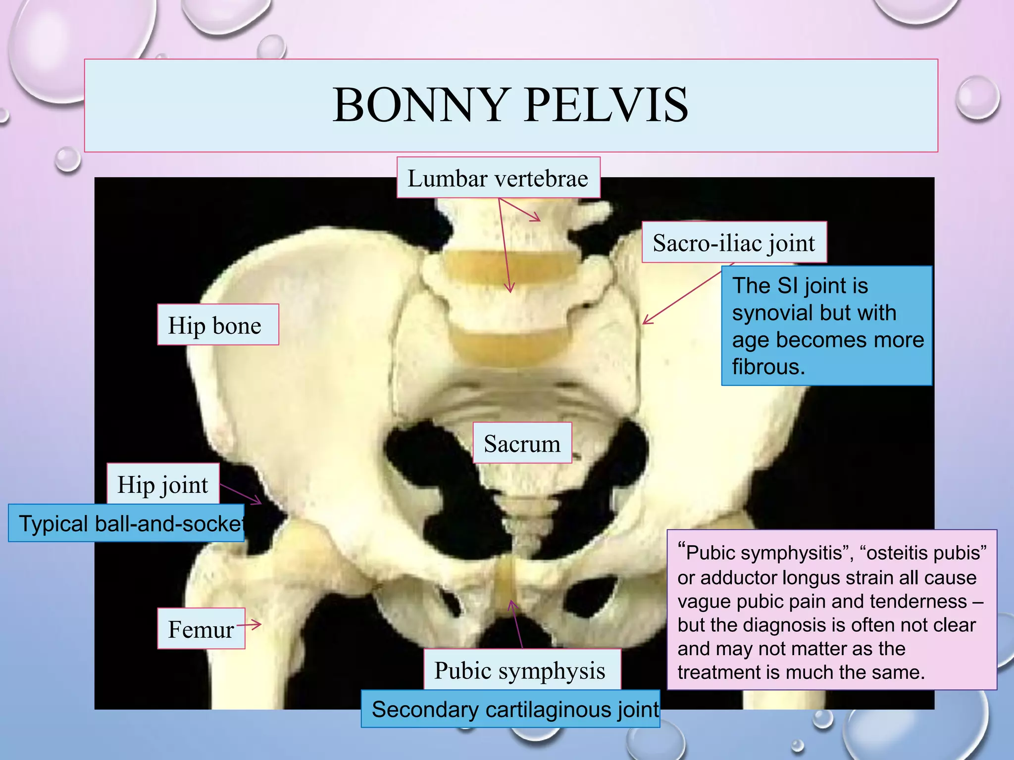
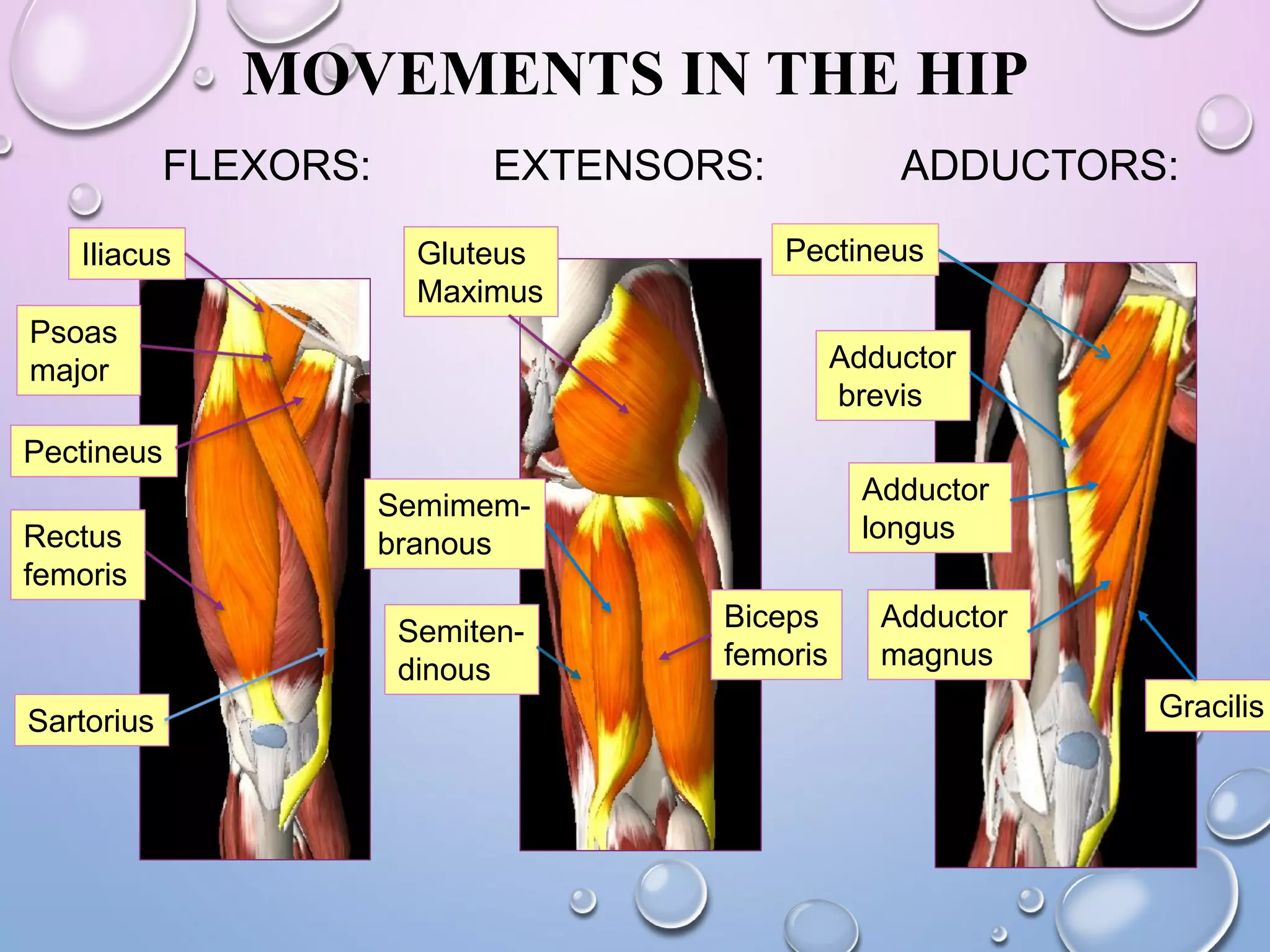
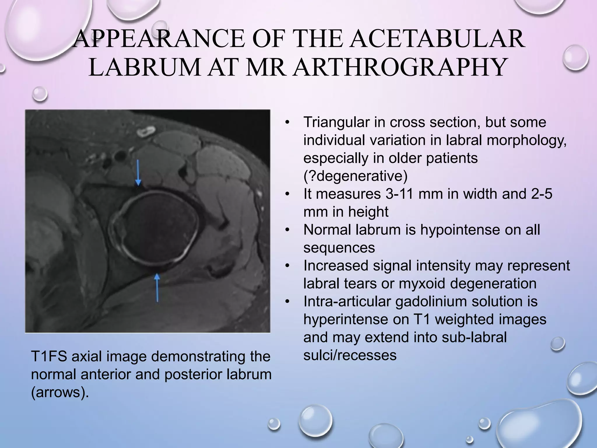
HIP GIRDLE:
FROM ANATOMY TO ORTHOPEDICS
PRESENTED BY
DR. MARYNA KORNIEIEVA
ASST. OF ANATOMY
BONNY PELVIS
Hip bone
Femur
Sacrum
Lumbar vertebrae
Hip joint
Sacro-iliac joint
Pubic symphysis
Secondary cartilaginous joint
Typical ball-and-socket
“Pubic symphysitis”, “osteitis pubis”
or adductor longus strain all cause
vague pubic pain and tenderness –
but the diagnosis is often not clear
and may not matter as the
treatment is much the same.
The SI joint is
synovial but with
age becomes more
fibrous.
HIP BONE
Ilium
Pubis
Iliac crest
Iliac fossa
Internal lip of iliac crest
External lip of iliac crest
Ischium
Anterior
Superior
iliac spine
Anterior
inferior
iliac spine
Auricular
surface
Ischial
tuberosity
Ischial spine
Greater sciatic notch
Lesser sciatic notch
Pubic
tubercle
Pubic body
Body of
ischium
Superior pubic ramus
Inferior pubic ramus
Pubic crest
Ischial ramus
Obturator foramen
Acetabulum
RADIOGRAPHIC APPEARANCES
OF THE BONY PELVIS
Anteroposterior radiograph of the male pelvis.
Representation of the radiograph of the pelvis.
SACROILIAC JOINT (SI)
Articulation: Adjacent
auricular articular surfaces
of the Ilium and sacrum;
Motion is limited with a mean
of 2.5 (0.8-3.6) degree.
Type: bicondylar synovial
joint, undergoes to gradual
sclerosis trough the age;
The interosseous sacro-iliac joint is probably the strongest in the body – if it is
disrupted it is indicative of a very high energy impact.
Capsule: along the
margins of articular
surfaces;
LIGAMENTS OF SI JOINT
During pregnancy, the ovaries and placenta produce the hormone relaxin which
increases flexibility of the ligaments that hold the sacroiliac joint together, resulting in
a looser joint and increased range of motion to accommodate the head of the fetus
passing through the birth canal.
OTHER LIGAMENTS OF HIP GIRDLE
Sacrotuberous lig.
Sacrospinous lig.
Greater sciatic foramen
Lesser sciatic foramen
Boundaries:
Antero-lateral: greater sciatic
notch of the illium;
Postero-medial: sacrotuberous
ligament;
Inferior: sacrospinous lig and
the ischial spine;
Superior: anterior sacroilliac
ligament.
Boundaries:
Anterior: the tuberosity of the ischium.
Superior: the spine of the ischium and
sacrospinous ligament.
Posterior: the sacrotuberous ligament.
TRAUMAS OF SI JOINT
Dysfunction in the sacroiliac joint, or SI joint, is thought to cause low back and/or leg
pain. The leg pain can be particularly difficult, and may feel similar to sciatica or pain
caused by a lumbar disc herniation.
Pain in the SI joint occurs as result of
excessive motion and in case of excessive
immobilization (as in ankylosing spondylitis).
SI PROCEDURES
SI joint injection
X-ray pictures (or ultrasound) are taken
throughout to ensure the needle is in the
correct area.
Once the needle is
correctly
positioned into the
joint a mixture of
local anaesthetic
with or without a
locally acting
steroid is injected.
HIP JOINT
Acetabular fossa
Labrum acetabulare
Acetabular
notch Transverse
acetabular
ligament
Ligament of the head
of the femur
Articular surfaces: the head of the femur
and the acetabulum of the pelvic bone.
Lunate surface
Kind of joint: synovial
ball and socket joint.
HIP JOINT
Synovial membrane: lines
fibrous capsule from inside
and forms a tubular covering
around the ligamentum teres.
Attachment of the fibrous
capsule:
medially – along the margins of
the acetabulum and transverse
acetabular ligament;
laterally – along the
intertrochanteric line and just
proximal to intertrochanteric
crest.
Acetabular margin
Trochanteric line
Acetabular margin
Neck of the femur
FEMUR
Proximal epiphysis of the femur:
1.Head (with the fovea capitis, for the
attachment of the ligament of the head),
2. Neck, 3. Greater trochanter, 4. Lesser
trochanter, 5. Intertrochanteric line, 6.
Intertrochanteric crest, 6. Gluteal
tuberosity, 7. Linea aspera; 8. Orthopedic
tubercle.
The issue to establish in fractured neck
of femur is whether the head is at risk of
avascular necrosis and therefore will need
to be replaced; avascular necrosis will most
likely occur when the fracture is proximal to
the capsular attachment tearing the
retinacular arteries that run up the femoral
neck to the head.
Capsular
attachment
CLINICAL SOLUTIONS
Radiograph (anteroposterior view)
of a displaced femoral neck fracture
treated by way of femoral head
replacement with a bipolar
prosthetic device.
The most common internal
fixation device used today is the
sliding screw-plate device.
(A) Femoral neck fracture treated by
way of internal fixation with 3 parallel
cannulated lag screws. (B) Schematic
representation of screw configuration
as viewed from the side.
BLOOD SUPPLY OF THE HIP
PERTHE’S DISEASE
CAUSES: SYMPTOMS:
MOVEMENTS IN THE HIP
LATERAL
ROTATORS:
ABDUCTORS:
Gluteus
medius
Gluteus minimus
Piriformis
Obturator
externus
Obturator
internus
Superior
and
Inferior
gemelli
Quadratus
femoris
MOVEMENTS IN THE HIP
FLEXORS: EXTENSORS:
Sartorius
Iliacus
Psoas
major
Rectus
femoris
Pectineus
Gluteus
Maximus
Biceps
femoris
Semiten-
dinous
Semimem-
branous
ADDUCTORS:
Adductor
magnus
Adductor
brevis
Gracilis
Adductor
longus
Pectineus
HIP DISLOCATIONS
Dislocated hip usually occurs
posteriorly due to anatomy (the
acetabulum is directed posteriorly).
Central dislocation through the acetabulum
is also possible.
LIGAMENTS OF THE HIP JOINT
BURSAE AROUND HIP
Trochanteric
bursitis
pain pattern
Hip arthroscopy for trochanteric
bursitis – bursectomy.
X-RAY OF NORMAL HIP
AN ADULT A CHILD
DEVELOPMENTAL DYSPLASIA OF HIP
The cause is unknown.
Low levels of amniotic fluid in the womb
during pregnancy and immaturity can increase
baby's risk of DDH.
DDH is a disruption in the normal
relationship between the head of the
femur and the acetabulum.
Symptoms:
DIAGNOSTICS
Pelvis X-Ray (AP view) showing left
sided dysplastic hip with femur head lying
in the upper outer quadrant and disrupted
Shenton's line.
Shenton's line
The goal of treatment is to keep the femoral
head in good contact with the acetabulum.
ANGLE BETWEEN THE NECK AND
SHAFT OF THE FEMUR
COXA VALGA COXA VARANormal adult:
The normal NS angle is about 160° in the young child.
› 130 ‹ 120
CROSS-SECTION THROUGH THE HIP
Trochanteric bursa
Obturator externus
Gluteus medius
Iliopsoas
Tensor fasciae lata
Rectus femoris
SartoriusFemoral vessels
Femoral nerve
Ilio-psoas bursa
Obturator internus
Gemellus inferior
Sciatic nerve
T1FS CORONAL IMAGE OF A NORMAL HIP.
T1FS AXIAL IMAGE OF A NORMAL HIP
T1FS SAGITTAL IMAGE OF A NORMAL HIP
APPEARANCE OF THE ACETABULAR
LABRUM AT MR ARTHROGRAPHY
T1FS axial image demonstrating the
normal anterior and posterior labrum
(arrows).
• Triangular in cross section, but some
individual variation in labral morphology,
especially in older patients
(?degenerative)
• It measures 3-11 mm in width and 2-5
mm in height
• Normal labrum is hypointense on all
sequences
• Increased signal intensity may represent
labral tears or myxoid degeneration
• Intra-articular gadolinium solution is
hyperintense on T1 weighted images
and may extend into sub-labral
sulci/recesses
SUBLABRAL SULCI
• Normal recesses
adjacent to labrum that
fill with contrast
• May be mistaken for
labral tears/detachments
• Anterosuperior
• Posterinferior
• Anteroinferior
• Posterosuperior
• A tear generally has
irregular edges and
extends more than 50%
across the depth of the
labrum
PD sagittal image demonstrating the
anterior labrum (arrowhead),
acetabular notch (arrow) and
posterior sulcus (double arrow).
PD axial image demonstrating a
posterior sublabral recess
(arrow).
LABRAL PATHOLOGY - LABRAL TEARS
Imaging Features:
• Extension of contrast into the labral tissue
• Associated features include labral blunting, deformity and hypertrophy
• Look for bucket handle tears, especially in cases of labro-acetabular
separation
T1FS axial image demonstrating
labro-acetabular separation (arrow)
PD image demonstrating a tear at
the chondro-labral junction
Aetiology:
1) Post-traumatic
• hyperextension,
external rotation:
anterosuperior
labrum
• axial loading of
flexed hip:
posterior labrum
2) Degenerative
3) Associated with
acetabular dysplasia
4) Following
posterior hip
dislocation.
Hip girdle from anatomy to orthopedics

Hip girdle from anatomy to orthopedics

  • 1.
    HIP GIRDLE: FROM ANATOMYTO ORTHOPEDICS PRESENTED BY DR. MARYNA KORNIEIEVA ASST. OF ANATOMY
  • 2.
    BONNY PELVIS Hip bone Femur Sacrum Lumbarvertebrae Hip joint Sacro-iliac joint Pubic symphysis Secondary cartilaginous joint Typical ball-and-socket “Pubic symphysitis”, “osteitis pubis” or adductor longus strain all cause vague pubic pain and tenderness – but the diagnosis is often not clear and may not matter as the treatment is much the same. The SI joint is synovial but with age becomes more fibrous.
  • 3.
    HIP BONE Ilium Pubis Iliac crest Iliacfossa Internal lip of iliac crest External lip of iliac crest Ischium Anterior Superior iliac spine Anterior inferior iliac spine Auricular surface Ischial tuberosity Ischial spine Greater sciatic notch Lesser sciatic notch Pubic tubercle Pubic body Body of ischium Superior pubic ramus Inferior pubic ramus Pubic crest Ischial ramus Obturator foramen Acetabulum
  • 4.
    RADIOGRAPHIC APPEARANCES OF THEBONY PELVIS Anteroposterior radiograph of the male pelvis. Representation of the radiograph of the pelvis.
  • 5.
    SACROILIAC JOINT (SI) Articulation:Adjacent auricular articular surfaces of the Ilium and sacrum; Motion is limited with a mean of 2.5 (0.8-3.6) degree. Type: bicondylar synovial joint, undergoes to gradual sclerosis trough the age; The interosseous sacro-iliac joint is probably the strongest in the body – if it is disrupted it is indicative of a very high energy impact. Capsule: along the margins of articular surfaces;
  • 6.
    LIGAMENTS OF SIJOINT During pregnancy, the ovaries and placenta produce the hormone relaxin which increases flexibility of the ligaments that hold the sacroiliac joint together, resulting in a looser joint and increased range of motion to accommodate the head of the fetus passing through the birth canal.
  • 7.
    OTHER LIGAMENTS OFHIP GIRDLE Sacrotuberous lig. Sacrospinous lig. Greater sciatic foramen Lesser sciatic foramen Boundaries: Antero-lateral: greater sciatic notch of the illium; Postero-medial: sacrotuberous ligament; Inferior: sacrospinous lig and the ischial spine; Superior: anterior sacroilliac ligament. Boundaries: Anterior: the tuberosity of the ischium. Superior: the spine of the ischium and sacrospinous ligament. Posterior: the sacrotuberous ligament.
  • 8.
    TRAUMAS OF SIJOINT Dysfunction in the sacroiliac joint, or SI joint, is thought to cause low back and/or leg pain. The leg pain can be particularly difficult, and may feel similar to sciatica or pain caused by a lumbar disc herniation. Pain in the SI joint occurs as result of excessive motion and in case of excessive immobilization (as in ankylosing spondylitis).
  • 9.
    SI PROCEDURES SI jointinjection X-ray pictures (or ultrasound) are taken throughout to ensure the needle is in the correct area. Once the needle is correctly positioned into the joint a mixture of local anaesthetic with or without a locally acting steroid is injected.
  • 10.
    HIP JOINT Acetabular fossa Labrumacetabulare Acetabular notch Transverse acetabular ligament Ligament of the head of the femur Articular surfaces: the head of the femur and the acetabulum of the pelvic bone. Lunate surface Kind of joint: synovial ball and socket joint.
  • 11.
    HIP JOINT Synovial membrane:lines fibrous capsule from inside and forms a tubular covering around the ligamentum teres. Attachment of the fibrous capsule: medially – along the margins of the acetabulum and transverse acetabular ligament; laterally – along the intertrochanteric line and just proximal to intertrochanteric crest. Acetabular margin Trochanteric line Acetabular margin Neck of the femur
  • 12.
    FEMUR Proximal epiphysis ofthe femur: 1.Head (with the fovea capitis, for the attachment of the ligament of the head), 2. Neck, 3. Greater trochanter, 4. Lesser trochanter, 5. Intertrochanteric line, 6. Intertrochanteric crest, 6. Gluteal tuberosity, 7. Linea aspera; 8. Orthopedic tubercle. The issue to establish in fractured neck of femur is whether the head is at risk of avascular necrosis and therefore will need to be replaced; avascular necrosis will most likely occur when the fracture is proximal to the capsular attachment tearing the retinacular arteries that run up the femoral neck to the head. Capsular attachment
  • 13.
    CLINICAL SOLUTIONS Radiograph (anteroposteriorview) of a displaced femoral neck fracture treated by way of femoral head replacement with a bipolar prosthetic device. The most common internal fixation device used today is the sliding screw-plate device. (A) Femoral neck fracture treated by way of internal fixation with 3 parallel cannulated lag screws. (B) Schematic representation of screw configuration as viewed from the side.
  • 14.
  • 15.
  • 16.
    MOVEMENTS IN THEHIP LATERAL ROTATORS: ABDUCTORS: Gluteus medius Gluteus minimus Piriformis Obturator externus Obturator internus Superior and Inferior gemelli Quadratus femoris
  • 17.
    MOVEMENTS IN THEHIP FLEXORS: EXTENSORS: Sartorius Iliacus Psoas major Rectus femoris Pectineus Gluteus Maximus Biceps femoris Semiten- dinous Semimem- branous ADDUCTORS: Adductor magnus Adductor brevis Gracilis Adductor longus Pectineus
  • 18.
    HIP DISLOCATIONS Dislocated hipusually occurs posteriorly due to anatomy (the acetabulum is directed posteriorly). Central dislocation through the acetabulum is also possible.
  • 19.
  • 20.
    BURSAE AROUND HIP Trochanteric bursitis painpattern Hip arthroscopy for trochanteric bursitis – bursectomy.
  • 21.
    X-RAY OF NORMALHIP AN ADULT A CHILD
  • 22.
    DEVELOPMENTAL DYSPLASIA OFHIP The cause is unknown. Low levels of amniotic fluid in the womb during pregnancy and immaturity can increase baby's risk of DDH. DDH is a disruption in the normal relationship between the head of the femur and the acetabulum. Symptoms:
  • 23.
    DIAGNOSTICS Pelvis X-Ray (APview) showing left sided dysplastic hip with femur head lying in the upper outer quadrant and disrupted Shenton's line. Shenton's line The goal of treatment is to keep the femoral head in good contact with the acetabulum.
  • 24.
    ANGLE BETWEEN THENECK AND SHAFT OF THE FEMUR COXA VALGA COXA VARANormal adult: The normal NS angle is about 160° in the young child. › 130 ‹ 120
  • 25.
    CROSS-SECTION THROUGH THEHIP Trochanteric bursa Obturator externus Gluteus medius Iliopsoas Tensor fasciae lata Rectus femoris SartoriusFemoral vessels Femoral nerve Ilio-psoas bursa Obturator internus Gemellus inferior Sciatic nerve
  • 26.
    T1FS CORONAL IMAGEOF A NORMAL HIP.
  • 27.
    T1FS AXIAL IMAGEOF A NORMAL HIP
  • 28.
    T1FS SAGITTAL IMAGEOF A NORMAL HIP
  • 29.
    APPEARANCE OF THEACETABULAR LABRUM AT MR ARTHROGRAPHY T1FS axial image demonstrating the normal anterior and posterior labrum (arrows). • Triangular in cross section, but some individual variation in labral morphology, especially in older patients (?degenerative) • It measures 3-11 mm in width and 2-5 mm in height • Normal labrum is hypointense on all sequences • Increased signal intensity may represent labral tears or myxoid degeneration • Intra-articular gadolinium solution is hyperintense on T1 weighted images and may extend into sub-labral sulci/recesses
  • 30.
    SUBLABRAL SULCI • Normalrecesses adjacent to labrum that fill with contrast • May be mistaken for labral tears/detachments • Anterosuperior • Posterinferior • Anteroinferior • Posterosuperior • A tear generally has irregular edges and extends more than 50% across the depth of the labrum PD sagittal image demonstrating the anterior labrum (arrowhead), acetabular notch (arrow) and posterior sulcus (double arrow). PD axial image demonstrating a posterior sublabral recess (arrow).
  • 31.
    LABRAL PATHOLOGY -LABRAL TEARS Imaging Features: • Extension of contrast into the labral tissue • Associated features include labral blunting, deformity and hypertrophy • Look for bucket handle tears, especially in cases of labro-acetabular separation T1FS axial image demonstrating labro-acetabular separation (arrow) PD image demonstrating a tear at the chondro-labral junction Aetiology: 1) Post-traumatic • hyperextension, external rotation: anterosuperior labrum • axial loading of flexed hip: posterior labrum 2) Degenerative 3) Associated with acetabular dysplasia 4) Following posterior hip dislocation.