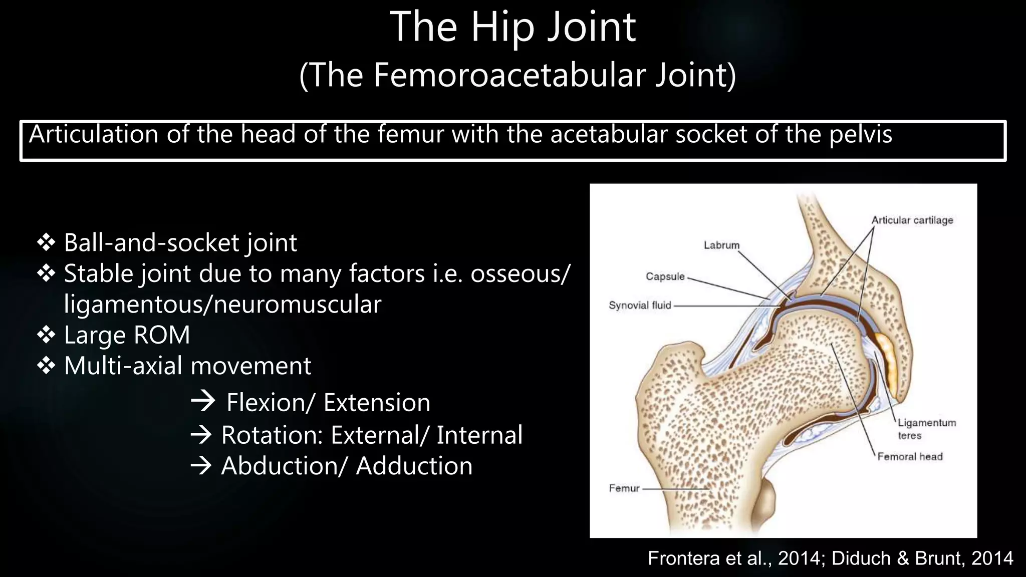
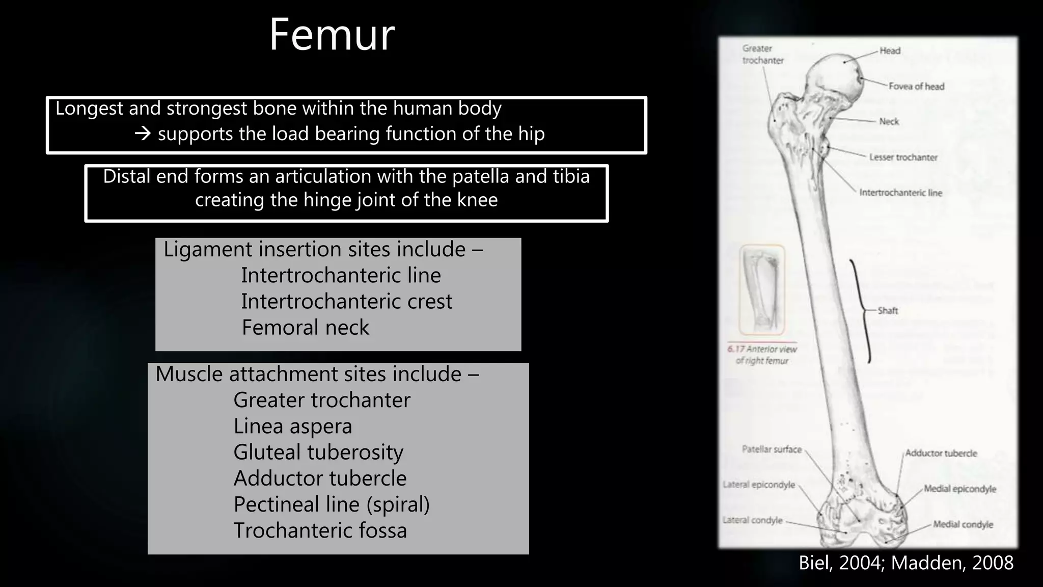
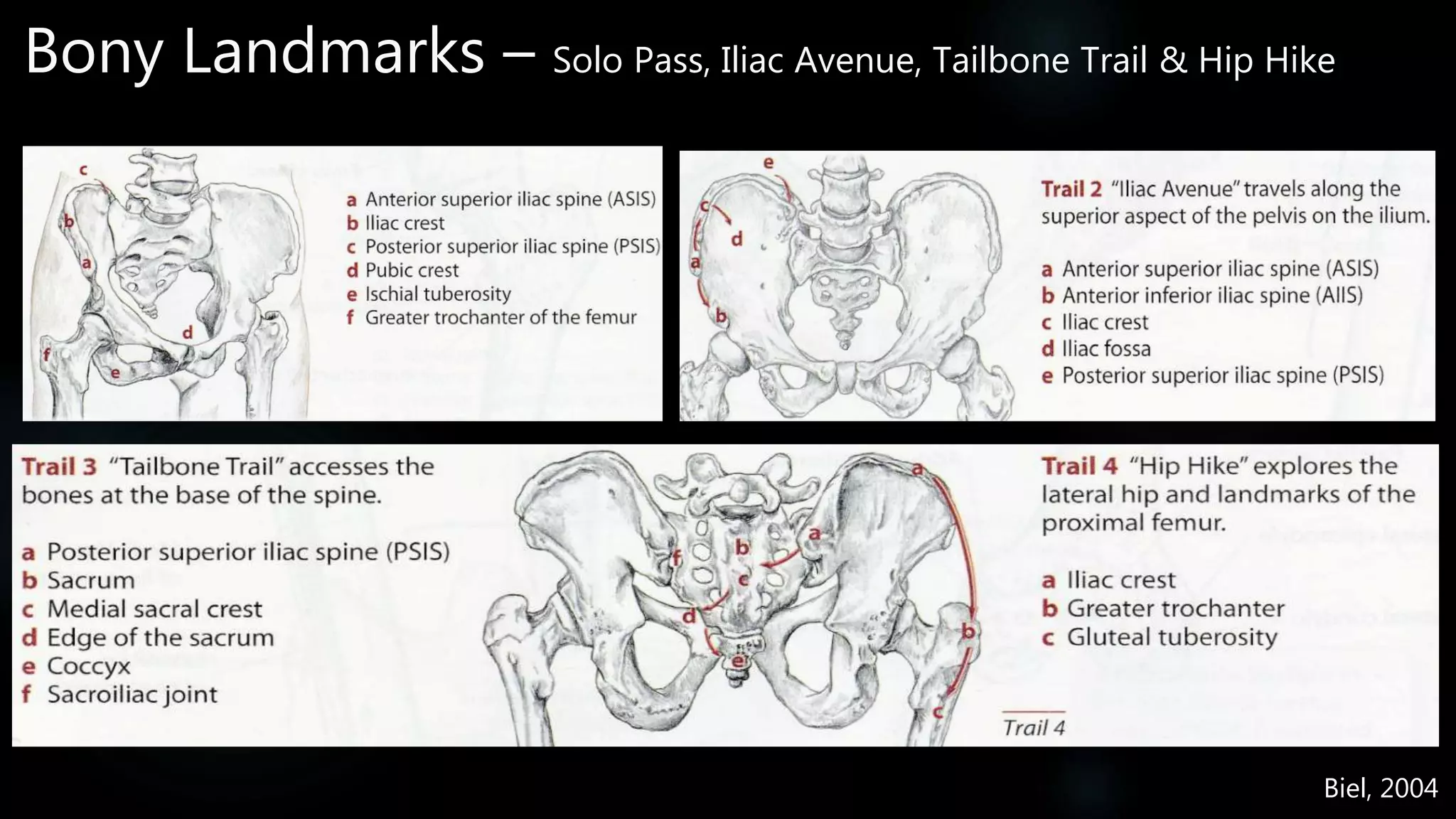
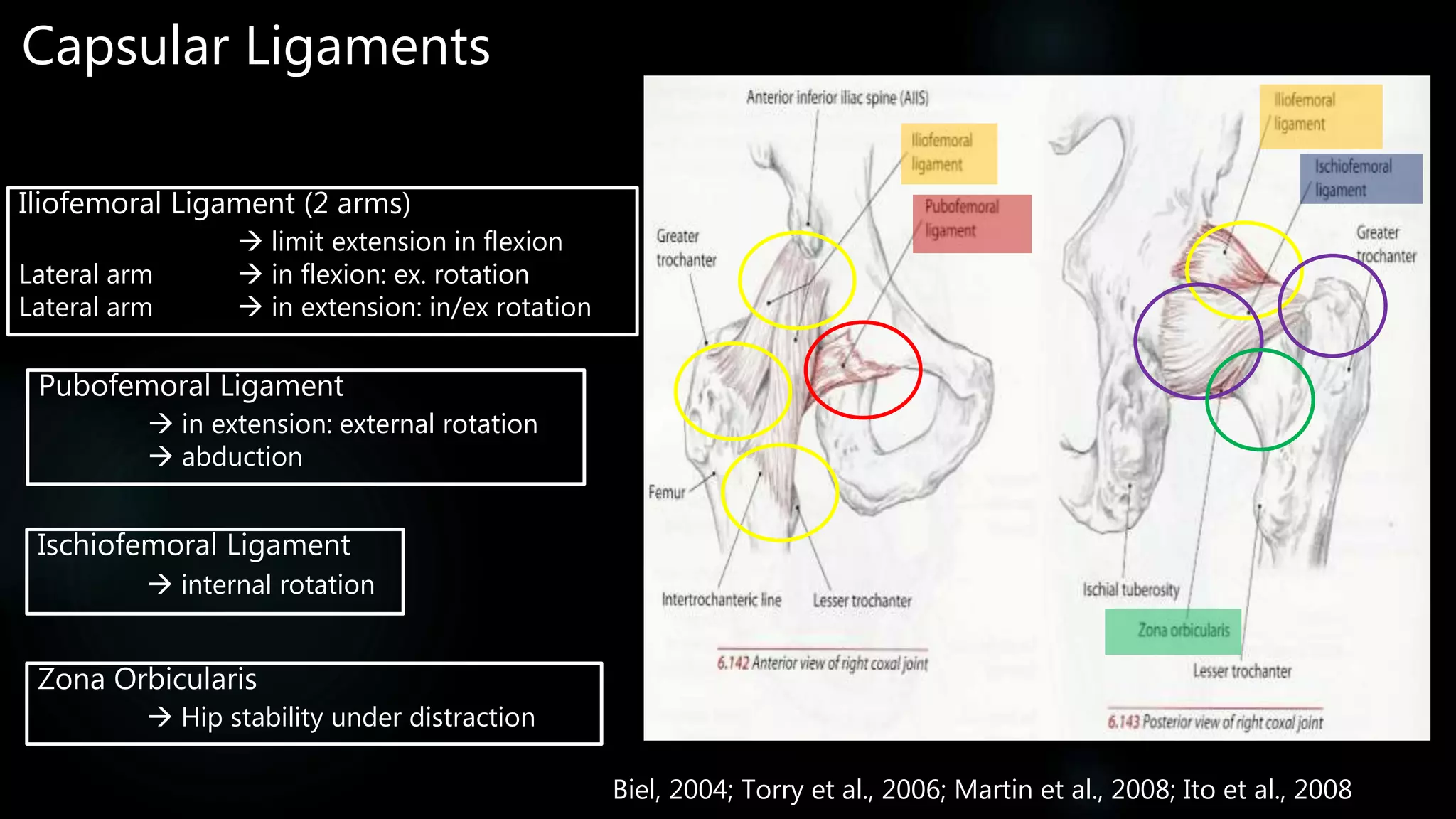
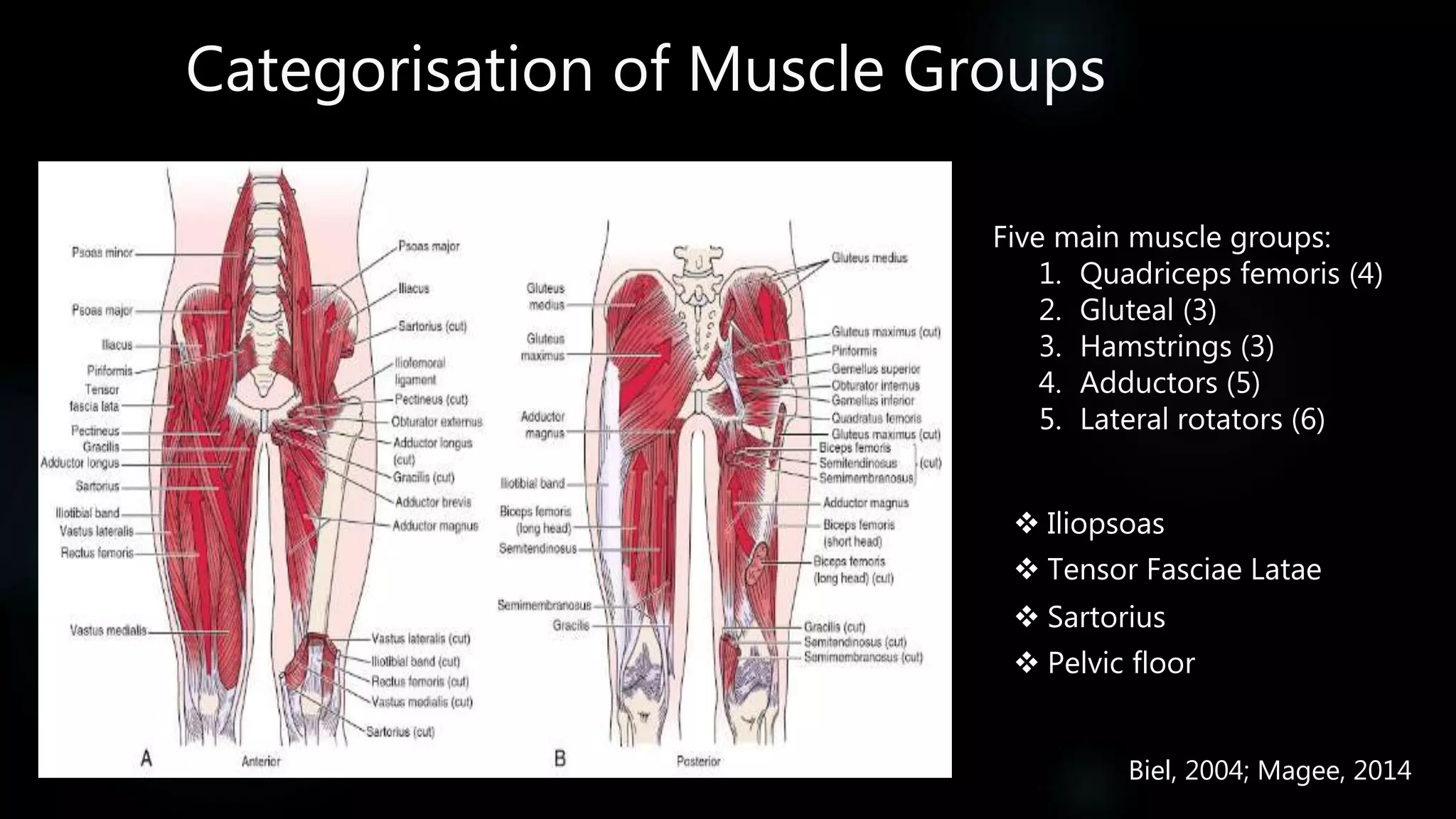
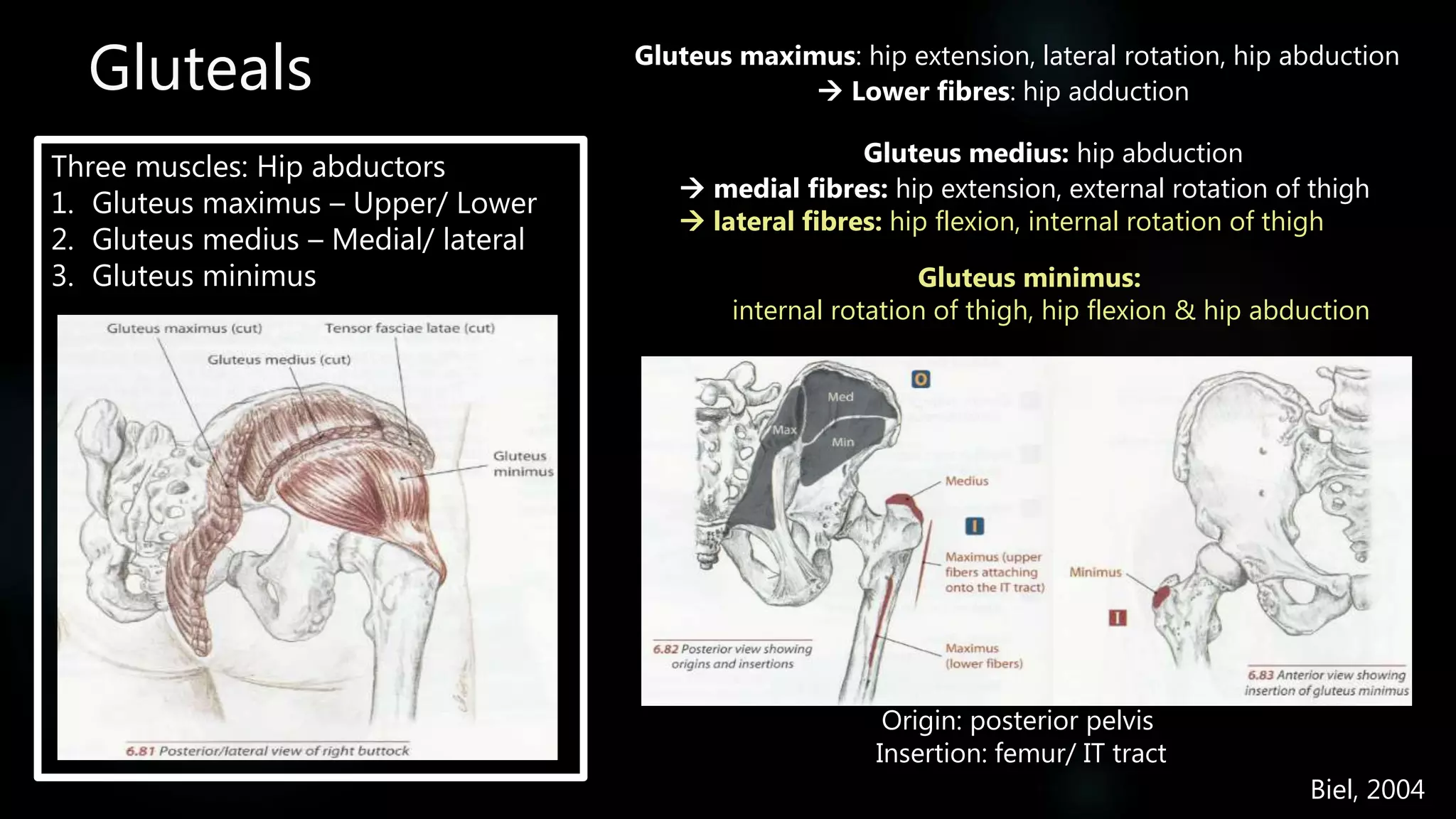
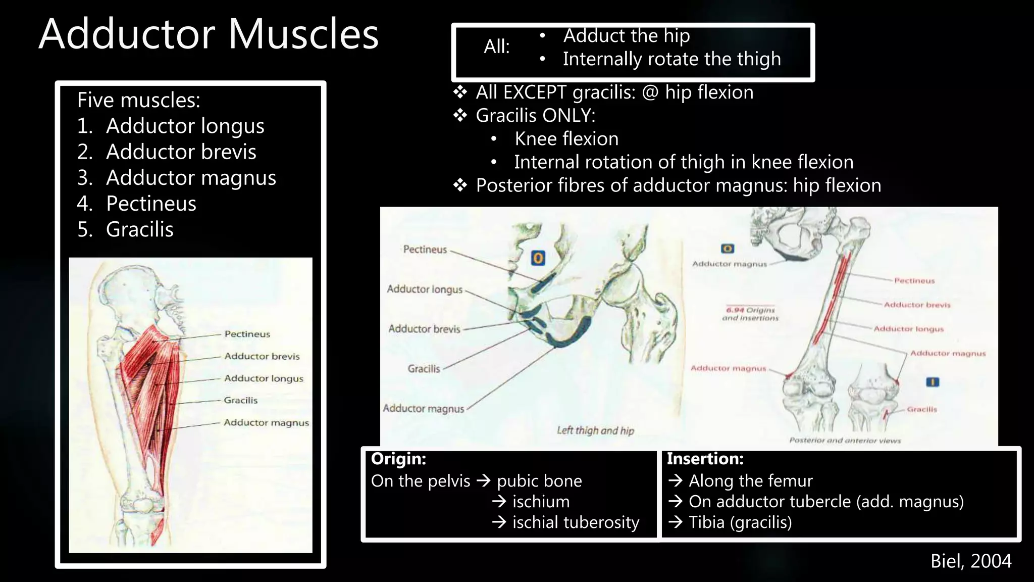
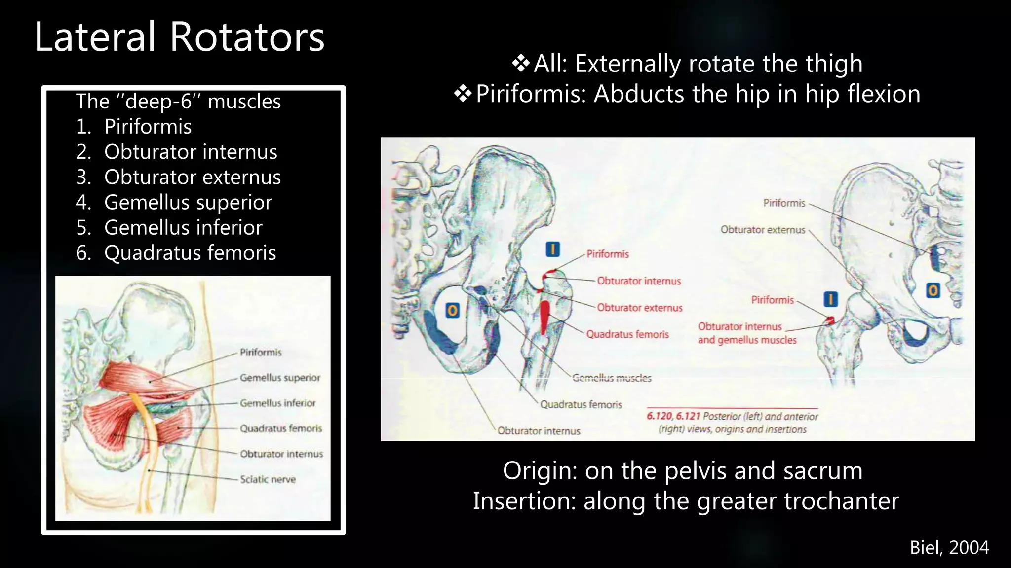
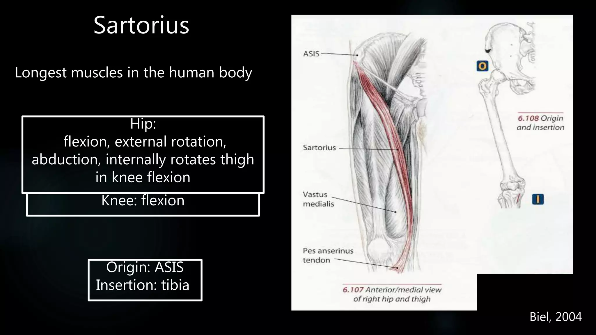
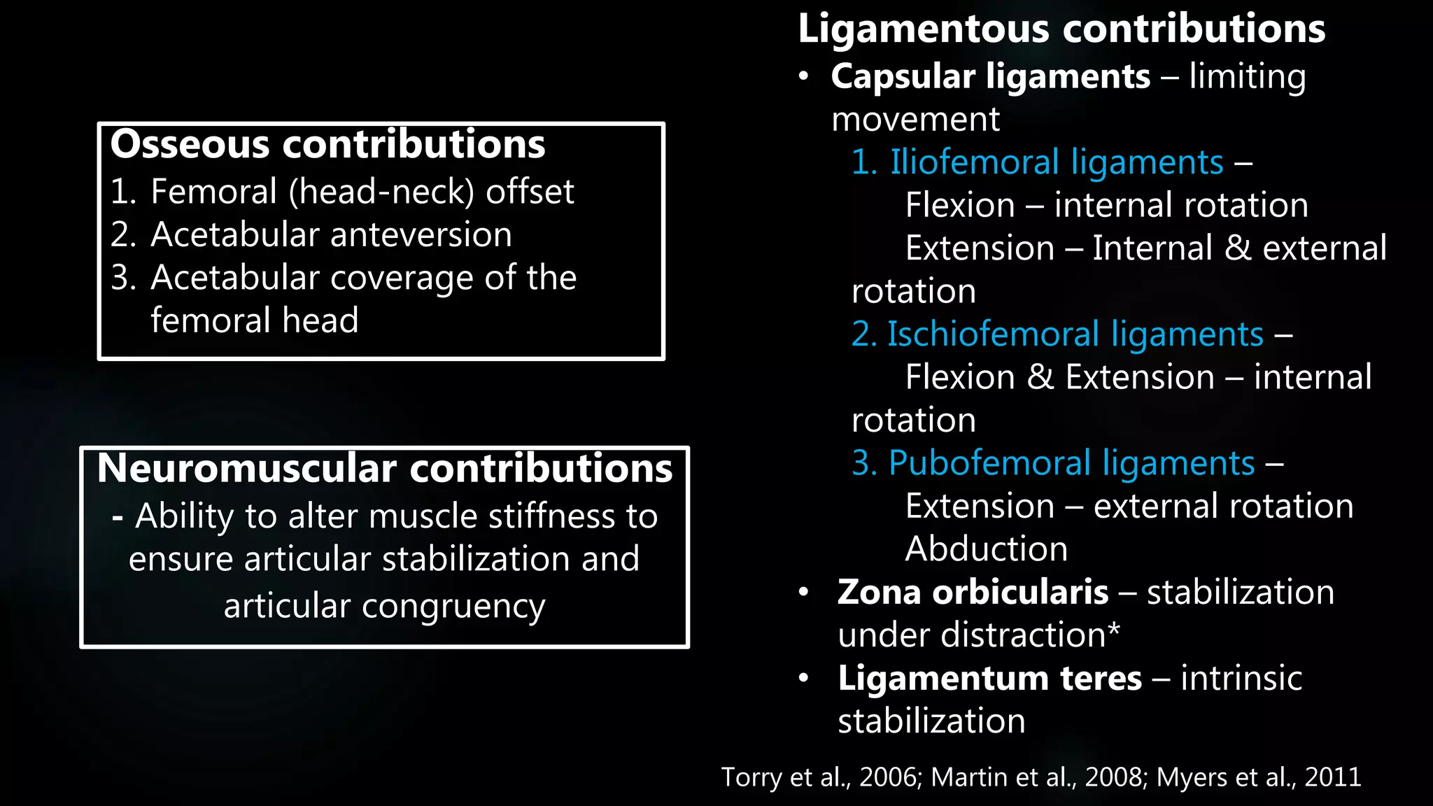
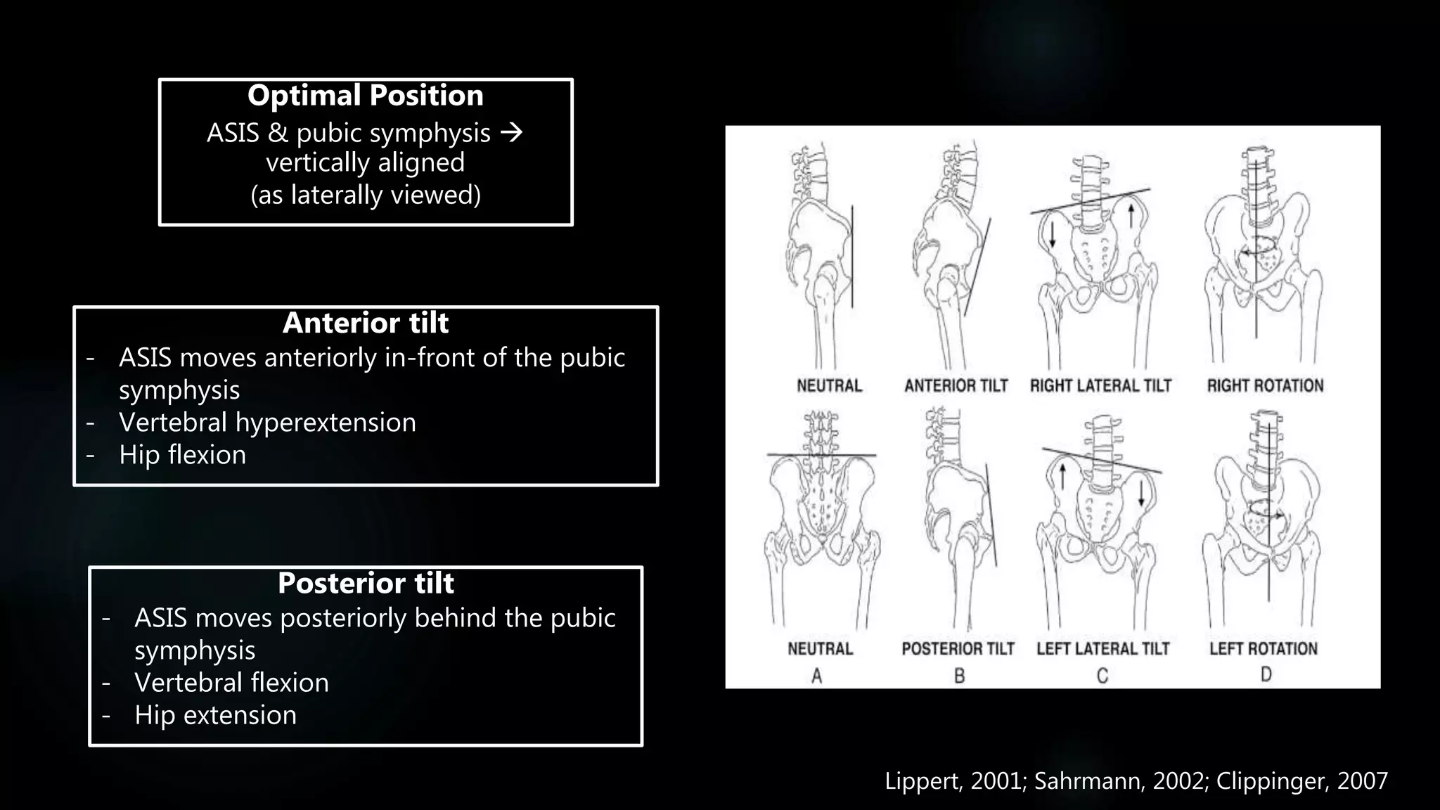
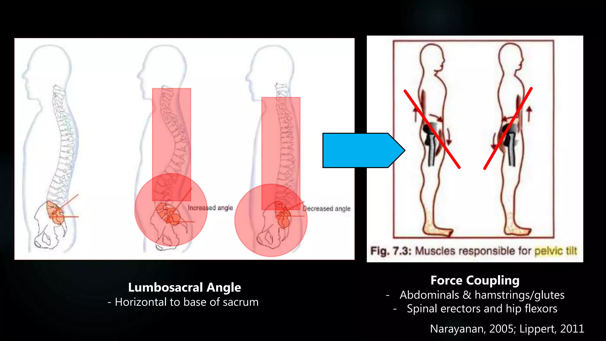
The Hip
ANATOMY AND FUNCTION
with Jill Costley
Overview
 Summary of the pelvis
 Hip & thigh anatomy
 Ligaments
 Muscles
 Functions of the structures associated with the hip
 Movement impairments of the hip/ groin and associated
functional tests
 How can we use this knowledge to inform our practice?
 Application to athletes
The Pelvis
Two coxal bones, the sacrum and the coccyx
• Protects organs
• Childbirth in females
• Supports the spine and upper body
• Supports movement
• Provides attachment sites for muscles and
ligaments of the hip and trunk
Coxal bone: ilium,
ischium (posterior) &
pubis (anterior)
Triradiate cartilage - Not
fully fused until
approximately 18 yrs old
Includes the acetabulum (socket) of the hip joint
Functions of the Pelvis:
Agur & Dalley, 2009
The Hip Joint
Anatomy
The Hip Joint
(The Femoroacetabular Joint)
 Ball-and-socket joint
 Stable joint due to many factors i.e. osseous/
ligamentous/neuromuscular
 Large ROM
 Multi-axial movement
 Flexion/ Extension
 Rotation: External/ Internal
 Abduction/ Adduction
Frontera et al., 2014; Diduch & Brunt, 2014
Articulation of the head of the femur with the acetabular socket of the pelvis
American College of Sports Medicine, 2014; Swartz, 2014
The Acetabulum
Depth of the acetabulum is
increased through the
labrum
Articular (lunate surface)
Non-articular - acetabular fossa
Transverse acetabular
ligament crossing the
acetabular notch
Singh, 2008
The Labrum
• Fibrocartilaginous rim surrounding the acetabulum
• Transverse acetabular ligament  acetabular notch
• Thickest posteriorly
• Tears common in acetabular dysplasia
Reflected
tendon of the
rectus
Anterior lunate
cartilage
Posterior lunate
cartilage
Acetabular
fossa
Acetabular
fossa
Transverse
acetabular
ligament
Lunate cartilage
Ligamentum
teres
Acetabular
labrum
Femoral headObturator
foramen
Transverse
acetabular
ligament
Byrd, 2013
Femur
Longest and strongest bone within the human body
 supports the load bearing function of the hip
Biel, 2004; Madden, 2008
Distal end forms an articulation with the patella and tibia
creating the hinge joint of the knee
Muscle attachment sites include –
Greater trochanter
Linea aspera
Gluteal tuberosity
Adductor tubercle
Pectineal line (spiral)
Trochanteric fossa
Ligament insertion sites include –
Intertrochanteric line
Intertrochanteric crest
Femoral neck
Biel, 2004
Bony Landmarks – Solo Pass, Iliac Avenue, Tailbone Trail & Hip Hike
Biel, 2004
Capsular LIGAMENTS
The Hip
Anatomy
Biel, 2004; Torry et al., 2006; Martin et al., 2008; Ito et al., 2008
Iliofemoral Ligament (2 arms)
 limit extension in flexion
Lateral arm  in flexion: ex. rotation
Lateral arm  in extension: in/ex rotation
Capsular Ligaments
Zona Orbicularis
 Hip stability under distraction
Ischiofemoral Ligament
 internal rotation
Pubofemoral Ligament
 in extension: external rotation
 abduction
The Hip & Thigh
Anatomy
MUSCLES &
FUNCTION
Categorisation of Muscle Groups
 Iliopsoas
Five main muscle groups:
1. Quadriceps femoris (4)
2. Gluteal (3)
3. Hamstrings (3)
4. Adductors (5)
5. Lateral rotators (6)
 Tensor Fasciae Latae
 Sartorius
Biel, 2004; Magee, 2014
 Pelvic floor
Quadriceps Femoris
Four muscles:
1. Vastus Lateralis
2. Rectus Femoris (biarticular)
3. Vastus Medialis
4. Vastus Intermedius
Biel, 2004
 All: extend the knee
 Rectus femoris: flexes the hip
Origin: AIIS (rectus femoris)/ along femur (all others)
Insertion: Tibial tuberosity
Gluteals
Three muscles: Hip abductors
1. Gluteus maximus – Upper/ Lower
2. Gluteus medius – Medial/ lateral
3. Gluteus minimus
Biel, 2004
Origin: posterior pelvis
Insertion: femur/ IT tract
Gluteus maximus: hip extension, lateral rotation, hip abduction
 Lower fibres: hip adduction
Gluteus medius: hip abduction
 medial fibres: hip extension, external rotation of thigh
 lateral fibres: hip flexion, internal rotation of thigh
Gluteus minimus:
internal rotation of thigh, hip flexion & hip abduction
Hamstrings
Biel, 2004
Origin: Ischial tuberosity/ femur (BFSH)
Insertion: fibula (BF)/ tibia (semi’s)
Three muscles:
1. Biceps femoris (BFLH & BFSH)
2. Semitendinosus
3. Semimembranosus
Semi’s: internally rotates a flexed knee, hip extension, @
Internal rotation of thigh,
BFLH: hip extension/ @ external rotation of the thigh
ALL: Posterior pelvic tilt, knee flexion
Adductor Muscles
Five muscles:
1. Adductor longus
2. Adductor brevis
3. Adductor magnus
4. Pectineus
5. Gracilis
• Adduct the hip
• Internally rotate the thigh
 All EXCEPT gracilis: @ hip flexion
 Gracilis ONLY:
• Knee flexion
• Internal rotation of thigh in knee flexion
 Posterior fibres of adductor magnus: hip flexion
Biel, 2004
Origin:
On the pelvis  pubic bone
 ischium
 ischial tuberosity
Insertion:
 Along the femur
 On adductor tubercle (add. magnus)
 Tibia (gracilis)
All:
Lateral Rotators
All: Externally rotate the thigh
Piriformis: Abducts the hip in hip flexion
Biel, 2004
Origin: on the pelvis and sacrum
Insertion: along the greater trochanter
The ‘’deep-6’’ muscles
1. Piriformis
2. Obturator internus
3. Obturator externus
4. Gemellus superior
5. Gemellus inferior
6. Quadratus femoris
Gluteus minimus
Sciatic nerve
Gluteus medius
Piriformis
Inferior gemellus
Quadratus femoris
Superior gemellus
Internus obturator
Byrd, 2013
Others
1. Iliopsoas (Iliacus and psoas major)
2. Sartorius
3. Tensor Fasciae Latae (TFL)
4. Perineum
Iliopsoas
Biel, 2004
Two muscles:
1. Psoas major
2. Iliacus
Fixed origin:
hip flexion/ external
rotation?
Origin: Iliac fossa
Insertion: Lesser trochanter
Fixed insertion:
trunk flexion towards thigh/
anterior pelvic tilt
Iliacus
Psoas major
Fixed origin:
hip flexion/ external
rotation?
Fixed insertion:
trunk flexion towards thigh/
anterior pelvic tilt
Unilateral:
@ lateral lumbar flexion
Origin: lumbar processes
Insertion: lesser trochanter
Sartorius
Hip:
flexion, external rotation,
abduction, internally rotates thigh
in knee flexion
Origin: ASIS
Insertion: tibia
Biel, 2004
Knee: flexion
Longest muscles in the human body
Tensor Fasciae Latae
Biel, 2004
Stabilisation of knee and hip
Iliotibial Tract - Fascia
Hip flexion, internal rotation and abduction
Origin: Iliac crest
Insertion: Iliotibial tract
Origin: Gluteal fascia
Insertion: Tibial tubercle
Perineum &
Pelvic Floor
Biel, 2004
Perineum
Diamond shaped area found
inferiorly within pelvis
Bordered by the two ischial
tuberosities, the pubis symphysis
and the coccyx
Pelvic floor
Supports the abdominal and pelvic
viscera
 2 levator ani
 2 coccygeus
 fascia
Singh, 2008
The Hip & Thigh
STABILITY OF THE HIP
Contributions to hip stabilization –
Osseous Structures
Ligamentous Structures
Neuromuscular Structures
ROM  needs large degree of stability
Osseous contributions
1. Femoral (head-neck) offset
2. Acetabular anteversion
3. Acetabular coverage of the
femoral head
Ligamentous contributions
• Capsular ligaments – limiting
movement
1. Iliofemoral ligaments –
Flexion – internal rotation
Extension – Internal & external
rotation
2. Ischiofemoral ligaments –
Flexion & Extension – internal
rotation
3. Pubofemoral ligaments –
Extension – external rotation
Abduction
• Zona orbicularis – stabilization
under distraction*
• Ligamentum teres – intrinsic
stabilization
Neuromuscular contributions
- Ability to alter muscle stiffness to
ensure articular stabilization and
articular congruency
Torry et al., 2006; Martin et al., 2008; Myers et al., 2011
The Hip & Thigh
ANTERIOR & POSTERIOR TILT
Lippert, 2001; Sahrmann, 2002; Clippinger, 2007
Anterior tilt
- ASIS moves anteriorly in-front of the pubic
symphysis
- Vertebral hyperextension
- Hip flexion
Posterior tilt
- ASIS moves posteriorly behind the pubic
symphysis
- Vertebral flexion
- Hip extension
Optimal Position
ASIS & pubic symphysis 
vertically aligned
(as laterally viewed)
Force Coupling
- Abdominals & hamstrings/glutes
- Spinal erectors and hip flexors
Lumbosacral Angle
- Horizontal to base of sacrum
Narayanan, 2005; Lippert, 2011
The Hip & Thigh
MOVEMENT TESTS
Movement Tests
Thomas Test: indicates tightness within the rectus femoris or the iliopsoas
- Positive result = testing limb lying above surface of plinth while flexed or extended
FABER’s (Patrick) Test: may indicate the presence of hip pathology or SI joint dysfunction
- Positive result = pain within the back region of testing side/ restricted movement in
testing side above that of the opposite limb
 tight hip adductors/ flexors/ joint capsule?
Ober Test: indicates tensor fasciae latae (TFL) or iliotibial band (ITB) tightness
- Positive result = remaining off the table upon adduction
Tightness:
Piriformis Test: tests the cause of buttock pain i.e. tight piriformis muscle or as a result of referred
pain due to the sciatic nerve
- Positive result = gluteal pain or radiating pain from sciatic nerve
Palmer et al., 1998; Martin et al., 2010
Scouring (Quadrant) Test: may indicate the presence of a non-specific joint pathology
Craig’s Test: Test for femoral anteversion/ retroversion
Sign of the Buttock: indicates if gluteal pain is localized or being referred from problems
within the hip, sciatic nerve or hamstring
Trendelenburg Test: a unilateral weight-bearing test that may indicate gluteus
maximus weakness
- Positive result = a drop of the pelvis as seen from behind
Weakness:
Other:
Palmer et al., 1998; Martin et al., 2010
FADIR Test
(Impingement Test)
Flexion, ADduction, Internal Rotation
High Sensitivity
Wilson & Furukawa (2014)
Positive test: Pain
e.g. hip labral tears, loose material, chondral
lesions, femoral acetabular impingements
Gold-standard for checking overall hip function
Low Specificity
The Hip & Thigh
HIP PROBLEMS –
TO BE AWARE OF
1. Femoroacetabular impingements (FAI)
 FABER test is most sensitive
 CAM and PINCHER deformities  causes labral tears
2. Piriformis Syndromes
 Log roll test is most sensitive
 Gluteal region pain especially when sitting or walking
3. Tumours
4. Femoral anteversion
 Craig’s Test
 Acetabular dysplasia  usually shows hypertrophied labrum  gymnasts
5. Labral tears
 FADIR test is most sensitive
 Can occur without FAI
 Dull or sharp groin pain (may radiate)
6. Cartilage loss
 wear and tear  hyaline articular
Wilson & Furukawa, 2014; Imhoff et al., 2015
Case Study 1 –
Athlete 1
Cam and pincher deformities –
congenital?/genetic?
Griffin et al., 2016
Soft tissue damage through:
• Repetition
• Abnormal femur/ acetabular shape
• Excessive movements close to outer range
 abnormal contact/ collision
Triad of symptoms, clinical signs & imaging findings
1. FADIR Test – sensitive but not specific
2. Limited hip ROM?  internal rotation in flexion
3. FABER Test ?
4. Hip muscles commonly weak in FAI patients
UEFA injury study: A prospective study of
hip and groin injuries in professional
football over seven consecutive seasons
FAI’s going to become more common
through advancements in improvements of
MRI and arthroscopic techniques as well as
increased awareness and knowledge of this
condition
Lavigne et al., 2004; Werner et al., 2009; Khan et al., 2016
Hockey, football, soccer, rugby,
martial arts, and tennis
Cam-type: men
Pincer-type: women
Associated with other pathologies
such as adductor and hip flexors
injuries, ACL injuries etc.
Limited ROM may increase stress
placed upon other joints e.g
knees/ spine
Case Study 2 – Gaelic Groin
Second most common injury to hamstring strains
Werner et al., 2009; Glasgow et al., 2011
Groin – ‘’junction between the anteromedial part of the
thigh, including the proximal part of the adductor
muscle bellies, and the lower abdomen’’
Glasgow et al., 2011
Implications for S&C
Werner et al., 2009; Glasgow et al., 2011
Field based games:
2 months off-season period  complete strength, flexibility and stability training
 Increase ROM, strengthen core and groin/glutes
High groin injury rates in:
• Soccer
• AFL
• Rugby League
Work with the coach i.e. match play exposure
 EASIER SAID THAN DONE
Athlete 2
1) Rehab can be a long process!!
2) Commitment – athlete and therapist/ coach
3) Cooperation between physio’s & S&C.
Summary
• Structure: Acetabulum and femur
• Muscles: Quadriceps (4), gluteals (3), hamstrings (3), adductors (5), lateral rotators (6)
• Capsule ligaments  ischiofemoral, iliofemoral, pubofemoral
• Movement: Hip flexion/ extension, adduction/ abduction, internal/ external rotation
• Stability: Osseous, ligamentous & neuromuscular factors
• Tests: FABER, FADIR, Thomas, Ober, Piriformis, Trendelenburg, Craig’s, Scouring, Sign of the Butt.
• Conditions: FAI, tumours, femoral anteversion, labral tears, cartilage loss……..
• FAI – triad of symptoms, signs & radiological feature  increase in cases in future?
• Gaelic groin – chronic high load  history taking, clinical examination, observation, tests
 2 month off-season: strength, flexibility & stability training
 Strengthen core and groin/gluteal muscles
 Applies to other field sports
References
Provided via Email
Thanks for Listening!!

The Pelvis and Hip: Function and Anatomy

  • 1.
    The Hip ANATOMY ANDFUNCTION with Jill Costley
  • 2.
    Overview  Summary ofthe pelvis  Hip & thigh anatomy  Ligaments  Muscles  Functions of the structures associated with the hip  Movement impairments of the hip/ groin and associated functional tests  How can we use this knowledge to inform our practice?  Application to athletes
  • 3.
    The Pelvis Two coxalbones, the sacrum and the coccyx • Protects organs • Childbirth in females • Supports the spine and upper body • Supports movement • Provides attachment sites for muscles and ligaments of the hip and trunk Coxal bone: ilium, ischium (posterior) & pubis (anterior) Triradiate cartilage - Not fully fused until approximately 18 yrs old Includes the acetabulum (socket) of the hip joint Functions of the Pelvis: Agur & Dalley, 2009
  • 4.
  • 5.
    The Hip Joint (TheFemoroacetabular Joint)  Ball-and-socket joint  Stable joint due to many factors i.e. osseous/ ligamentous/neuromuscular  Large ROM  Multi-axial movement  Flexion/ Extension  Rotation: External/ Internal  Abduction/ Adduction Frontera et al., 2014; Diduch & Brunt, 2014 Articulation of the head of the femur with the acetabular socket of the pelvis
  • 6.
    American College ofSports Medicine, 2014; Swartz, 2014
  • 7.
    The Acetabulum Depth ofthe acetabulum is increased through the labrum Articular (lunate surface) Non-articular - acetabular fossa Transverse acetabular ligament crossing the acetabular notch Singh, 2008 The Labrum • Fibrocartilaginous rim surrounding the acetabulum • Transverse acetabular ligament  acetabular notch • Thickest posteriorly • Tears common in acetabular dysplasia
  • 8.
    Reflected tendon of the rectus Anteriorlunate cartilage Posterior lunate cartilage Acetabular fossa Acetabular fossa Transverse acetabular ligament Lunate cartilage Ligamentum teres Acetabular labrum Femoral headObturator foramen Transverse acetabular ligament Byrd, 2013
  • 9.
    Femur Longest and strongestbone within the human body  supports the load bearing function of the hip Biel, 2004; Madden, 2008 Distal end forms an articulation with the patella and tibia creating the hinge joint of the knee Muscle attachment sites include – Greater trochanter Linea aspera Gluteal tuberosity Adductor tubercle Pectineal line (spiral) Trochanteric fossa Ligament insertion sites include – Intertrochanteric line Intertrochanteric crest Femoral neck
  • 10.
  • 11.
    Bony Landmarks –Solo Pass, Iliac Avenue, Tailbone Trail & Hip Hike Biel, 2004
  • 12.
  • 13.
    Biel, 2004; Torryet al., 2006; Martin et al., 2008; Ito et al., 2008 Iliofemoral Ligament (2 arms)  limit extension in flexion Lateral arm  in flexion: ex. rotation Lateral arm  in extension: in/ex rotation Capsular Ligaments Zona Orbicularis  Hip stability under distraction Ischiofemoral Ligament  internal rotation Pubofemoral Ligament  in extension: external rotation  abduction
  • 14.
    The Hip &Thigh Anatomy MUSCLES & FUNCTION
  • 15.
    Categorisation of MuscleGroups  Iliopsoas Five main muscle groups: 1. Quadriceps femoris (4) 2. Gluteal (3) 3. Hamstrings (3) 4. Adductors (5) 5. Lateral rotators (6)  Tensor Fasciae Latae  Sartorius Biel, 2004; Magee, 2014  Pelvic floor
  • 16.
    Quadriceps Femoris Four muscles: 1.Vastus Lateralis 2. Rectus Femoris (biarticular) 3. Vastus Medialis 4. Vastus Intermedius Biel, 2004  All: extend the knee  Rectus femoris: flexes the hip Origin: AIIS (rectus femoris)/ along femur (all others) Insertion: Tibial tuberosity
  • 17.
    Gluteals Three muscles: Hipabductors 1. Gluteus maximus – Upper/ Lower 2. Gluteus medius – Medial/ lateral 3. Gluteus minimus Biel, 2004 Origin: posterior pelvis Insertion: femur/ IT tract Gluteus maximus: hip extension, lateral rotation, hip abduction  Lower fibres: hip adduction Gluteus medius: hip abduction  medial fibres: hip extension, external rotation of thigh  lateral fibres: hip flexion, internal rotation of thigh Gluteus minimus: internal rotation of thigh, hip flexion & hip abduction
  • 18.
    Hamstrings Biel, 2004 Origin: Ischialtuberosity/ femur (BFSH) Insertion: fibula (BF)/ tibia (semi’s) Three muscles: 1. Biceps femoris (BFLH & BFSH) 2. Semitendinosus 3. Semimembranosus Semi’s: internally rotates a flexed knee, hip extension, @ Internal rotation of thigh, BFLH: hip extension/ @ external rotation of the thigh ALL: Posterior pelvic tilt, knee flexion
  • 19.
    Adductor Muscles Five muscles: 1.Adductor longus 2. Adductor brevis 3. Adductor magnus 4. Pectineus 5. Gracilis • Adduct the hip • Internally rotate the thigh  All EXCEPT gracilis: @ hip flexion  Gracilis ONLY: • Knee flexion • Internal rotation of thigh in knee flexion  Posterior fibres of adductor magnus: hip flexion Biel, 2004 Origin: On the pelvis  pubic bone  ischium  ischial tuberosity Insertion:  Along the femur  On adductor tubercle (add. magnus)  Tibia (gracilis) All:
  • 20.
    Lateral Rotators All: Externallyrotate the thigh Piriformis: Abducts the hip in hip flexion Biel, 2004 Origin: on the pelvis and sacrum Insertion: along the greater trochanter The ‘’deep-6’’ muscles 1. Piriformis 2. Obturator internus 3. Obturator externus 4. Gemellus superior 5. Gemellus inferior 6. Quadratus femoris
  • 21.
    Gluteus minimus Sciatic nerve Gluteusmedius Piriformis Inferior gemellus Quadratus femoris Superior gemellus Internus obturator Byrd, 2013
  • 22.
    Others 1. Iliopsoas (Iliacusand psoas major) 2. Sartorius 3. Tensor Fasciae Latae (TFL) 4. Perineum
  • 23.
    Iliopsoas Biel, 2004 Two muscles: 1.Psoas major 2. Iliacus Fixed origin: hip flexion/ external rotation? Origin: Iliac fossa Insertion: Lesser trochanter Fixed insertion: trunk flexion towards thigh/ anterior pelvic tilt Iliacus Psoas major Fixed origin: hip flexion/ external rotation? Fixed insertion: trunk flexion towards thigh/ anterior pelvic tilt Unilateral: @ lateral lumbar flexion Origin: lumbar processes Insertion: lesser trochanter
  • 24.
    Sartorius Hip: flexion, external rotation, abduction,internally rotates thigh in knee flexion Origin: ASIS Insertion: tibia Biel, 2004 Knee: flexion Longest muscles in the human body
  • 25.
    Tensor Fasciae Latae Biel,2004 Stabilisation of knee and hip Iliotibial Tract - Fascia Hip flexion, internal rotation and abduction Origin: Iliac crest Insertion: Iliotibial tract Origin: Gluteal fascia Insertion: Tibial tubercle
  • 26.
    Perineum & Pelvic Floor Biel,2004 Perineum Diamond shaped area found inferiorly within pelvis Bordered by the two ischial tuberosities, the pubis symphysis and the coccyx Pelvic floor Supports the abdominal and pelvic viscera  2 levator ani  2 coccygeus  fascia
  • 27.
  • 28.
    The Hip &Thigh STABILITY OF THE HIP
  • 29.
    Contributions to hipstabilization – Osseous Structures Ligamentous Structures Neuromuscular Structures ROM  needs large degree of stability
  • 30.
    Osseous contributions 1. Femoral(head-neck) offset 2. Acetabular anteversion 3. Acetabular coverage of the femoral head Ligamentous contributions • Capsular ligaments – limiting movement 1. Iliofemoral ligaments – Flexion – internal rotation Extension – Internal & external rotation 2. Ischiofemoral ligaments – Flexion & Extension – internal rotation 3. Pubofemoral ligaments – Extension – external rotation Abduction • Zona orbicularis – stabilization under distraction* • Ligamentum teres – intrinsic stabilization Neuromuscular contributions - Ability to alter muscle stiffness to ensure articular stabilization and articular congruency Torry et al., 2006; Martin et al., 2008; Myers et al., 2011
  • 31.
    The Hip &Thigh ANTERIOR & POSTERIOR TILT
  • 32.
    Lippert, 2001; Sahrmann,2002; Clippinger, 2007 Anterior tilt - ASIS moves anteriorly in-front of the pubic symphysis - Vertebral hyperextension - Hip flexion Posterior tilt - ASIS moves posteriorly behind the pubic symphysis - Vertebral flexion - Hip extension Optimal Position ASIS & pubic symphysis  vertically aligned (as laterally viewed)
  • 33.
    Force Coupling - Abdominals& hamstrings/glutes - Spinal erectors and hip flexors Lumbosacral Angle - Horizontal to base of sacrum Narayanan, 2005; Lippert, 2011
  • 34.
    The Hip &Thigh MOVEMENT TESTS
  • 35.
    Movement Tests Thomas Test:indicates tightness within the rectus femoris or the iliopsoas - Positive result = testing limb lying above surface of plinth while flexed or extended FABER’s (Patrick) Test: may indicate the presence of hip pathology or SI joint dysfunction - Positive result = pain within the back region of testing side/ restricted movement in testing side above that of the opposite limb  tight hip adductors/ flexors/ joint capsule? Ober Test: indicates tensor fasciae latae (TFL) or iliotibial band (ITB) tightness - Positive result = remaining off the table upon adduction Tightness: Piriformis Test: tests the cause of buttock pain i.e. tight piriformis muscle or as a result of referred pain due to the sciatic nerve - Positive result = gluteal pain or radiating pain from sciatic nerve Palmer et al., 1998; Martin et al., 2010
  • 36.
    Scouring (Quadrant) Test:may indicate the presence of a non-specific joint pathology Craig’s Test: Test for femoral anteversion/ retroversion Sign of the Buttock: indicates if gluteal pain is localized or being referred from problems within the hip, sciatic nerve or hamstring Trendelenburg Test: a unilateral weight-bearing test that may indicate gluteus maximus weakness - Positive result = a drop of the pelvis as seen from behind Weakness: Other: Palmer et al., 1998; Martin et al., 2010
  • 37.
    FADIR Test (Impingement Test) Flexion,ADduction, Internal Rotation High Sensitivity Wilson & Furukawa (2014) Positive test: Pain e.g. hip labral tears, loose material, chondral lesions, femoral acetabular impingements Gold-standard for checking overall hip function Low Specificity
  • 38.
    The Hip &Thigh HIP PROBLEMS – TO BE AWARE OF
  • 39.
    1. Femoroacetabular impingements(FAI)  FABER test is most sensitive  CAM and PINCHER deformities  causes labral tears 2. Piriformis Syndromes  Log roll test is most sensitive  Gluteal region pain especially when sitting or walking 3. Tumours 4. Femoral anteversion  Craig’s Test  Acetabular dysplasia  usually shows hypertrophied labrum  gymnasts 5. Labral tears  FADIR test is most sensitive  Can occur without FAI  Dull or sharp groin pain (may radiate) 6. Cartilage loss  wear and tear  hyaline articular Wilson & Furukawa, 2014; Imhoff et al., 2015
  • 40.
    Case Study 1– Athlete 1 Cam and pincher deformities – congenital?/genetic? Griffin et al., 2016 Soft tissue damage through: • Repetition • Abnormal femur/ acetabular shape • Excessive movements close to outer range  abnormal contact/ collision Triad of symptoms, clinical signs & imaging findings 1. FADIR Test – sensitive but not specific 2. Limited hip ROM?  internal rotation in flexion 3. FABER Test ? 4. Hip muscles commonly weak in FAI patients
  • 41.
    UEFA injury study:A prospective study of hip and groin injuries in professional football over seven consecutive seasons FAI’s going to become more common through advancements in improvements of MRI and arthroscopic techniques as well as increased awareness and knowledge of this condition Lavigne et al., 2004; Werner et al., 2009; Khan et al., 2016 Hockey, football, soccer, rugby, martial arts, and tennis Cam-type: men Pincer-type: women Associated with other pathologies such as adductor and hip flexors injuries, ACL injuries etc. Limited ROM may increase stress placed upon other joints e.g knees/ spine
  • 42.
    Case Study 2– Gaelic Groin Second most common injury to hamstring strains Werner et al., 2009; Glasgow et al., 2011 Groin – ‘’junction between the anteromedial part of the thigh, including the proximal part of the adductor muscle bellies, and the lower abdomen’’
  • 43.
  • 44.
    Implications for S&C Werneret al., 2009; Glasgow et al., 2011 Field based games: 2 months off-season period  complete strength, flexibility and stability training  Increase ROM, strengthen core and groin/glutes High groin injury rates in: • Soccer • AFL • Rugby League Work with the coach i.e. match play exposure  EASIER SAID THAN DONE Athlete 2 1) Rehab can be a long process!! 2) Commitment – athlete and therapist/ coach 3) Cooperation between physio’s & S&C.
  • 45.
    Summary • Structure: Acetabulumand femur • Muscles: Quadriceps (4), gluteals (3), hamstrings (3), adductors (5), lateral rotators (6) • Capsule ligaments  ischiofemoral, iliofemoral, pubofemoral • Movement: Hip flexion/ extension, adduction/ abduction, internal/ external rotation • Stability: Osseous, ligamentous & neuromuscular factors • Tests: FABER, FADIR, Thomas, Ober, Piriformis, Trendelenburg, Craig’s, Scouring, Sign of the Butt. • Conditions: FAI, tumours, femoral anteversion, labral tears, cartilage loss…….. • FAI – triad of symptoms, signs & radiological feature  increase in cases in future? • Gaelic groin – chronic high load  history taking, clinical examination, observation, tests  2 month off-season: strength, flexibility & stability training  Strengthen core and groin/gluteal muscles  Applies to other field sports
  • 46.
  • 47.

Editor's Notes

  • #4 The pelvic consists of two coxal bones, the sacrum (which is situated between the two coxal bones) and the coccyx (which projects inferiorly from the sacrum)
  • #8 TAL  helps prevent widening of acetabulum (anteroposteriorly)
  • #9 Reflected tendon of the rectus
  • #11 Acetabulum and femoral head
  • #12 Iliac crest ASIS PSIS AIIS Sacrum Greater trochanter Gluteal tuberosity Coccyx
  • #14 Iliofemoral ligament  AIIS & acetabular rim  intertrochanteric line Pubofemoral  superior pubic ramis and acetabular rim  intertrochanteric crest Ischiofemoral  acetabular rim  femoral neck
  • #17 Hip pointer  rect fem pulling from pelvis All insertion: tibial tuberosity Origin – rect fem – AIIS - vast lat. – greater trochanter, gluteal tuberosity, linea aspera - vast med. – linea aspera - vast int. – femur (like lateralis)
  • #18 Max – coccyx, sacrum, posterior iliac crest & pelvic lig  ITB & gluteal tuberosity Med – gluteal surface  greater trochanter Min – gluteal surface  greater trochanter (anteriorly)
  • #19 ALL: posterior pelvic tilt, knee flexion & hip extension (except BFSH) BF’s: (external rot of flexed knee) BFLH: ischial tuberosity  fibular head BFSH: linea aspera  fibular head (Semi’s: @ internal rotation of thigh extended and with flexed knee) ST: ischial tuberosity  per anserinus tendon (tibia) (anteriolateral) SM: ischial tuberosity  tibia (posterior)
  • #20 All: hip adduction WITHOUT GRACILIS: @ hip flexion Gracilis: knee flexion (with internal rotation) Mag: posterior fibres: hip extension Pectineus, Longus, Brevis: pubic bone to pectineal line/ linea aspera Mag: inferior pubic ramus + ischium  linea aspera + adductor tubercle Gracilis: inferior pubic ramus  per anserinus tendon
  • #21 ALL: externally rotate Piriformis: anterior sacrum  greater trochanter (abducts in hip flexion) All others  pelvis to region around greater trochanter
  • #22 Muscles run into each other with fibres into each muscle
  • #24 SAME FUNCTION Fixed origin: hip flexion/ external rotation Fised insertion: trunk flexion towards thigh + anterior pelvic tilt Psoas major: Unilateral: lateral trunk flexion  Lumbar spine  lesser trochanter Iliacus: Iliac fossa  lesser trochanter
  • #25 Flexion, abduction, external rotation, internal rotation with flexed knee  knee flexion ASIS  per anserinus tendon
  • #26 TFL - hip adductor, flexor and internal rotator TFL to ITB  fascia running from glutealfascia to tibial tubercle
  • #31 Shoulder position impacts how muscle works  same with hip  leads on to anterior and posterior tilt Congruency  argreement/ harmony
  • #33 Force couples  dynamic  ensure fibres are in correct position to run Optimal lumbosacral angle is @ 30*
  • #34 Lordosis, kyphosis
  • #36 Ober: lateral  leg to table Piriformis stretch  lasenge’s manoeuvre
  • #37 Scouring  flexed knee  adductors in pressure  through to abduction  pain? Sign of buttock  straight leg raise  flex knee if limited & check
  • #41 Footballers  12-13 yrs old  FAI’s Suspect FAI – examine gait , single leg control, muscle tenderness around the hip and hip ROM including internal rotation in flexion and FABER distance. Impingement test and must reproduce pain similar to patients to be positive  examine the groin to ensure that pain is not coming from other structure
  • #42 B  reduced femoral head and neck offset C  excessive over coverage of the femoral head by the acetabulum D  B and C together
  • #43 Groin can involve the trunk, pelvis & lower limbs so large range of symptoms 1) dec. hip internal rotation 2) dec. hip stabilty 3) dec. rnage of short hip adductors 4) dec. hip adductor strength 5) altered muscle recruitment First while able to struggle on but symptoms become worse and then harder to treat Strategies for gaelic groin  need a mutli-factoral approach which treats all impairments/dysfunctions  relationship between contra & ipslatral hip adductors and abdominal musculature Adductor longus, rectus abdominis, obliquus interis, transverse abdominis strength  important for the stability and force transmission through groin  therefore deficiencies should involve and multi-factorial approach Overuse  force imbalance over the pubis symphysis  chronic changes affects ofther surrounding musculature
  • #44 Treatment – high occurrence rate! 1 out of every 6 Footballers – adductor injuruies (64%)  hip flexor/iliopsoas injuries 8%  reinjury takes longer to recover!
  • #45 Common in soccer – uefa injury study – a prospective study of hip and groin injureies in professional football over seven consecutive seasons Work with the coach: Consider match play exposure I.e. months were match play is high  low-intensity training
  • #48 Hip distraction Tests  what to do to fix these… What is the impact of interventions that you put into