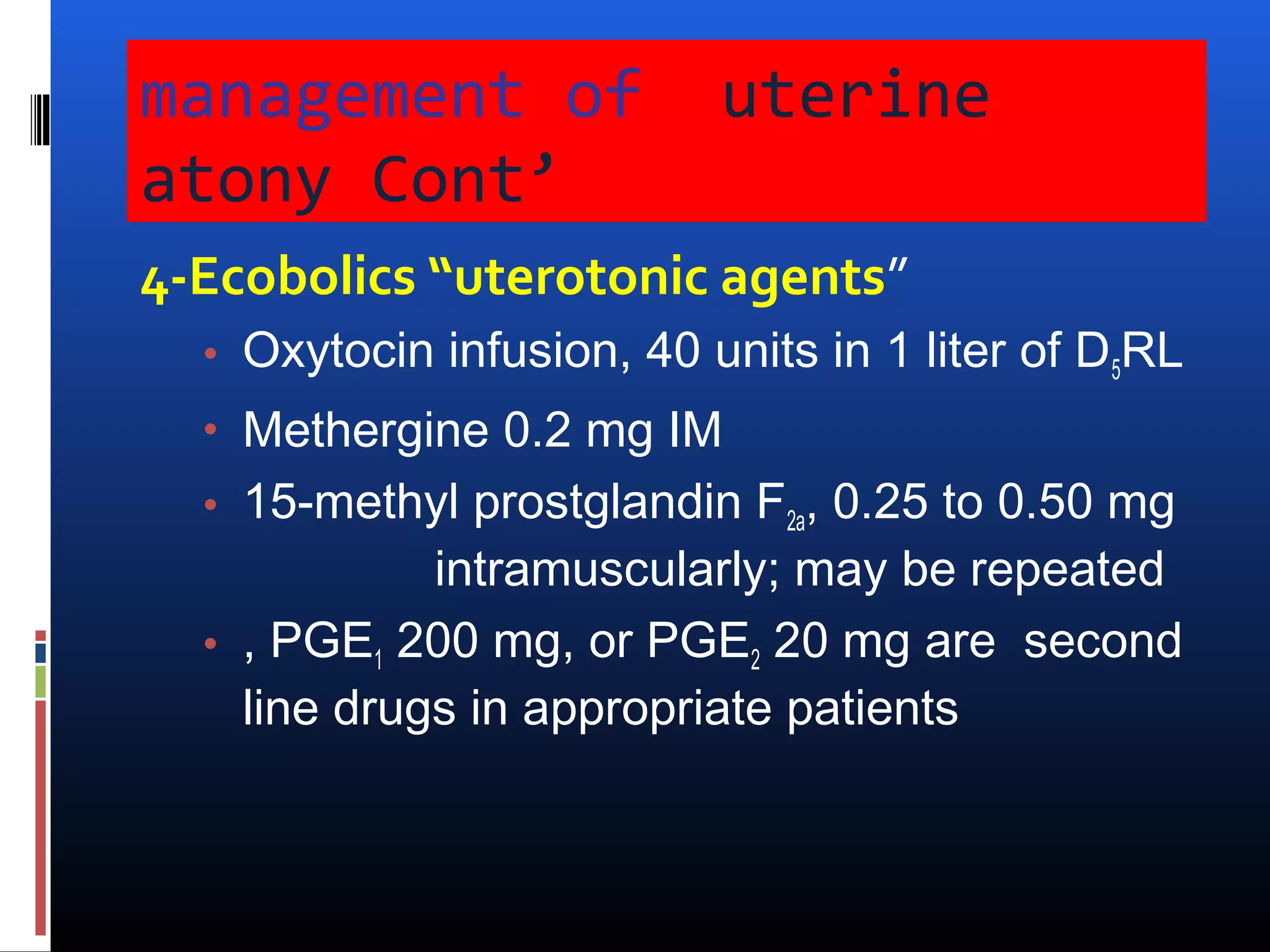
Postpartum
Hemorrhage
Definition
Blood loss         > 500 ml at vaginal delivery
                   > 1000 ml at Cesarean
ACOG               10% drop in hematocrit
                   Need for blood transfusion
Severe PPH         > 1000 ml loss at vaginal delivery

Any amount of blood loss causes
S/O Hypovolemic Hemorrhagic Shock
- Tachycardia - Hypotension - Reduced urine out put
Why it is important?

 PPH remained one of the top 3 causes of
 direct maternal deaths.

 Incidence 4% after vaginal delivery
           6,5% after CS delivery
We have 4 problems

 Problem 1: almost 50% of deliveries lose >500
  ml of blood.
 Problem 2: estimated blood loss is often less
  than half the actual blood loss.
 Problem 3: Most of the serious causes of
  “PPH” have origins prior to the end of the 3rd
  Stage of labor.
 Problem 4: PPH, as defined, is technically
  misdiagnosed and clinically irrelevant.
Measuring Blood Loss
 A key step to EFFECTIVE
 TREATMENT…..
 Underestimation leads to delayed intervention.


 Visual estimated amounts of blood loss are far from accurate
  by as much as 30-50%: especially for very large amounts.

 Old methods for estimating blood loss tend to be complex.


 (include weighing soaked clothes and pads, collection into
  pans etc., Acid haematin techniques, Spectrophometric
  technics and measuring plasma volume changes)
Measuring Blood Loss in PPH
    THE BRASSS-V DRAPE
Advantages of Brasss-V

 Simple and practical
 Low cost: ( Plastic)
 Accurate:
 Objective


 Can be used in a wide
range of settings

 Provides a hygienic delivery surface
CAUSES OF PPH
 FOUR “ T”s

TONE
TRUAMA
TISSUE RETENSION
THROMBIN

    BUT MOST IMPORTANT IS
Tone”Uterine Atony”90%of
causes
  - Uterine over distension
    Polyhydramnios, Multiple gestations,
       Macrosomia
    Prolonged labor: “uterine fatigue”
    Precipitory labor
    High parity
    Chorioamnionitis
    Retained product of conception
    Halogenated anesthetic
TRUMA “Obstetric OR OPERATIVE”
    “7% of causes”
  7% of causes
  Obstetric Trauma
  - Uterine Rupture
  - Lacerations of the Birth Canal
   - Operative Trauma
      Cesarean sections
      Episiotomies
      Forceps, Vacuums, Rotations
Tissue retension “Abnormal
placentaion”
   - PlacentaPrevia
    - Abruptio Placentae
    - Accreta, increta, percreta
    - Vasa previa
Thrombin “Coagulation
Defects
  - Sepsis
  - Amniotic Fluid Embolism
  - Abruptio Placentae associated
   coagulopathy
  - HELLP Syndrome
  - Dilutional Coagulopathy
  - Inherited Clotting Disorders
  - Anticoagulant Therapy
Thrombin “Coagulation
Defects
2-3% of causes
  - Sepsis
  - Amniotic Fluid Embolism
  - Abruptio Placentae associated
   coagulopathy
  - HELLP Syndrome
  - Dilutional Coagulopathy
  - Inherited Clotting Disorders
  - Anticoagulant Therapy
Prevention of Postpartum
Hemorrhage
   Oxytocin
   With or soon after delivery
   Cord traction


  Continues tension
   Gentle pull with contraction
   Uterine massage after placental delivery
Goals of Therapy
•Maintain the following:
          Systolic pressure >90mm Hg
          Urine output >0.5 mL/kg/hr
          Normal mental status
•Eliminate the source of hemorrhage
•Avoid overzealous volume replacement
that may contribute to pulmonary edema
Management Protocol

 • Examine the uterus to rule out atony


 • Examine the vagina and cervix to rule
  out lacerations; repair if present

 • Explore the uterus and perform
  curettage to rule out retained placenta
On recognition of
Hemorrhage
1. Initiate volume replacement with
     lactated ringers or normal saline.
2.   Alert blood bank and surgical team.
3.   Control the blood loss.
4.   Initiate decisive therapy.
5.   Monitor for complications.
MANAGEMENT of Uterine
   atony uterus for retained placental
1. Explore
   tissue.

2. Uterine massage
3Firm bimanual compression
management of              uterine
atony Cont’
4-Ecobolics “uterotonic agents”
  • Oxytocin infusion, 40 units in 1 liter of D5RL
  • Methergine 0.2 mg IM
  • 15-methyl prostglandin F2a, 0.25 to 0.50 mg
             intramuscularly; may be repeated
  • , PGE1 200 mg, or PGE2 20 mg are second
    line drugs in appropriate patients
Vaginal exploration
 General anesthesia usually best
 Uterine cavity manual exploration for retained
  placenta / uterine rupture
Vaginal exploration cont’
Uterine inversion restitution
Vaginal exploration cont’
Intrauterine balloon Cather
Bakri Balloon is a tamponade
technique that can be used for
PPH.


                                 24
When medical managament fails


 SURGICAL MANAGEMENT

  Uterus conserving : NEED OF TIME
  Definitive - Hysterectomy
MANAGEMENT”cont’”

  If Hemorrhage is not controlled by
   medications, massage, manual uterine
   exploration, or suturing lacerations in the
   birth canal,
 then surgical or radiological options must
   be considered. At this time, start:
  1. Packed red blood cell transfusion
  2. Foley catheter and monitor urine output
Selective Artertial Embolization


   If the patient is stable
   and bleeding is not “torrential”,
    and if interventional radiology is available,
    then pelvic arteriography may show the
    site of blood loss and therapeutic arterial
    embolization may suffice to stop the
    bleeding.
Uterine artery embolization
 Real time X-Ray (Fluoroscopy)

   Gelatin Sponges are injected
   into the bleeding vessel until
   stasis of flow in target vessel is
   achieved.
   Acess via RTfemorals
   to internal iliac and
   subsequently the uterine arteries
Pre embolization vs. .post embolization


 Pre Embolization      Post Embolization
Laparotomy for Obstetric
Hemorrhage
:
    - Bleeding at Cesarean section
    - “Torrential” Hemorrhage
    - Pelvic hematoma (expanding)
    - Bleeding uncontroled by other
      means
AT laparotomy

 Consider vertical abdominal incision
 General anesthesia usually best
 Get Help!
 Avoid compounding problems by making major
    mistakes
   Direct manual uterine compression / uterotonics
   Direct aortic compression
   Modified B-Lynch Suture for atony: #2 chromic
   Ligation of uterine and utero-ovarian vessels: #1
    chromic
B-Lynch suture vs Modified B-Lynch
Suture
Anterior view of   posterior view of
uterus showing     uterus showing
modified B-Lynch   modified B-Lynch
Technique          Technique
OTHER COMPRESSION SUTURES
 Hayman Uterine
  Compression Suture          Cho’s Multiple Square Suture


          Global Stitch By
          Dr. Gunasheela Bangalore
COMPLICATIONS

NIL   - IF DONE PROPERLY

TOO TIGHT COMPRESSION --

          CUT THROUGH STITCH

          UTERINE NECROSIS

          INTRAPERITONEAL BLEED
Uterine artery ligation

 http://t3.gstatic.com/i
  mages?
  q=tbn:ANd9GcQOaGG
  cLP1wYmyIsIQ8fyhFBB
  whABO3K3uFHL4V7Df
  d51ePIddvGg
Sutures are placed to ligate the
ascending uterine artery and the
anastomotic branch of the ovarian
artery.


                                    37
Internal iliac (hypogastric) artery
ligation

  50% success rate
  Desirous of children
  Experience of surgeon
  Steps:
  Palpate common iliac
 bifurcation
  Ligate at least 2-3 cm
  from bifurcation
  #1 silk. Do not divide
  vessel
Repaire of cervical laceration
 Palpate uterine cavity to assure its integrity


 Full thickness mucosal repair above the apex


 Contionous interlocking absorbable sutures


 Hematoma incised,clot removed,bleeding vessels
  ligated ,oblitrate defect with interlocking sutures
 Antibiotics& vaginal pack for 24 hours .

40
Uterine Rupture

  Prior Cesarean section = 1-2%


  Modern obstetrics = 1/10,000 to
   1/20,000 in unscarred uterus
  In “Neglected labors”, this accounts
   for many maternal deaths where
   modern obstetrical care is not available.
Classic Symptoms of Uterine
Rupture
   Fetal distress
   Vaginal bleeding
   Cessation of labor
   Shock
   Easily palpable fetal parts
   Loss of uterine catheter pressure
Management of Uterine Rupture

 Laparotomy
  Debride and repair in 2-3 layers
   of Maxon/PDS
  Subtotal Hysterectomy
  Total Hysterectomy
Management of Abnormal Placentation

 Diagnosis of exclusion after adressing tone and
    truma
   Curettage of uterine cavity
   Localized resection and uterine repair:
    (Vasopressin 1cc/10cc N.S-sub endometrial)
   Leave placenta in situ
      If not bleeding: Methotrexate
      Uterus will not be normal size by 8 weeks
   Uterine, utero-ovarian, hypogastric artery
    ligation
   Subtotal/ total abdominal hysterectomy
Post-Hysterectomy Bleeding
 Patient usually has DIC – Rx with whole blood,
  FFP, platelets, etc.
 Transvaginal or transabdominal
 (pelvic) pressure pack

   Bowel bag with opening pulled through
    vagina cuff/ abd. Wall

   Stuff with 4 inch gauze tied end-to-end until
    pelvis packed tight
Military Anti-Shock Trousers
(MAST)
  Increases pelvic and abdominal pressure to
   reduce bleeding
  Can use at any point in the procedure
  Used when exploration is to be avoided
Secondary PPH

 Defined as excessive bleeding 24 hrs to 12
  weeks postpartum.
 Incidence is about 1 percent of women.
 Theory is that thought to be atony or
  subinvolution of placental site from retained
  products or infection.


                                                  47
Management of Secondary PPH

 Evaluate for underlying disorders
  (coagulopathies).
 For atony give uterotonics.
 If large amount of bleeding, fever uterine
  tenderness, or foul smelling discharge treat
  for endometritis.
 Consider suction currettage.


                                                 48
Case 1

 A 22y/o G1P0 was delivered by vaccum
  assisted vaginal delivery approximately 2
  hours ago. She was induced for mild
  preeclampsia at 37 weeks and required
  pitocin augmentation for several hours prior
  to needing an operative vaginal delivery for
  fetal distress. She had a second degree
  laceration that was repaired, but she has
  soaked a whole pad in the last 15 minutes and
  the nurse would like you to evaluate her.
                                                  49
Case 2

 A 22 yo G4P3 approximately 4 days s/p
  delivery presents at OB triage and mentions
  to you that she feels lightheaded and has
  been having bleeding at about a pad an hour
  for the last 2 days.


                                                50
Case3

 A 34yo G6P6 patient at term has just delivered
  a 4000gm infant after second stage of labor
  lasting 3 ½ hours. The placenta delivered
  spontaneously and the patient is bleeding
  briskly.
 What is most probable cause?
 What the next step?
Postpartum hemorrhage for undergraduate

Postpartum hemorrhage for undergraduate

  • 1.
  • 2.
    Definition Blood loss > 500 ml at vaginal delivery > 1000 ml at Cesarean ACOG 10% drop in hematocrit Need for blood transfusion Severe PPH > 1000 ml loss at vaginal delivery Any amount of blood loss causes S/O Hypovolemic Hemorrhagic Shock - Tachycardia - Hypotension - Reduced urine out put
  • 3.
    Why it isimportant?  PPH remained one of the top 3 causes of direct maternal deaths.  Incidence 4% after vaginal delivery  6,5% after CS delivery
  • 4.
    We have 4problems  Problem 1: almost 50% of deliveries lose >500 ml of blood.  Problem 2: estimated blood loss is often less than half the actual blood loss.  Problem 3: Most of the serious causes of “PPH” have origins prior to the end of the 3rd Stage of labor.  Problem 4: PPH, as defined, is technically misdiagnosed and clinically irrelevant.
  • 5.
    Measuring Blood Loss A key step to EFFECTIVE TREATMENT…..  Underestimation leads to delayed intervention.  Visual estimated amounts of blood loss are far from accurate by as much as 30-50%: especially for very large amounts.  Old methods for estimating blood loss tend to be complex.  (include weighing soaked clothes and pads, collection into pans etc., Acid haematin techniques, Spectrophometric technics and measuring plasma volume changes)
  • 6.
    Measuring Blood Lossin PPH THE BRASSS-V DRAPE
  • 7.
    Advantages of Brasss-V Simple and practical  Low cost: ( Plastic)  Accurate:  Objective  Can be used in a wide range of settings  Provides a hygienic delivery surface
  • 8.
    CAUSES OF PPH FOUR “ T”s TONE TRUAMA TISSUE RETENSION THROMBIN BUT MOST IMPORTANT IS
  • 9.
    Tone”Uterine Atony”90%of causes - Uterine over distension  Polyhydramnios, Multiple gestations, Macrosomia  Prolonged labor: “uterine fatigue”  Precipitory labor  High parity  Chorioamnionitis  Retained product of conception  Halogenated anesthetic
  • 10.
    TRUMA “Obstetric OROPERATIVE” “7% of causes”  7% of causes  Obstetric Trauma - Uterine Rupture - Lacerations of the Birth Canal - Operative Trauma Cesarean sections Episiotomies Forceps, Vacuums, Rotations
  • 11.
    Tissue retension “Abnormal placentaion”  - PlacentaPrevia - Abruptio Placentae - Accreta, increta, percreta - Vasa previa
  • 12.
    Thrombin “Coagulation Defects - Sepsis - Amniotic Fluid Embolism - Abruptio Placentae associated coagulopathy - HELLP Syndrome - Dilutional Coagulopathy - Inherited Clotting Disorders - Anticoagulant Therapy
  • 13.
    Thrombin “Coagulation Defects 2-3% ofcauses - Sepsis - Amniotic Fluid Embolism - Abruptio Placentae associated coagulopathy - HELLP Syndrome - Dilutional Coagulopathy - Inherited Clotting Disorders - Anticoagulant Therapy
  • 14.
    Prevention of Postpartum Hemorrhage  Oxytocin  With or soon after delivery  Cord traction Continues tension  Gentle pull with contraction  Uterine massage after placental delivery
  • 15.
    Goals of Therapy •Maintainthe following: Systolic pressure >90mm Hg Urine output >0.5 mL/kg/hr Normal mental status •Eliminate the source of hemorrhage •Avoid overzealous volume replacement that may contribute to pulmonary edema
  • 16.
    Management Protocol •Examine the uterus to rule out atony • Examine the vagina and cervix to rule out lacerations; repair if present • Explore the uterus and perform curettage to rule out retained placenta
  • 17.
    On recognition of Hemorrhage 1.Initiate volume replacement with lactated ringers or normal saline. 2. Alert blood bank and surgical team. 3. Control the blood loss. 4. Initiate decisive therapy. 5. Monitor for complications.
  • 18.
    MANAGEMENT of Uterine atony uterus for retained placental 1. Explore tissue. 2. Uterine massage 3Firm bimanual compression
  • 19.
    management of uterine atony Cont’ 4-Ecobolics “uterotonic agents” • Oxytocin infusion, 40 units in 1 liter of D5RL • Methergine 0.2 mg IM • 15-methyl prostglandin F2a, 0.25 to 0.50 mg intramuscularly; may be repeated • , PGE1 200 mg, or PGE2 20 mg are second line drugs in appropriate patients
  • 20.
    Vaginal exploration  Generalanesthesia usually best  Uterine cavity manual exploration for retained placenta / uterine rupture
  • 21.
  • 22.
  • 23.
  • 24.
    Bakri Balloon isa tamponade technique that can be used for PPH. 24
  • 25.
    When medical managamentfails SURGICAL MANAGEMENT  Uterus conserving : NEED OF TIME  Definitive - Hysterectomy
  • 26.
    MANAGEMENT”cont’”  IfHemorrhage is not controlled by medications, massage, manual uterine exploration, or suturing lacerations in the birth canal,  then surgical or radiological options must be considered. At this time, start: 1. Packed red blood cell transfusion 2. Foley catheter and monitor urine output
  • 27.
    Selective Artertial Embolization  If the patient is stable  and bleeding is not “torrential”,  and if interventional radiology is available,  then pelvic arteriography may show the site of blood loss and therapeutic arterial embolization may suffice to stop the bleeding.
  • 28.
    Uterine artery embolization Real time X-Ray (Fluoroscopy)  Gelatin Sponges are injected  into the bleeding vessel until  stasis of flow in target vessel is  achieved.  Acess via RTfemorals  to internal iliac and  subsequently the uterine arteries
  • 29.
    Pre embolization vs..post embolization Pre Embolization Post Embolization
  • 30.
    Laparotomy for Obstetric Hemorrhage : - Bleeding at Cesarean section - “Torrential” Hemorrhage - Pelvic hematoma (expanding) - Bleeding uncontroled by other means
  • 31.
    AT laparotomy  Considervertical abdominal incision  General anesthesia usually best  Get Help!  Avoid compounding problems by making major mistakes  Direct manual uterine compression / uterotonics  Direct aortic compression  Modified B-Lynch Suture for atony: #2 chromic  Ligation of uterine and utero-ovarian vessels: #1 chromic
  • 32.
    B-Lynch suture vsModified B-Lynch Suture
  • 33.
    Anterior view of posterior view of uterus showing uterus showing modified B-Lynch modified B-Lynch Technique Technique
  • 34.
    OTHER COMPRESSION SUTURES Hayman Uterine Compression Suture Cho’s Multiple Square Suture Global Stitch By Dr. Gunasheela Bangalore
  • 35.
    COMPLICATIONS NIL - IF DONE PROPERLY TOO TIGHT COMPRESSION -- CUT THROUGH STITCH UTERINE NECROSIS INTRAPERITONEAL BLEED
  • 36.
    Uterine artery ligation http://t3.gstatic.com/i mages? q=tbn:ANd9GcQOaGG cLP1wYmyIsIQ8fyhFBB whABO3K3uFHL4V7Df d51ePIddvGg
  • 37.
    Sutures are placedto ligate the ascending uterine artery and the anastomotic branch of the ovarian artery. 37
  • 38.
    Internal iliac (hypogastric)artery ligation  50% success rate  Desirous of children  Experience of surgeon  Steps:  Palpate common iliac bifurcation  Ligate at least 2-3 cm from bifurcation  #1 silk. Do not divide vessel
  • 39.
    Repaire of cervicallaceration  Palpate uterine cavity to assure its integrity  Full thickness mucosal repair above the apex  Contionous interlocking absorbable sutures  Hematoma incised,clot removed,bleeding vessels ligated ,oblitrate defect with interlocking sutures  Antibiotics& vaginal pack for 24 hours . 
  • 40.
  • 41.
    Uterine Rupture Prior Cesarean section = 1-2% Modern obstetrics = 1/10,000 to 1/20,000 in unscarred uterus In “Neglected labors”, this accounts for many maternal deaths where modern obstetrical care is not available.
  • 42.
    Classic Symptoms ofUterine Rupture  Fetal distress  Vaginal bleeding  Cessation of labor  Shock  Easily palpable fetal parts  Loss of uterine catheter pressure
  • 43.
    Management of UterineRupture  Laparotomy  Debride and repair in 2-3 layers of Maxon/PDS  Subtotal Hysterectomy  Total Hysterectomy
  • 44.
    Management of AbnormalPlacentation  Diagnosis of exclusion after adressing tone and truma  Curettage of uterine cavity  Localized resection and uterine repair: (Vasopressin 1cc/10cc N.S-sub endometrial)  Leave placenta in situ  If not bleeding: Methotrexate  Uterus will not be normal size by 8 weeks  Uterine, utero-ovarian, hypogastric artery ligation  Subtotal/ total abdominal hysterectomy
  • 45.
    Post-Hysterectomy Bleeding  Patientusually has DIC – Rx with whole blood, FFP, platelets, etc.  Transvaginal or transabdominal  (pelvic) pressure pack  Bowel bag with opening pulled through vagina cuff/ abd. Wall  Stuff with 4 inch gauze tied end-to-end until pelvis packed tight
  • 46.
    Military Anti-Shock Trousers (MAST)  Increases pelvic and abdominal pressure to reduce bleeding  Can use at any point in the procedure  Used when exploration is to be avoided
  • 47.
    Secondary PPH  Definedas excessive bleeding 24 hrs to 12 weeks postpartum.  Incidence is about 1 percent of women.  Theory is that thought to be atony or subinvolution of placental site from retained products or infection. 47
  • 48.
    Management of SecondaryPPH  Evaluate for underlying disorders (coagulopathies).  For atony give uterotonics.  If large amount of bleeding, fever uterine tenderness, or foul smelling discharge treat for endometritis.  Consider suction currettage. 48
  • 49.
    Case 1  A22y/o G1P0 was delivered by vaccum assisted vaginal delivery approximately 2 hours ago. She was induced for mild preeclampsia at 37 weeks and required pitocin augmentation for several hours prior to needing an operative vaginal delivery for fetal distress. She had a second degree laceration that was repaired, but she has soaked a whole pad in the last 15 minutes and the nurse would like you to evaluate her. 49
  • 50.
    Case 2  A22 yo G4P3 approximately 4 days s/p delivery presents at OB triage and mentions to you that she feels lightheaded and has been having bleeding at about a pad an hour for the last 2 days. 50
  • 51.
    Case3 A 34yoG6P6 patient at term has just delivered a 4000gm infant after second stage of labor lasting 3 ½ hours. The placenta delivered spontaneously and the patient is bleeding briskly.  What is most probable cause?  What the next step?

Editor's Notes

  • #6 About 5% of women will loose more than 1000 mls of blood during normal vaginal delivery. ICD-10 Describes PPH as blood loss of more than 500ml for vaginal delivery and 750 mls for Cesarean section. Old methods for estimating blood loss include visual, gravimetric, collection into pans etc. Acid haematin techniques, spectrophometric, plasma volume changes.