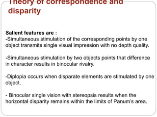
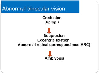
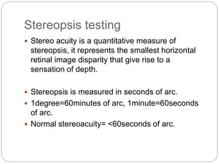
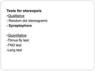
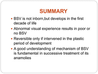
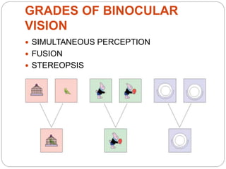
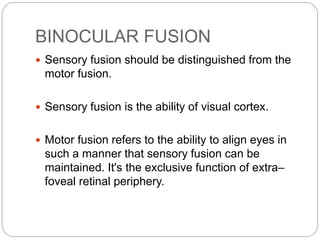
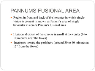
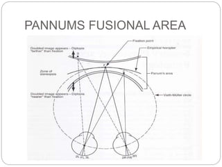
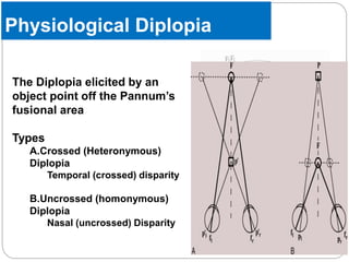
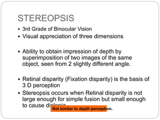
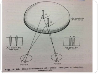
2. DEFINITION
State of simultaneous vision with two seeing
eyes (neither of which needs necessarily be
normal) that occurs when an individual fixes
his visual attention on an object of regard.
3. Why to study binocular vision?
The basic laws of binocular vision forms the very foundation
on which our current understanding of strabismus and its
symptoms and sensory consequences is based.
Knowledge on BSV forms basis to orthoptic treatments and
many physiological processes in the eye.
4. Types of Vision
1.Monocular
Vision with single eye
2.Bi-ocular
no co-ordination between two eyes
3.Binocular
Co-ordinated vision from two eyes
5. Pre-requisites for development of
BSV
Motor Mechanism:
•correct neuromuscular development so that the visual axes
are directed at the object
•overlap of visual fields
Sensory Mechanism:
•approximately equal image clarity and size in the two eyes
•corresponding retinal areas so that the eyes are cyclopean
•normal visual pathways
Mental Process:
•ability of visual cortex to promote binocular single vision
10. Visual space vs physical space
Visual space is the subjective space that is based
of anatomic and physiological systems,visual
clues and on learning.
Physical space is the actual space of the object.
Anatomic distribution of retinal elements does not
coincide with physiological distribution of spatial
values – Kundst- Munsterberg illusion
11. VISUAL DIRECTION
1. OCULOCENTRIC –
- Monocular
- Laws of oculocentric visual direction
2. EGOCENTRIC –
- Cyclopean eye
- Hering’s law of identical visual direction
13. RETINAL CORRESPONDENCE
Retinal rceptors in both eyes that dictate a
common visual direction under binocular
conditions are called corrresponding retinal
points.
Corresponding retinal points are areas of zero
disparity.
Normal retinal correspondence
Abnormal retinal correspondence(ARC)
Harmonius arc
Un harmonius arc
HARMONIUS angle of anamoly= angle of strabismus
UNHARMONIUS angle of anamoly< angle of
15. HOROPTER
It is the sum total of all the points in the physical
space that stimulate corresponding retinal
elements of the two eyes.
1. Vieth muller horopter / theoritical / geometric
horopter
2. Empirical horopter
18. BINOCULAR FUSION
Sensory fusion should be distinguished from the
motor fusion.
Sensory fusion is the ability of visual cortex.
Motor fusion refers to the ability to align eyes in
such a manner that sensory fusion can be
maintained. It's the exclusive function of extra–
foveal retinal periphery.
19. PANNUMS FUSIONAL AREA
Region in front and back of the horopter in which single
vision is present is known as Panum’s area of single
binocular vision or Panum’s fusional area
Horizontal extent of these areas is small at the center (6 to
10 minutes near the fovea)
Increases toward the periphery (around 30 to 40 minutes at
12° from the fovea)
21. Physiological Diplopia
The Diplopia elicited by an
object point off the Pannum’s
fusional area
Types
A.Crossed (Heteronymous)
Diplopia
Temporal (crossed) disparity
B.Uncrossed (homonymous)
Diplopia
Nasal (uncrossed) Disparity
22. Fixation Disparity
It is the minute image displacement, rarely
exceeding several minutes of arc of angle, occurs
within Panum’s space while fusion is maintained.
• Due to presence of pannum’s fusional area
– A physiological variation in placement of retinal
image displacement from corresponding retinal
points
• Even Allow fusion
• Displacement of retinal images in two eyes
– Retinal disparity
23.
24. Theories of binocular fusion
1. Physiologic hypothesis – most accepted
2. Synergy hypothesis
3. Local sign hypothesis
4. Eye movement hypothesis
5. Suppression hypothesis
26. Dichoptic stimulation
• Refers to different stimulation in the two eyes
which occurs when binocular stimuli fall on non
corresponding points on the two retinae.
• Depending on the degree of non correspondence
five classes of percepts are obtained.
• Depth with fusion
• Depth with diplopia
• Diplopia without depth
• Binocular rivalry and suppression
• Binocular lustre.
27. Retinal rivalry and suppression
• When dissimilar contours are presented to the
corresponding retinal areas fusion becomes
impossible.
• This conflict which results when such signals are
transmitted to the visual cortex is termed as retinal
rivalry.
• When presented with such signals,signal presented
from one eye is actively suppressed. This results on
perception of single signal at a time. This is known
as suppression
29. Binocular lustre
• The lustrous appearance of surfaces like a waxed
surface,table top is due to binocular lustre.
• It results from the different position of partially
reflected objects in the surface by virtue of the
different positions of the two eyes.
30. STEREOPSIS
3rd Grade of Binocular Vision
Visual appreciation of three dimensions
Ability to obtain impression of depth by
superimposition of two images of the same
object, seen from 2 slightly different angle.
Retinal disparity (Fixation disparity) is the basis of
3 D perception
Stereopsis occurs when Retinal disparity is not
large enough for simple fusion but small enough
to cause diplopiaNot similar to depth perception.
31.
32. PHYSIOLOGIC BASIS OF STEREOPSIS
• Horizontal disparity produces stereopsis.
•Local stereopsis – square by square matching
•Global stereopsis - to resolve ambiguities
•Fine stereopsis - specific pattern matching < 0.5
degrees
•Coarse stereopsis - act beyond the range of retinal
rivalry , dissimilar images can be simultaneously
perceived
-Fusion is not absolutely required for stereopsis .
-But similar parts of stereogram reaching corresponding
points must fuse to form reference points.
33. •Stereoscopic acuity
-Minimal disparity beyond which no stereoscopic effect
is produced characterizes the person’s stereoscopic
acuity.
- normal = depth of 10 arc sec
-Closest fixation of 10 inches = depth of thousandth of
an inch
-Beyond 600 mts there is no true stereopsis.
34. PERCEPTION OF DEPTH
Perception of distance of objects from each other
or from the observer.
Several clues contribute-
A] BINOCULAR CLUE: Stereopsis.
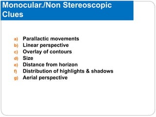
B] MONOCULAR CLUES:
35. Monocular./Non Stereoscopic
Clues
a) Parallactic movements
b) Linear perspective
c) Overlay of contours
d) Size
e) Distance from horizon
f) Distribution of highlights & shadows
g) Aerial perspective
36. PARALLACTIC
MOVEMENTS
Most important in depth perception next to
stereopsis
Slight shift of head while fixation is maintained
results in change of relative position of objects in
gaze
Objects beyond fixation point – move in same
direction
Objects closer – move in opposite direction
39. THEORIES OF BINOCULAR
VISION
Theory of correspondence and disparity
Neurophsiological theory
Older theories
Alternation theory
Projection theory
Motor theory
Isomorphism theory
40. Theory of correspondence and
disparity
Salient features are :
-Simultaneous stimulation of the corresponding points by one
object transmits single visual impression with no depth quality.
-Simultaneous stimulation by two objects points that difference
in character results in binocular rivalry.
-Diplopia occurs when disparate elements are stimulated by one
object.
- Binocular single vision with stereopsis results when the
horizontal disparity remains within the limits of Panum’s area.
41. Neurophysiology of binocular vision
Optic chiasma.
•First structure that contributes for
BSV
•Partial decussation at chiasm
•serves to project to same place
in visual cortex
2 neuronal pathways
1. Parvocellular
2. Magnocellular
42. Milestone of BSV Development
2 to 3 weeks – the infant turns his head to fixate an object.
4 to 5 weeks - infant can sustain monocular fixation of large
near objects
First 1 to 3 months -superimpose images.
3 months- binocular fusion.
3 to 6 months – stereopsis.
Development of horopter & vergence is also influenced by
dramatic changes in eyeball size & orbital position during
44. Anamolies of binocularity
Confusion
When squinting occurs the two foveas view two different
objects that are physically separated in objective space, and
send two different images to a single cortical perceptual area.
This leads to confusion.
Diplopia
When squinting occurs an object in space is perceived by the
fovea of one eye and some other extra-foveal point of the
other eye, which has a different projection or localization value
in space. Thus an object would be localized twice in space
causing diplopia.
45.
46. INVESTIGATIONS
For retinal correspondence
For supression
1.Red filter test
2.Worth FDT
3.Bagolinis striate glass test
4.After image testing
51. Stereopsis testing
Stereo acuity is a quantitative measure of
stereopsis, it represents the smallest horizontal
retinal image disparity that give rise to a
sensation of depth.
Stereopsis is measured in seconds of arc.
1degree=60minutes of arc, 1minute=60seconds
of arc.
Normal stereoacuity= <60seconds of arc.
57. MACULAR BV
EXTRAMACULAR BV
BEGINS AT 2-3 MONTHS MATURE AT BIRTH
IF DEPRIVED NEVER DEVELOPS WILL NOT DEPRIVE
POOR VN AND NYSTAGMUS --
SEVERES FIXATION ---
ONCE DEVELOPED
REINFORCEMENT REQUIRE TILL 9
YRS AGE
---
OTHERWISE AMBLYOPIA
DEVELOPS
---
CONES AND PARVOCELLULAR
GANGLION CELLS
RODS AND MAGNOCELLULAR
ANGLION CELLS
14’ OF ARC OF STEREOPSIS 200’ ARC OF STEREOPSIS
58. BV MACULAR EXTRAMACULAR
SIMULTANEOUS
PERCEPTION
NONE EXCELLENT
FUSION EXCELLENT LIMITED
STEREOPSIS EXCELLENT LIMITED
SO THERE ARE NO ADAPTATIONS IF MACULAR
BINOCULAR VISION IS IMPAIRED AND
THERE WILL BE LIMITED FUSION AND
STEREOPSIS
IF EXTRAMACULAR BV IS IMPAIRED-
SUPPRESSION AND ARC TAKES PLACE
59. SUMMARY
BSV is not inborn,but develops in the first
decade of life
Abnormal visual experience results in poor or
no BSV
Reversible only if intervened in the plastic
period of development
A good understanding of mechanism of BSV
is fundamental in successive treatment of its
anamolies