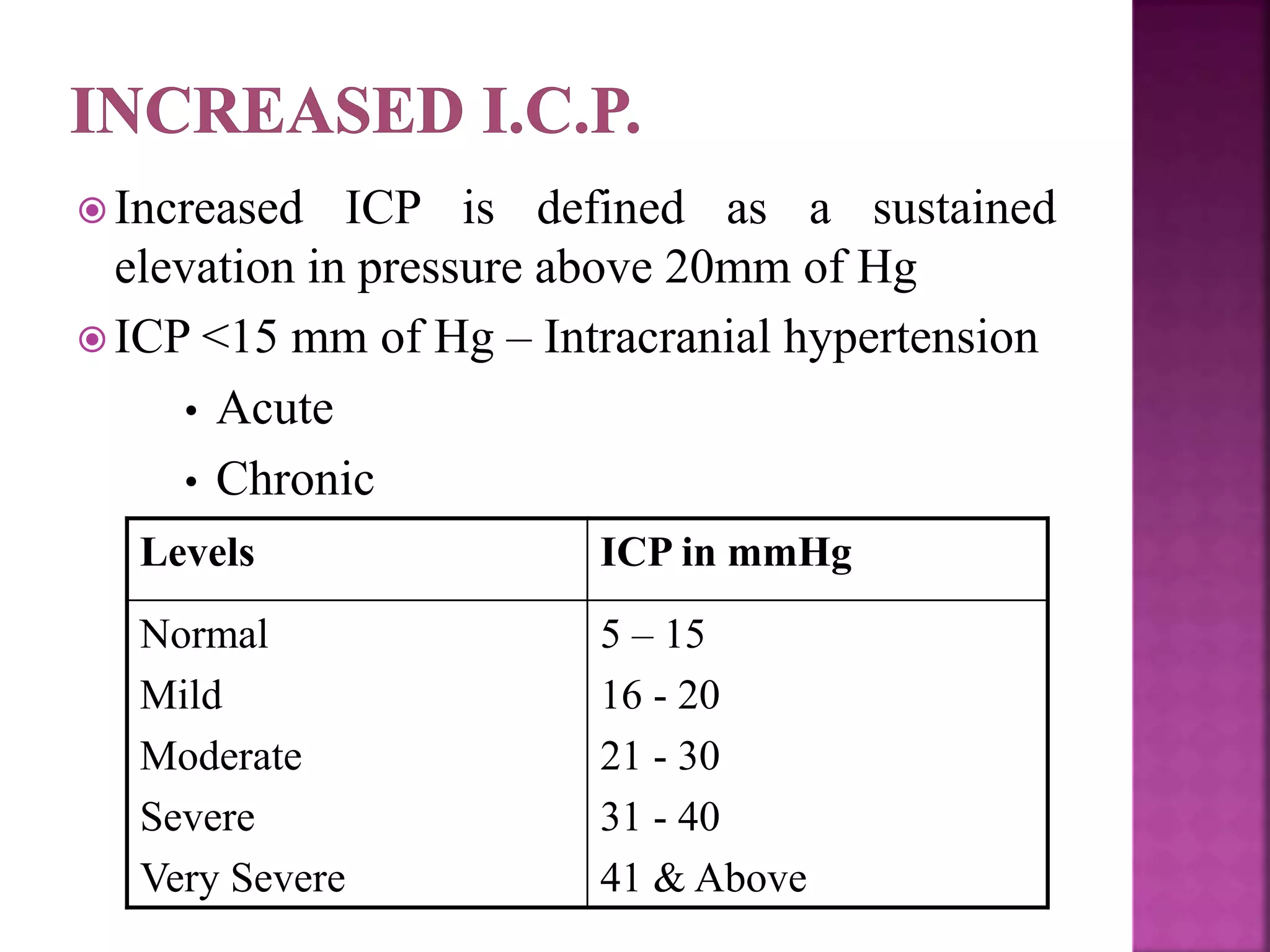
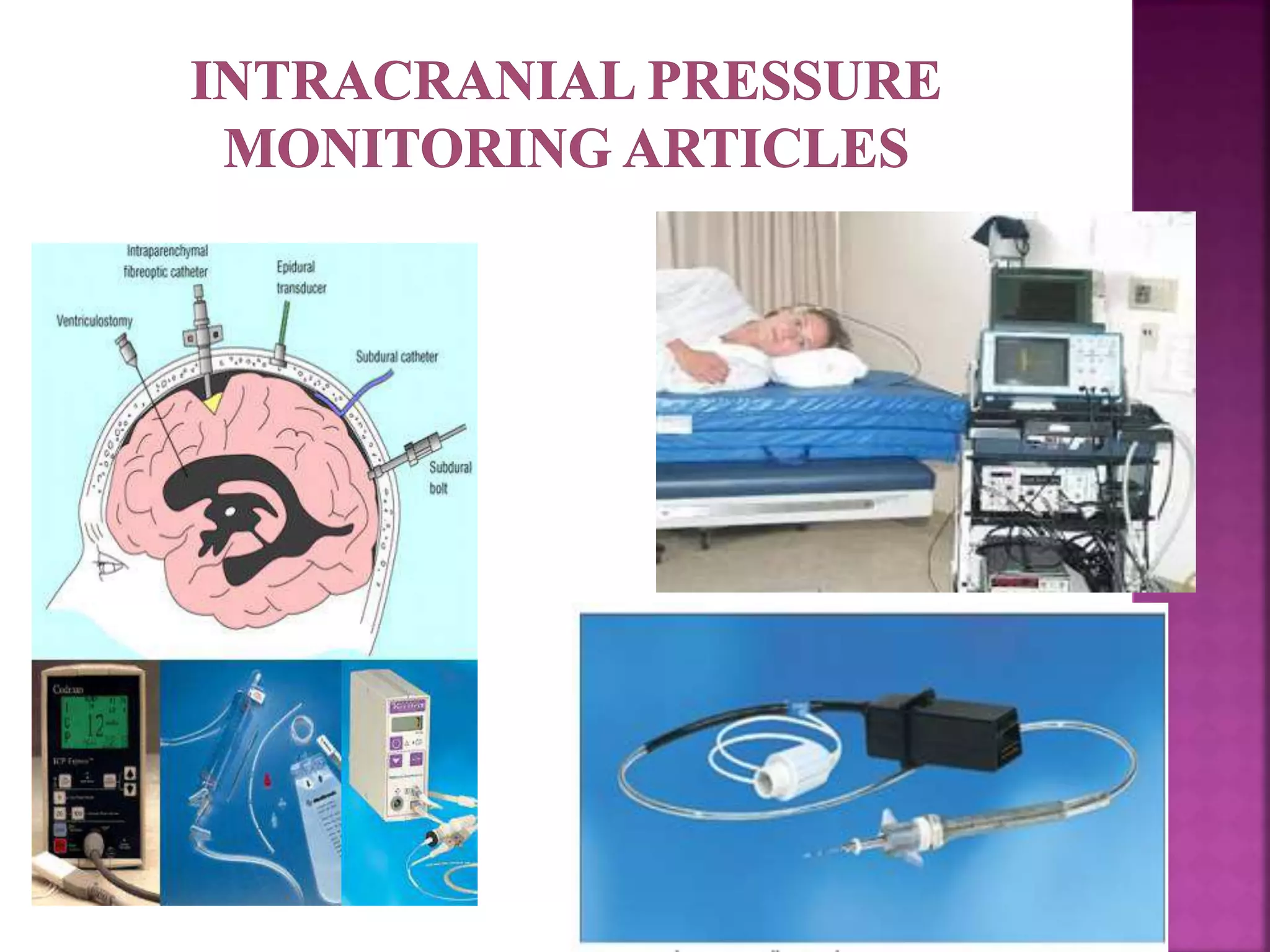
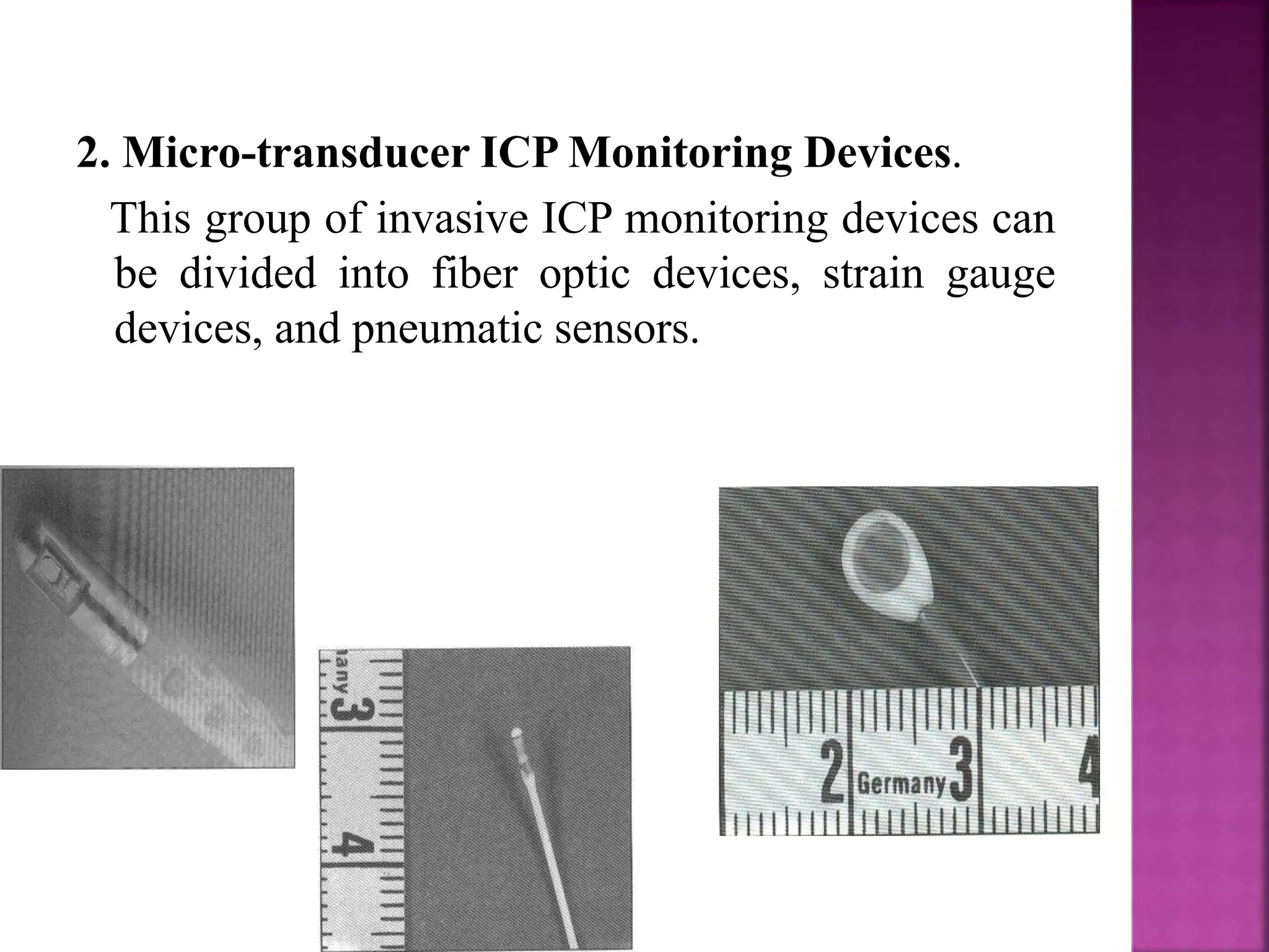
Presented To: Presented By:
Mrs. Vinay Kumari Amandeep Kaur
Associate Professor Msc. (N) 2nd year
MMCON 1915703
 The skull is a rigid structure.
 It contains:
 Brain
 Blood
 Cerebro spinal fluid (CSF)
 Any increase of theses contents and/or the addition
of any mass will lead to increase in the pressure
(the Monroe-Kellie doctrine).
 Brain 1300-1750 mls
 Tissue 300-400 mls.
 Intra-cellular fluid 900-1000 mls.
 Extra-cellular fluid 100-150 mls
 Blood 100-150 mls.
 CSF 100-150 mls.
 COMPENSATORY MECHANISMS FOR
EXPANDING MASSES:
 Immediate
 Decrease in CSF volume by movement of fluid to
the lumbar area.
 Decrease in the blood volume by squeezing blood
out of sinuses
 Delayed
 Decrease in the extra-cellular fluid.
 CEREBROSPINAL FLUID
 Secreted at the rate of 500 mls per day
 Secreted by the choroid plexus in the lateral ventricles
 Flows through the ventricular system
 Exits to the subarachnoid space through the foramina of
Magendie and Luschka
 Absorbed into the venous system via the arachnoid
granulations
 Any obstruction to the flow will lead to
HYDROCEPHALUS and increased pressure.
 BRAIN OEDEMA
 An excess of brain water may occur:
 Around lesions within the brain:
 Tumor
 Abscess
 In relation to traumatic damage
 In relation to ischemic brain insult
 Leads to increase in the pressure.
 Types of edema:
 Vasogenic (extra cellular): tumors
 Cytotoxic (intra cellular): metabolic states
 Interstitial (extra cellular): increased IVP
Pressure
Flow = ----------------
Resistance
Cerebral Perfusion Pressure
(systemic pressure – intracranial pressure)
Cerebral Blood Flow = --------------------------------------
Cerebral Vascular Resistance
 Brain 80 – 85 %
 CSF 8 – 12 %
 Cerebral blood volume 5 – 8 %
 Total Intra cranial volume 1500 ± 100ml
 Autoregulation of cerebral blood flow
 Regulation of CSF
 Regulation with help of metabolic changes
 Pathologic States that increase the volume of
one component necessitate decrease in the
volume of another to maintain normal Intra-
Cranial Pressure.
 Brain
 CSF
 Cerebral blood volume
 Glasgow Coma Scale (GCS ) < 8
 Posturing (extension, flexion)
 Bilateral or unilateral pupil dilation (except with
Epidural Hematomas)
 CT Scan results showing edema and/or mid-line
shift
 Physical assessment /neurological assessment
findings which indicate a need for monitoring
 Awake patient
 Coagulopathy
 Intracranial pressure (ICP) is the pressure
inside the skull and thus in the brain tissue and
cerebrospinal fluid (CSF)
 Normal range 5 – 15 mm Hg
 Normally 0-140 mm CSF (0-10 mm Hg)
 There are normal regular waves due to pulse and
respiration
 With increased pressure “pressure waves” appear
 With continued rise of ICP the PP falls
 When PP falls CBF is reduced
 Electrical cortical activity fails if CBF is 20ml/100gm/min
 When intracranial pressure reaches mean arterial pressure
circulation to the brain stops.
 Internal herniation:- Temporal lobe is pushed
down though Tentorium in cisura
 External herniation:- Cerebellar tonsills/
peduncle herniate through foramen magnum →
Compressing over IV ventricle → ↓CPP →
Death == “CONING”
 Effective pressure that allows the perfusion of
blood through the brain
 CPP = MAP – ICP
 Mean arterial pressure (MAP) = DP+ (SP-DP)
 DP + PP/3 3
 (ICP  CVP)
 CPP  MAP – CVP
 Normal CBF 45 – 50 ml / 100 gm / min
 Range 20 ml / kg / min to 70 ml / kg / min
 CBF Highest Frontal region
 CBF Medium Parietal region
 CBF Lowest Temporal area
 Cerebral Autoregulation
 Normal range MAP 50 – 150 mm Hg
 If Head injured ~~ Failure of autoregulation
 CBF = < 20ml / kg /min.
 Adverse effect on ICP
 Increased ICP is defined as a sustained
elevation in pressure above 20mm of Hg
 ICP <15 mm of Hg – Intracranial hypertension
• Acute
• Chronic
Levels ICP in mmHg
Normal
Mild
Moderate
Severe
Very Severe
5 – 15
16 - 20
21 - 30
31 - 40
41 & Above
 During slow increase in volume in a continuous mode,
the ICP rises to a plateau level at which the increase
level of CSF absorption keeps pace with the increase in
volume.
 Intermittent expansion causes only a transient rise in
ICP at first. When sufficient CSF has been absorbed to
accommodate the volume the ICP returns to normal.
 Expansion to a critical volume does however cause
persistent rise in ICP which thereafter increases
logarithmically with increasing volume ( volume -
pressure relationship).
 THE VOLUME PRESSURE RELATIOSHIP
 The ICP finally rises to the level of arterial pressure
which it self begins to increase, accompanied by
bradycardia or other disturbances of heart rhythm
(Cushing response). This is accompanied by dilatation
of small pial arteries and some slowing of venous flow
which is followed by pulsatile venous flow.
 CPP α CBF
 CPP α 1/ICP
 ↑ICP → ↓CBF, ↓Blood volume, ↑CO2
 Cerebral Oedema
 Vasogenic
 Cytotoxic oedema
 Hypoxemia
 Hyponatremia/ Water Intoxication
 Post-Cardiac Arrest
 Inflammatory—Meningitis/Encephalitis
 Interstitial oedema
 Intra Cranial Space Occupying Lesions
 Enlarged ventricular system
 Pneumocephalus
 Increase in C.B.F.
 Impaired cerebral venous drainage
Neonates/Infants
 Secondary cerebral oedema to peri-natal
hypoxia or trauma
 Congenital hydrocephalus
Older Infants/Toddlers/Children
 Meningitis
 Brain tumors (Infra tentorial)
 Pseudo tumor cerebrii
 Trauma
 Reye’s syndrome
 Thorough clinical assessment
 “WARNING SIGNS”:
Confusion, agitation, restlessness,
aggressiveness
Personality changes
Glasgow Coma Score (GCS)
Findings Score
1) Eye opening
Spontaneous
To voice
To Pain
None
4
3
2
1
2) Best verbal response
Oriented
Confused speech
Inappropriate words
Incomprehensible sounds
None
5
4
3
2
1
3)Best motor response
Obeys Commands
Localizes pain
Withdraws
Abnormal flexion
Extension
None
6
5
4
3
2
1
Prognostic value as per GCS
Score Percentage
GCS 3/ less
GCS 3 – 5
GCS 6 – 8
100
60 – 84
36 – 46
Meaning : The monitoring of intracranial pressure is
used in treating severe traumatic brain injury patients.
This process is called intracranial pressure monitoring.
All current clinical available measurement methods are
invasive and use various transducer systems.
 Ventricular system
 Sub-arachnoid space
 Epidural space
 Brain parenchyma
 Intraventricular catheter
 Subarachnoid screw
or bolt
 Epidural sensor
1. External Ventricular Drainage (EVD). Invasive
monitoring using the EVD technique, where a catheter
is placed into one of the ventricles through a burr hole,
is considered the gold of ICP monitoring. In addition to
measuring ICP, this technique can also be used for
drainage of CSF and administering of medicine intra-
thecally, for example, antibiotic administration in cases
of ventriculitis, possibly resulting from EVD
placement itself.
2. Micro-transducer ICP Monitoring Devices.
This group of invasive ICP monitoring devices can
be divided into fiber optic devices, strain gauge
devices, and pneumatic sensors.
 The idea of a noninvasive method of measuring
ICP is captivating, as complications seen in
relation to the invasive methods of ICP measuring,
that is, hemorrhage and infection, are avoidable.
Different techniues have been proposed.
1. Transcranial Doppler Ultrasonography (TCD).
The TCD technique applies ultrasound to initially
measure the blood flow velocity in the middle
cerebral artery. The difference between systolic
and diastolic flow velocity, divided by the mean
flow velocity, is called the pulsatility index (PI).
2. Tympanic Membrane Displacement (TMD). The
technique takes advantage of the communication of the
CSF and the perilymph via the perilymphatic duct.
Stimulation of the stapedial reflex causes a movement
of the tympanic membrane, which is shown to correlate
to ICP.
3. Fundoscopy and Papilledema. Papilledema, or optic
disc swelling, due to raised ICP can be visualized by
fundoscopy and graded by the Fris´en Scale into 5
categories depending on signs of disturbed axoplasmic
transport.
 Fundo-scopy and Papilledema
 A, B, & C waves
 Factors influencing waves
Systolic blood pressure
Alterations in respiration
Deteriorating neurological status
 Components of waves
P1 (upward spike)
P2 (tidal wave)
P3 (small notch)
A waves
 Plateau waves
 Most life threatening
 Seen in 5-20 min intervals
 Increased I.C.P.
 CPP compromised
 Amplitude 50 – 60 mm of Hg
B waves
 Saw toothed appearance
 Occur every 30 – 60 sec
 Amplitude 25 – 50 mm of Hg
 Indicates Unstable ICP& unconsciousness
 Stimulation ↑ amplitude
C waves
 Lowest amplitude
 Occur in 4 – 8 min intervals
 Never get elevated >20 – 25 mm of Hg
 Clinical significance unknown
 Infection
 intracranial hemorrhage or haematoma
 CSF Leakage
 Mechanical failure or blockage
 Over drainage of CSF
The Patient with Increased Intracranial Pressure
ASSESSMENT
 History
 Present Illness
 Obtain Subjective Data
 Neurologic examination
 Mental Status
 LOC
 Cranial Nerve Function
 Cerebral Function (balance and coordination)
 Reflexes
 Motor and Sensory Function
 Abnormal Respiratory Pattern
NURSING DIAGNOSIS
 Ineffective airway clearance related to diminished
protective reflexes
 Ineffective breathing patterns related to neurologic
dysfunction
 Ineffective cerebral tissue perfusion related to the
effects of increased ICP
 Deficient fluid volume related to fluid restriction
 Risk for infection related to ICP monitoring system
PLANNING AND GOALS
 Maintenance of patent airway
 Normalization of respiration
 Adequate cerebral tissue perfusion through
reduction in ICP
 Restoration of fluid balance
 Absence of infection
 Absence of complication
NURSING INTERVENTION
 Maintaining patent airway and adequate
ventilation
 Monitor vital signs
 Maintain fluid balance
 Position client with head of the bed elevated 30 to
45 degrees and neck in neutral position
 Maintain a quiet environment
 Avoid use of restraints
 Prevent straining at stool
 Prevent excessive cough and vomiting
 Prevent complication of immobility
 Preventing infection
 Administer medication as ordered
EVALUATION
 Maintained patent airway
 Attains optimal breathing pattern
 Attains desired fluid balance
 Infection prevented
 Complications prevented
 I.C.P. is an important parameter
 Physiology
 Pathology related to increased ICP
 Monitoring of ICP
 Interaction between ICP and anaesthetic agents
 Anaesthesiologist as Peri-operative Physician
 B.Mokri, “TheMonro-Kellie hypothesis: applications
in CSF volume depletion,” Neurology, vol. 56, no. 12,
pp. 1746–1748, 2001.
 A. Monro, Observations on Structure and Functions of
the Nervous System, Creech and Johnson, Edinbourg,
UK, 1783.
 G. Kellie, “Appearances observed in the dissection of
two individuals; death from cold and congestion of the
brain,” Transactions of the Medico-Chirurgical Society
of Edinburgh, vol. 1, article 84, 1824.
 F.Magendie, “Recherches anatomique et physiologique
sur le liquide c´ephalo-rachidien ou c´erebro-spinal,”
Paris, France,1842.
Intracranial pressure measurement

Intracranial pressure measurement

  • 1.
    Presented To: PresentedBy: Mrs. Vinay Kumari Amandeep Kaur Associate Professor Msc. (N) 2nd year MMCON 1915703
  • 2.
     The skullis a rigid structure.  It contains:  Brain  Blood  Cerebro spinal fluid (CSF)  Any increase of theses contents and/or the addition of any mass will lead to increase in the pressure (the Monroe-Kellie doctrine).
  • 3.
     Brain 1300-1750mls  Tissue 300-400 mls.  Intra-cellular fluid 900-1000 mls.  Extra-cellular fluid 100-150 mls  Blood 100-150 mls.  CSF 100-150 mls.
  • 4.
     COMPENSATORY MECHANISMSFOR EXPANDING MASSES:  Immediate  Decrease in CSF volume by movement of fluid to the lumbar area.  Decrease in the blood volume by squeezing blood out of sinuses  Delayed  Decrease in the extra-cellular fluid.
  • 5.
     CEREBROSPINAL FLUID Secreted at the rate of 500 mls per day  Secreted by the choroid plexus in the lateral ventricles  Flows through the ventricular system  Exits to the subarachnoid space through the foramina of Magendie and Luschka  Absorbed into the venous system via the arachnoid granulations  Any obstruction to the flow will lead to HYDROCEPHALUS and increased pressure.
  • 6.
     BRAIN OEDEMA An excess of brain water may occur:  Around lesions within the brain:  Tumor  Abscess  In relation to traumatic damage  In relation to ischemic brain insult  Leads to increase in the pressure.  Types of edema:  Vasogenic (extra cellular): tumors  Cytotoxic (intra cellular): metabolic states  Interstitial (extra cellular): increased IVP
  • 7.
    Pressure Flow = ---------------- Resistance CerebralPerfusion Pressure (systemic pressure – intracranial pressure) Cerebral Blood Flow = -------------------------------------- Cerebral Vascular Resistance
  • 8.
     Brain 80– 85 %  CSF 8 – 12 %  Cerebral blood volume 5 – 8 %  Total Intra cranial volume 1500 ± 100ml
  • 9.
     Autoregulation ofcerebral blood flow  Regulation of CSF  Regulation with help of metabolic changes
  • 10.
     Pathologic Statesthat increase the volume of one component necessitate decrease in the volume of another to maintain normal Intra- Cranial Pressure.  Brain  CSF  Cerebral blood volume
  • 11.
     Glasgow ComaScale (GCS ) < 8  Posturing (extension, flexion)  Bilateral or unilateral pupil dilation (except with Epidural Hematomas)  CT Scan results showing edema and/or mid-line shift  Physical assessment /neurological assessment findings which indicate a need for monitoring
  • 12.
  • 13.
     Intracranial pressure(ICP) is the pressure inside the skull and thus in the brain tissue and cerebrospinal fluid (CSF)  Normal range 5 – 15 mm Hg
  • 14.
     Normally 0-140mm CSF (0-10 mm Hg)  There are normal regular waves due to pulse and respiration  With increased pressure “pressure waves” appear  With continued rise of ICP the PP falls  When PP falls CBF is reduced  Electrical cortical activity fails if CBF is 20ml/100gm/min  When intracranial pressure reaches mean arterial pressure circulation to the brain stops.
  • 17.
     Internal herniation:-Temporal lobe is pushed down though Tentorium in cisura  External herniation:- Cerebellar tonsills/ peduncle herniate through foramen magnum → Compressing over IV ventricle → ↓CPP → Death == “CONING”
  • 18.
     Effective pressurethat allows the perfusion of blood through the brain  CPP = MAP – ICP  Mean arterial pressure (MAP) = DP+ (SP-DP)  DP + PP/3 3  (ICP  CVP)  CPP  MAP – CVP
  • 19.
     Normal CBF45 – 50 ml / 100 gm / min  Range 20 ml / kg / min to 70 ml / kg / min  CBF Highest Frontal region  CBF Medium Parietal region  CBF Lowest Temporal area
  • 20.
     Cerebral Autoregulation Normal range MAP 50 – 150 mm Hg  If Head injured ~~ Failure of autoregulation  CBF = < 20ml / kg /min.  Adverse effect on ICP
  • 21.
     Increased ICPis defined as a sustained elevation in pressure above 20mm of Hg  ICP <15 mm of Hg – Intracranial hypertension • Acute • Chronic Levels ICP in mmHg Normal Mild Moderate Severe Very Severe 5 – 15 16 - 20 21 - 30 31 - 40 41 & Above
  • 22.
     During slowincrease in volume in a continuous mode, the ICP rises to a plateau level at which the increase level of CSF absorption keeps pace with the increase in volume.  Intermittent expansion causes only a transient rise in ICP at first. When sufficient CSF has been absorbed to accommodate the volume the ICP returns to normal.
  • 23.
     Expansion toa critical volume does however cause persistent rise in ICP which thereafter increases logarithmically with increasing volume ( volume - pressure relationship).  THE VOLUME PRESSURE RELATIOSHIP
  • 24.
     The ICPfinally rises to the level of arterial pressure which it self begins to increase, accompanied by bradycardia or other disturbances of heart rhythm (Cushing response). This is accompanied by dilatation of small pial arteries and some slowing of venous flow which is followed by pulsatile venous flow.
  • 25.
     CPP αCBF  CPP α 1/ICP  ↑ICP → ↓CBF, ↓Blood volume, ↑CO2
  • 26.
     Cerebral Oedema Vasogenic  Cytotoxic oedema  Hypoxemia  Hyponatremia/ Water Intoxication  Post-Cardiac Arrest  Inflammatory—Meningitis/Encephalitis  Interstitial oedema  Intra Cranial Space Occupying Lesions  Enlarged ventricular system  Pneumocephalus  Increase in C.B.F.  Impaired cerebral venous drainage
  • 27.
    Neonates/Infants  Secondary cerebraloedema to peri-natal hypoxia or trauma  Congenital hydrocephalus Older Infants/Toddlers/Children  Meningitis  Brain tumors (Infra tentorial)  Pseudo tumor cerebrii  Trauma  Reye’s syndrome
  • 28.
     Thorough clinicalassessment  “WARNING SIGNS”: Confusion, agitation, restlessness, aggressiveness Personality changes Glasgow Coma Score (GCS)
  • 29.
    Findings Score 1) Eyeopening Spontaneous To voice To Pain None 4 3 2 1 2) Best verbal response Oriented Confused speech Inappropriate words Incomprehensible sounds None 5 4 3 2 1 3)Best motor response Obeys Commands Localizes pain Withdraws Abnormal flexion Extension None 6 5 4 3 2 1 Prognostic value as per GCS Score Percentage GCS 3/ less GCS 3 – 5 GCS 6 – 8 100 60 – 84 36 – 46
  • 33.
    Meaning : Themonitoring of intracranial pressure is used in treating severe traumatic brain injury patients. This process is called intracranial pressure monitoring. All current clinical available measurement methods are invasive and use various transducer systems.  Ventricular system  Sub-arachnoid space  Epidural space  Brain parenchyma
  • 34.
     Intraventricular catheter Subarachnoid screw or bolt  Epidural sensor
  • 35.
    1. External VentricularDrainage (EVD). Invasive monitoring using the EVD technique, where a catheter is placed into one of the ventricles through a burr hole, is considered the gold of ICP monitoring. In addition to measuring ICP, this technique can also be used for drainage of CSF and administering of medicine intra- thecally, for example, antibiotic administration in cases of ventriculitis, possibly resulting from EVD placement itself.
  • 38.
    2. Micro-transducer ICPMonitoring Devices. This group of invasive ICP monitoring devices can be divided into fiber optic devices, strain gauge devices, and pneumatic sensors.
  • 40.
     The ideaof a noninvasive method of measuring ICP is captivating, as complications seen in relation to the invasive methods of ICP measuring, that is, hemorrhage and infection, are avoidable. Different techniues have been proposed. 1. Transcranial Doppler Ultrasonography (TCD). The TCD technique applies ultrasound to initially measure the blood flow velocity in the middle cerebral artery. The difference between systolic and diastolic flow velocity, divided by the mean flow velocity, is called the pulsatility index (PI).
  • 41.
    2. Tympanic MembraneDisplacement (TMD). The technique takes advantage of the communication of the CSF and the perilymph via the perilymphatic duct. Stimulation of the stapedial reflex causes a movement of the tympanic membrane, which is shown to correlate to ICP. 3. Fundoscopy and Papilledema. Papilledema, or optic disc swelling, due to raised ICP can be visualized by fundoscopy and graded by the Fris´en Scale into 5 categories depending on signs of disturbed axoplasmic transport.
  • 42.
  • 44.
     A, B,& C waves  Factors influencing waves Systolic blood pressure Alterations in respiration Deteriorating neurological status  Components of waves P1 (upward spike) P2 (tidal wave) P3 (small notch)
  • 47.
    A waves  Plateauwaves  Most life threatening  Seen in 5-20 min intervals  Increased I.C.P.  CPP compromised  Amplitude 50 – 60 mm of Hg
  • 51.
    B waves  Sawtoothed appearance  Occur every 30 – 60 sec  Amplitude 25 – 50 mm of Hg  Indicates Unstable ICP& unconsciousness  Stimulation ↑ amplitude
  • 53.
    C waves  Lowestamplitude  Occur in 4 – 8 min intervals  Never get elevated >20 – 25 mm of Hg  Clinical significance unknown
  • 57.
     Infection  intracranialhemorrhage or haematoma  CSF Leakage  Mechanical failure or blockage  Over drainage of CSF
  • 58.
    The Patient withIncreased Intracranial Pressure
  • 59.
    ASSESSMENT  History  PresentIllness  Obtain Subjective Data  Neurologic examination  Mental Status  LOC  Cranial Nerve Function  Cerebral Function (balance and coordination)  Reflexes  Motor and Sensory Function  Abnormal Respiratory Pattern
  • 60.
    NURSING DIAGNOSIS  Ineffectiveairway clearance related to diminished protective reflexes  Ineffective breathing patterns related to neurologic dysfunction  Ineffective cerebral tissue perfusion related to the effects of increased ICP  Deficient fluid volume related to fluid restriction  Risk for infection related to ICP monitoring system
  • 61.
    PLANNING AND GOALS Maintenance of patent airway  Normalization of respiration  Adequate cerebral tissue perfusion through reduction in ICP  Restoration of fluid balance  Absence of infection  Absence of complication
  • 62.
    NURSING INTERVENTION  Maintainingpatent airway and adequate ventilation  Monitor vital signs  Maintain fluid balance  Position client with head of the bed elevated 30 to 45 degrees and neck in neutral position  Maintain a quiet environment  Avoid use of restraints  Prevent straining at stool  Prevent excessive cough and vomiting  Prevent complication of immobility  Preventing infection  Administer medication as ordered
  • 63.
    EVALUATION  Maintained patentairway  Attains optimal breathing pattern  Attains desired fluid balance  Infection prevented  Complications prevented
  • 64.
     I.C.P. isan important parameter  Physiology  Pathology related to increased ICP  Monitoring of ICP  Interaction between ICP and anaesthetic agents  Anaesthesiologist as Peri-operative Physician
  • 65.
     B.Mokri, “TheMonro-Kelliehypothesis: applications in CSF volume depletion,” Neurology, vol. 56, no. 12, pp. 1746–1748, 2001.  A. Monro, Observations on Structure and Functions of the Nervous System, Creech and Johnson, Edinbourg, UK, 1783.  G. Kellie, “Appearances observed in the dissection of two individuals; death from cold and congestion of the brain,” Transactions of the Medico-Chirurgical Society of Edinburgh, vol. 1, article 84, 1824.  F.Magendie, “Recherches anatomique et physiologique sur le liquide c´ephalo-rachidien ou c´erebro-spinal,” Paris, France,1842.