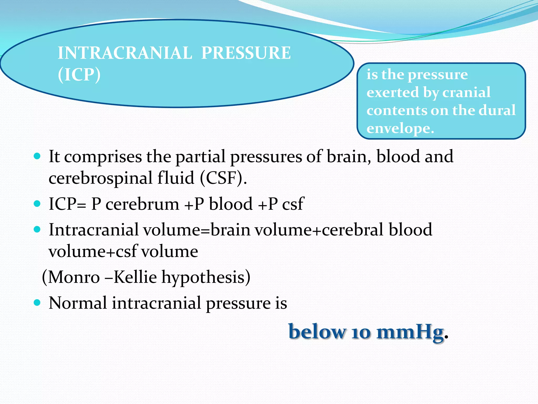
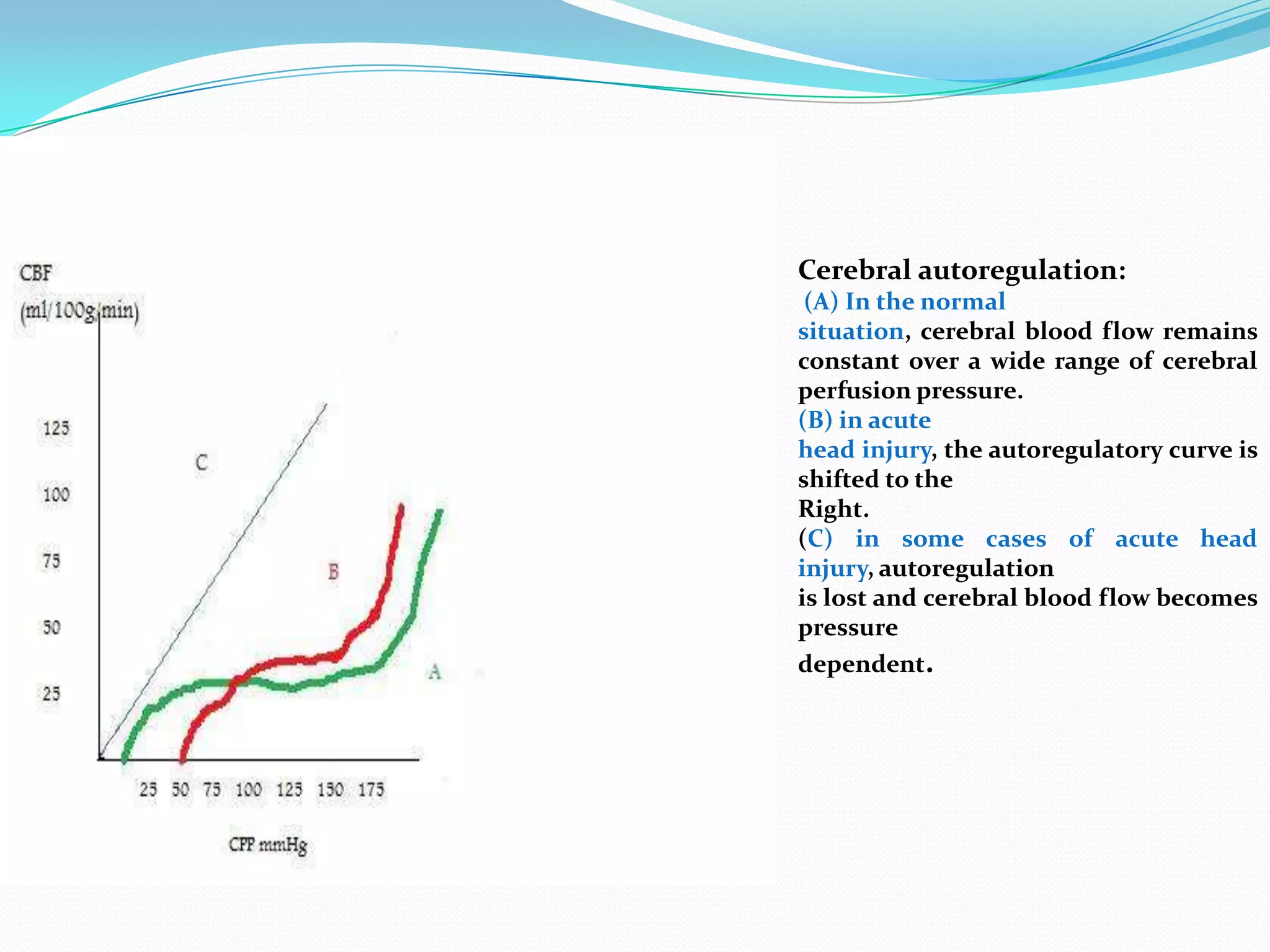
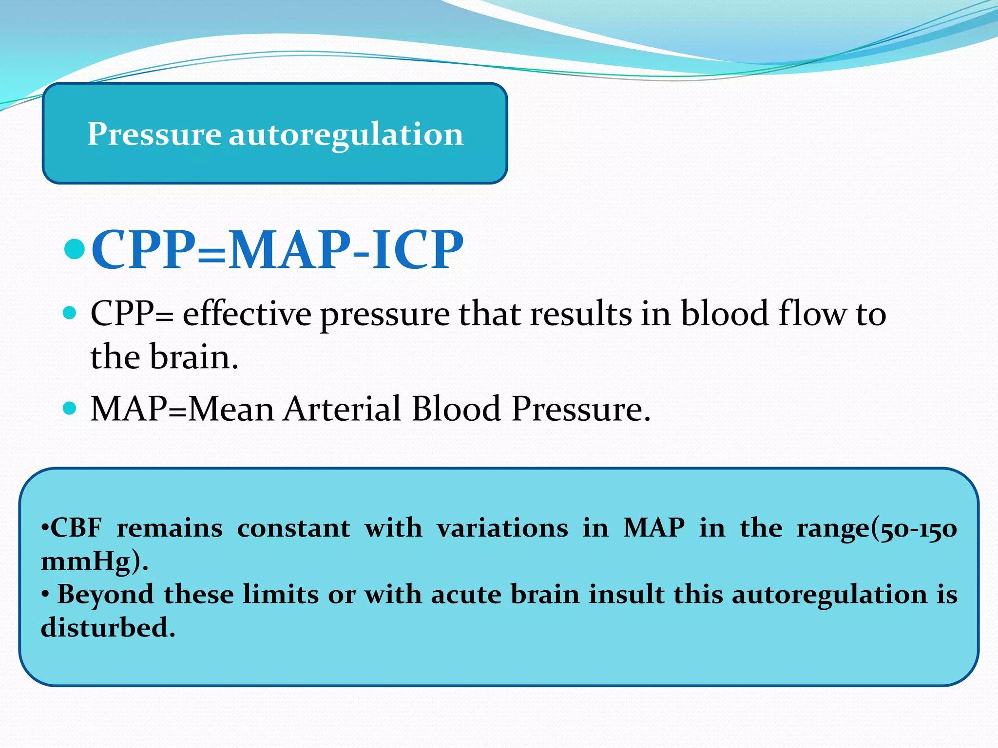
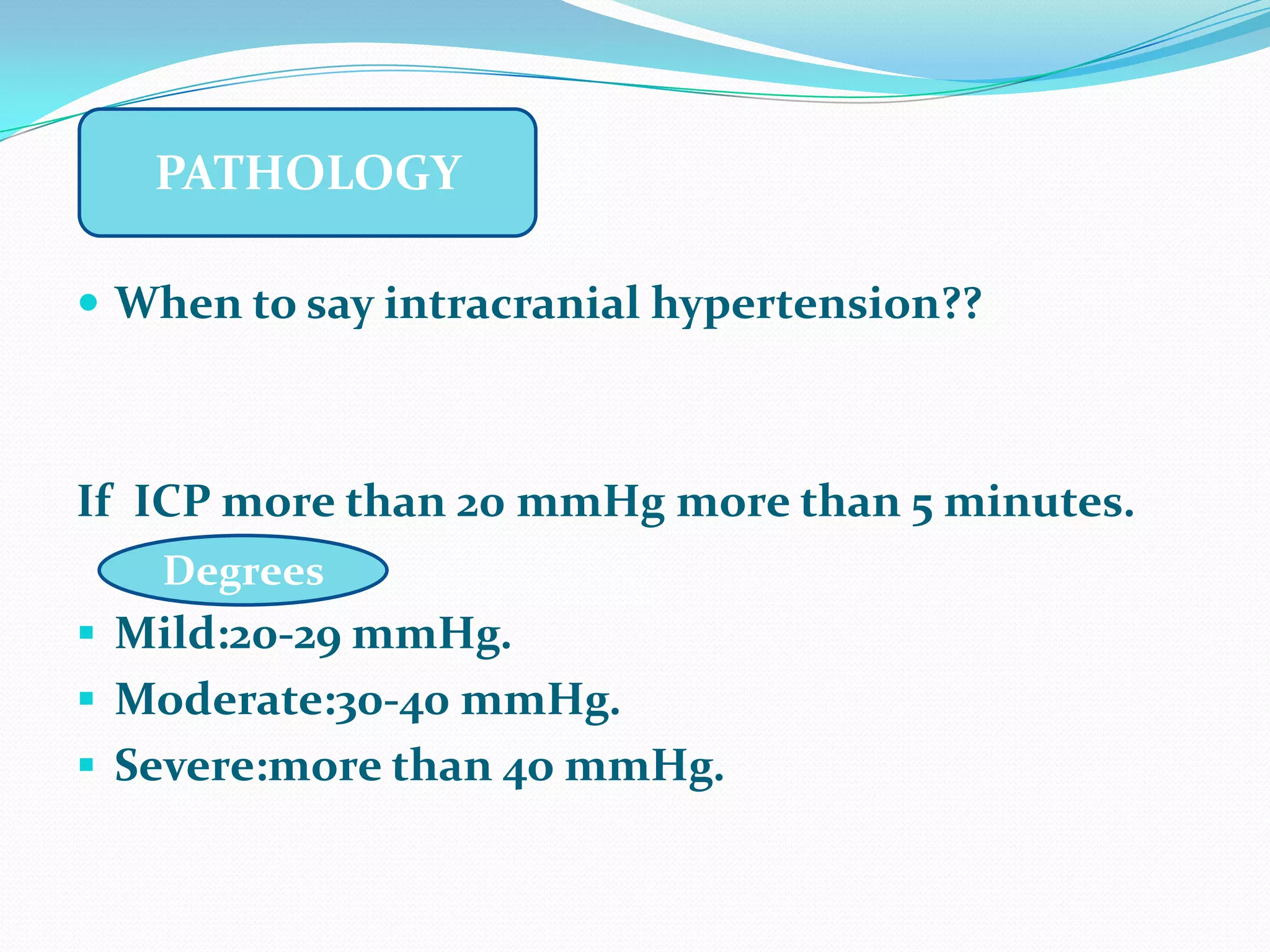
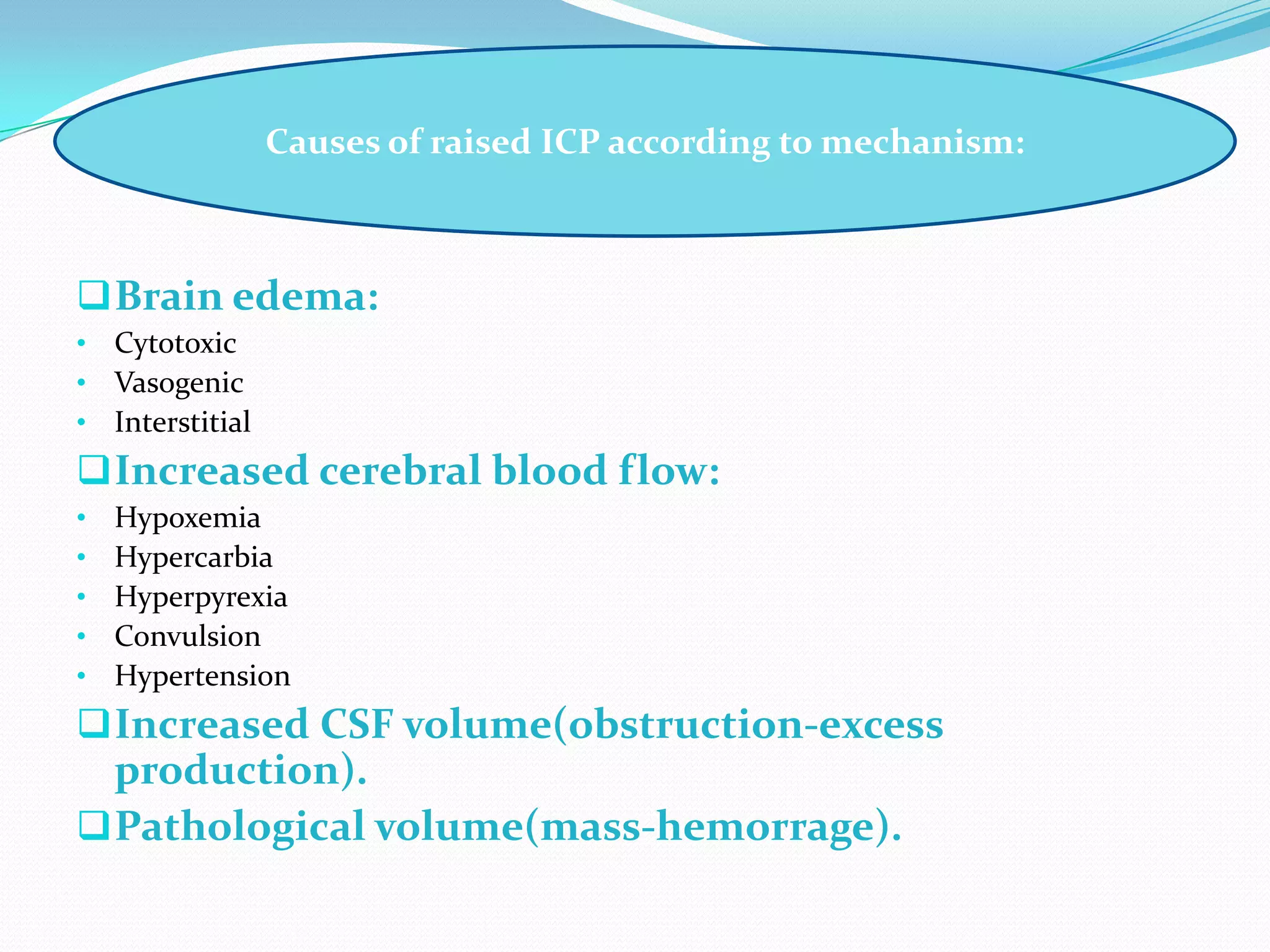
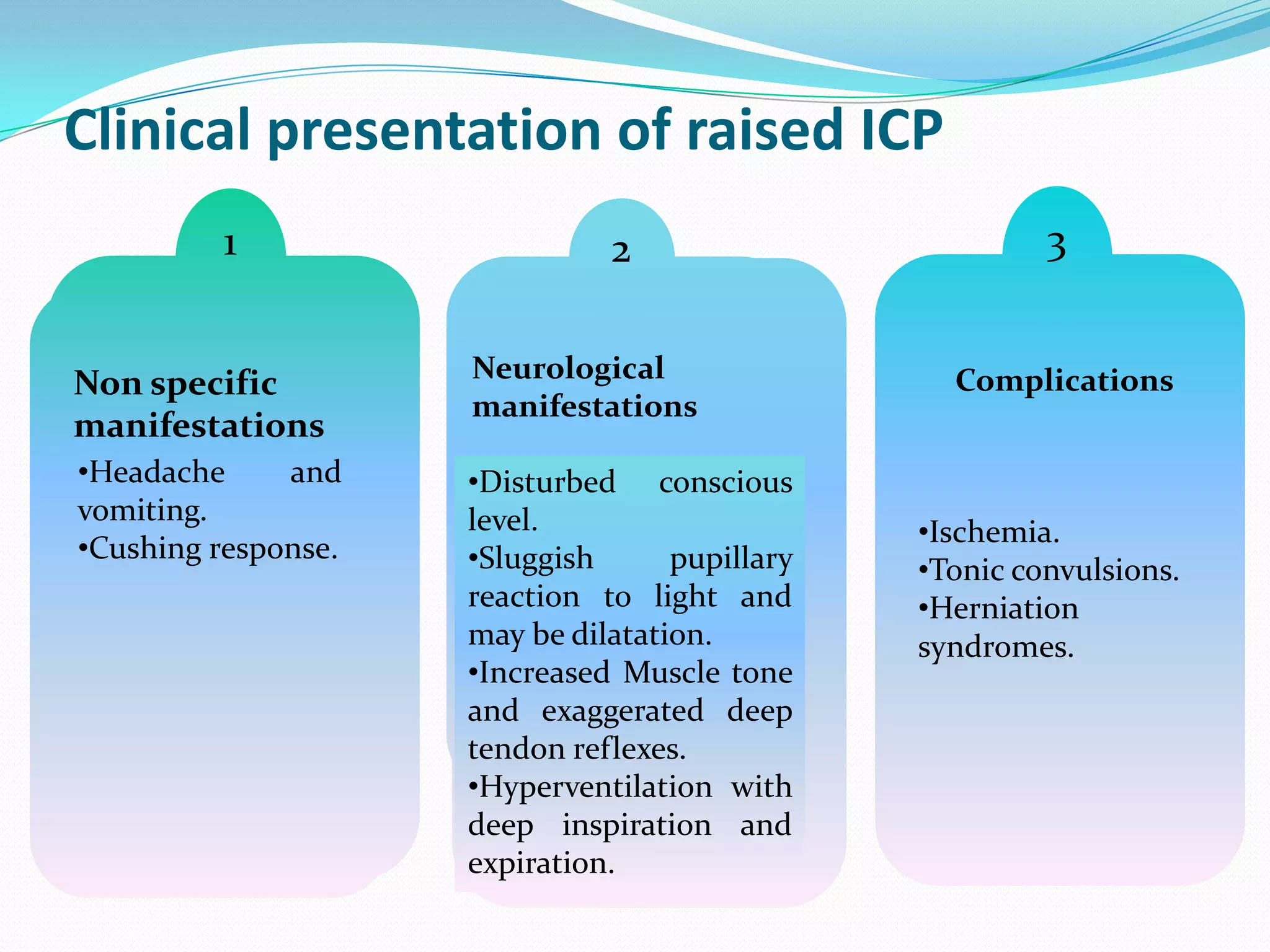
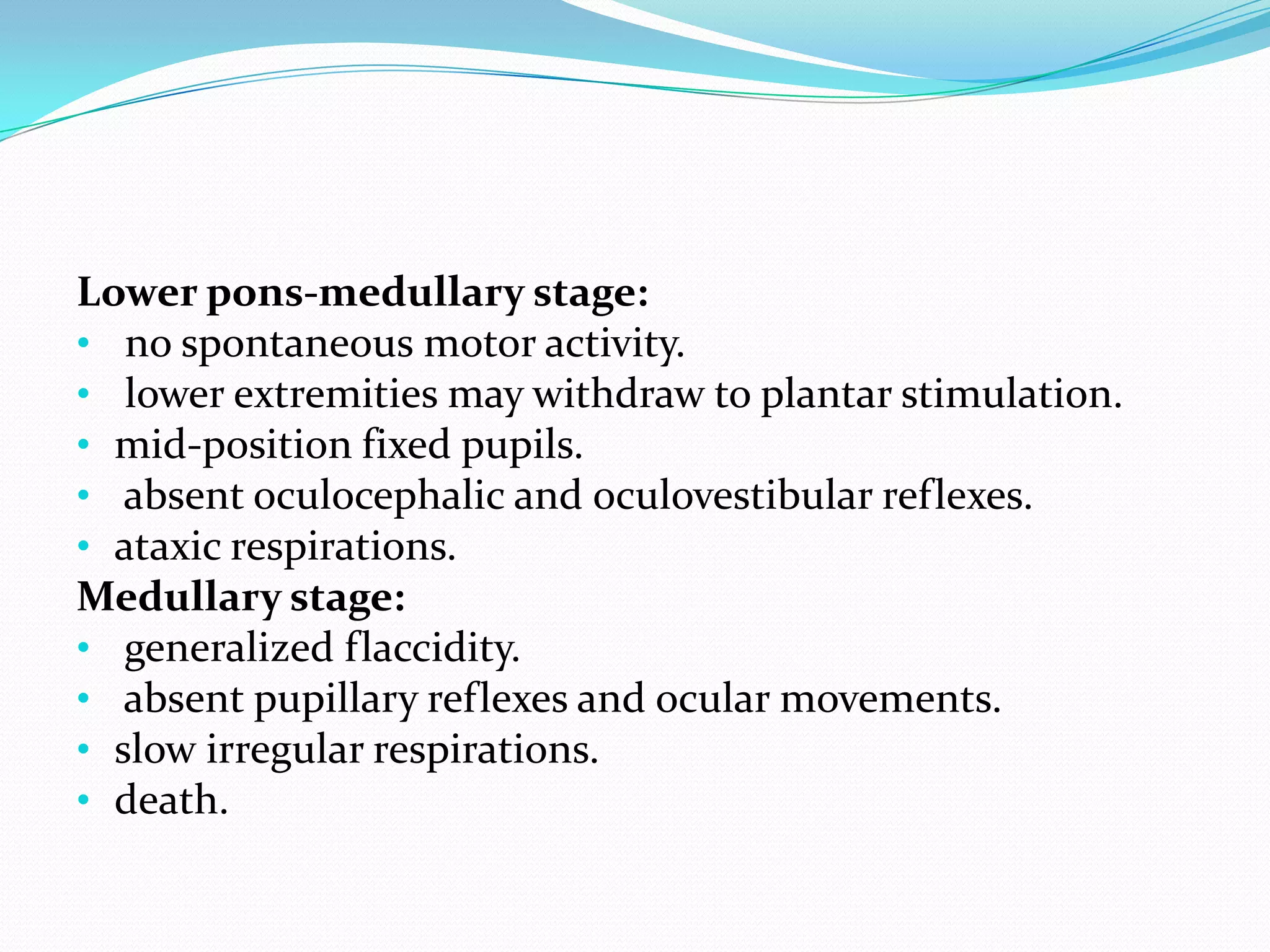
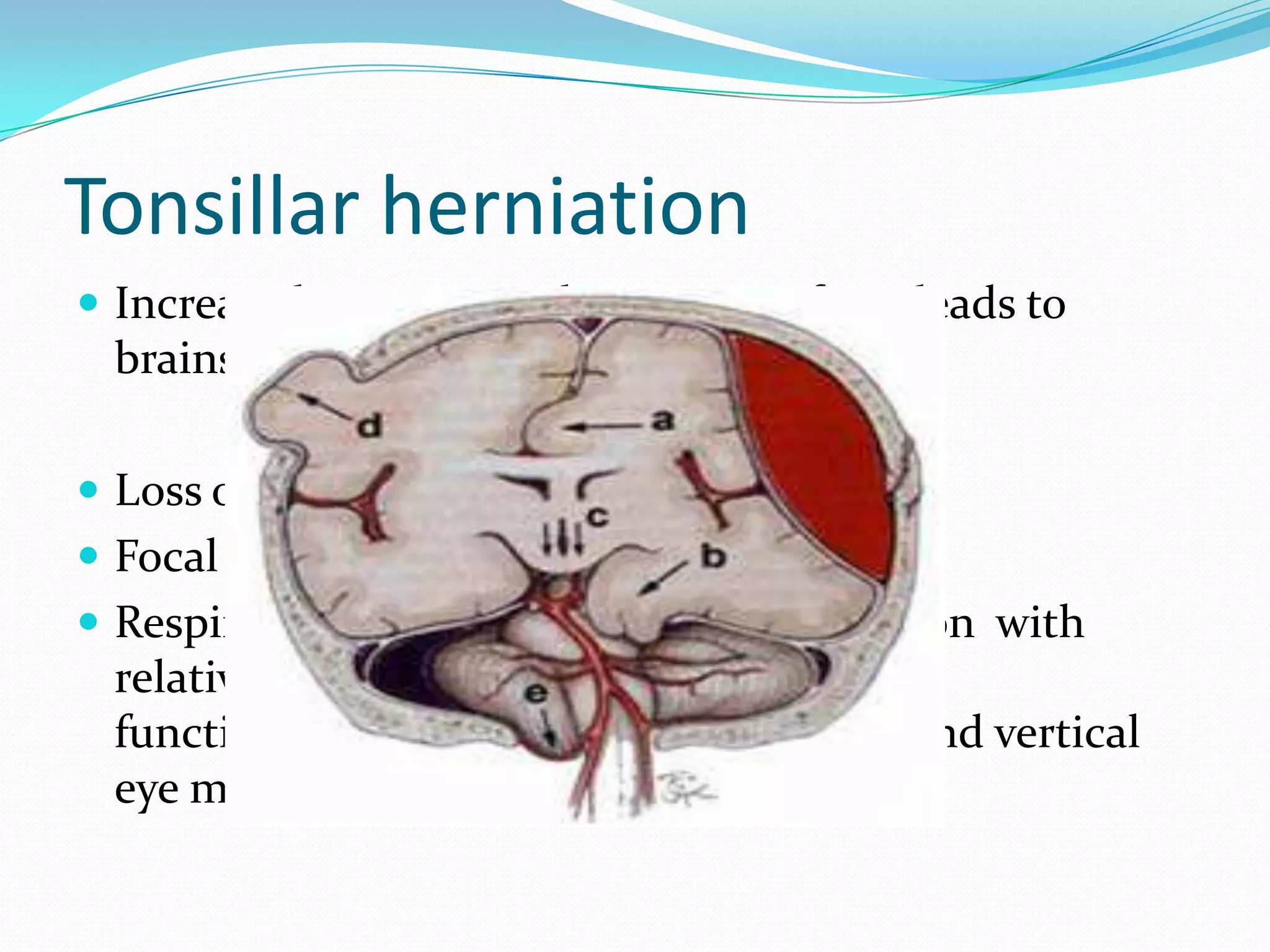
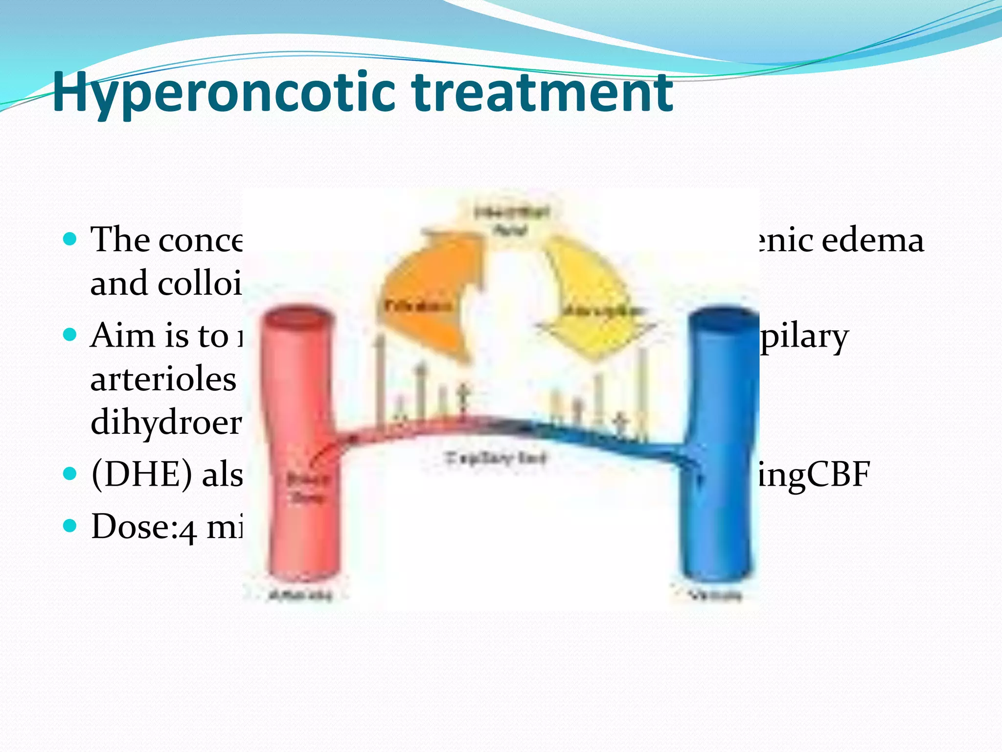
1) The document discusses raised intracranial pressure (ICP), including the definition of ICP, physiology of cerebral circulation, causes of raised ICP, clinical presentation, imaging, and management.
2) Causes of raised ICP include brain edema, increased cerebral blood flow, increased CSF volume, and pathological masses. Clinical manifestations range from non-specific signs to neurological deficits and herniation syndromes.
3) Management involves first-tier approaches like positioning, ventilation, hyperventilation, and hyperosmolar therapy as well as second-tier options of barbiturate coma, hypothermia, and decompressive craniectomy for refractory cases.
Intracerebral hemorhage Diagnosis and managementRamesh Babu
About ICH - Diagnosis and management, Discussed the clinical presentation, evaluation, radiological features and management including recent guidelines
Intracerebral hemorhage Diagnosis and managementRamesh Babu
About ICH - Diagnosis and management, Discussed the clinical presentation, evaluation, radiological features and management including recent guidelines
Management of Increased intracranial pressure in cerebellar strokeNeurology Residency
Brief presentation of medical and surgical treatment options for massive cerebellar stroke. Discussion between placement of an extraventricular drain or suboccipital decompressive craniectomy.
This is a lecture by Ruth S. Hwu, MD from the Ghana Emergency Medicine Collaborative. To download the editable version (in PPT), to access additional learning modules, or to learn more about the project, see http://openmi.ch/em-gemc. Unless otherwise noted, this material is made available under the terms of the Creative Commons Attribution Share Alike-3.0 License: http://creativecommons.org/licenses/by-sa/3.0/.
Defines intracranial pressure, cerebral perfusion pressure and mean arterial pressure. Depict formula for caculating ICP, CPP& MAP. Enumerate both pathological and non- pathological causes for increased ICP. Explain Monroe Kellie hypothesis, pathophysiology of increase Intracranial pressure medical, surgical and nursing management of Increased intracranial pressure.
Raised ICP: What are our option?
- Pathophysiology intracranial hypertension.
- Use Brain Trauma Foundation Guideline (first-tier and second-tier therapy).
- On going research is the effect of TH to decrease ICP.
Stroke is an acute neurologic condition resulting from a disruption in cerebral perfusion, either due to ischemia or hemorrhage.
Hemorrhagic stroke is the cerebral infarction due to hemorrhage.
Title: Sense of Taste
Presenter: Dr. Faiza, Assistant Professor of Physiology
Qualifications:
MBBS (Best Graduate, AIMC Lahore)
FCPS Physiology
ICMT, CHPE, DHPE (STMU)
MPH (GC University, Faisalabad)
MBA (Virtual University of Pakistan)
Learning Objectives:
Describe the structure and function of taste buds.
Describe the relationship between the taste threshold and taste index of common substances.
Explain the chemical basis and signal transduction of taste perception for each type of primary taste sensation.
Recognize different abnormalities of taste perception and their causes.
Key Topics:
Significance of Taste Sensation:
Differentiation between pleasant and harmful food
Influence on behavior
Selection of food based on metabolic needs
Receptors of Taste:
Taste buds on the tongue
Influence of sense of smell, texture of food, and pain stimulation (e.g., by pepper)
Primary and Secondary Taste Sensations:
Primary taste sensations: Sweet, Sour, Salty, Bitter, Umami
Chemical basis and signal transduction mechanisms for each taste
Taste Threshold and Index:
Taste threshold values for Sweet (sucrose), Salty (NaCl), Sour (HCl), and Bitter (Quinine)
Taste index relationship: Inversely proportional to taste threshold
Taste Blindness:
Inability to taste certain substances, particularly thiourea compounds
Example: Phenylthiocarbamide
Structure and Function of Taste Buds:
Composition: Epithelial cells, Sustentacular/Supporting cells, Taste cells, Basal cells
Features: Taste pores, Taste hairs/microvilli, and Taste nerve fibers
Location of Taste Buds:
Found in papillae of the tongue (Fungiform, Circumvallate, Foliate)
Also present on the palate, tonsillar pillars, epiglottis, and proximal esophagus
Mechanism of Taste Stimulation:
Interaction of taste substances with receptors on microvilli
Signal transduction pathways for Umami, Sweet, Bitter, Sour, and Salty tastes
Taste Sensitivity and Adaptation:
Decrease in sensitivity with age
Rapid adaptation of taste sensation
Role of Saliva in Taste:
Dissolution of tastants to reach receptors
Washing away the stimulus
Taste Preferences and Aversions:
Mechanisms behind taste preference and aversion
Influence of receptors and neural pathways
Impact of Sensory Nerve Damage:
Degeneration of taste buds if the sensory nerve fiber is cut
Abnormalities of Taste Detection:
Conditions: Ageusia, Hypogeusia, Dysgeusia (parageusia)
Causes: Nerve damage, neurological disorders, infections, poor oral hygiene, adverse drug effects, deficiencies, aging, tobacco use, altered neurotransmitter levels
Neurotransmitters and Taste Threshold:
Effects of serotonin (5-HT) and norepinephrine (NE) on taste sensitivity
Supertasters:
25% of the population with heightened sensitivity to taste, especially bitterness
Increased number of fungiform papillae
Report Back from SGO 2024: What’s the Latest in Cervical Cancer?bkling
Are you curious about what’s new in cervical cancer research or unsure what the findings mean? Join Dr. Emily Ko, a gynecologic oncologist at Penn Medicine, to learn about the latest updates from the Society of Gynecologic Oncology (SGO) 2024 Annual Meeting on Women’s Cancer. Dr. Ko will discuss what the research presented at the conference means for you and answer your questions about the new developments.
These simplified slides by Dr. Sidra Arshad present an overview of the non-respiratory functions of the respiratory tract.
Learning objectives:
1. Enlist the non-respiratory functions of the respiratory tract
2. Briefly explain how these functions are carried out
3. Discuss the significance of dead space
4. Differentiate between minute ventilation and alveolar ventilation
5. Describe the cough and sneeze reflexes
Study Resources:
1. Chapter 39, Guyton and Hall Textbook of Medical Physiology, 14th edition
2. Chapter 34, Ganong’s Review of Medical Physiology, 26th edition
3. Chapter 17, Human Physiology by Lauralee Sherwood, 9th edition
4. Non-respiratory functions of the lungs https://academic.oup.com/bjaed/article/13/3/98/278874
Recomendações da OMS sobre cuidados maternos e neonatais para uma experiência pós-natal positiva.
Em consonância com os ODS – Objetivos do Desenvolvimento Sustentável e a Estratégia Global para a Saúde das Mulheres, Crianças e Adolescentes, e aplicando uma abordagem baseada nos direitos humanos, os esforços de cuidados pós-natais devem expandir-se para além da cobertura e da simples sobrevivência, de modo a incluir cuidados de qualidade.
Estas diretrizes visam melhorar a qualidade dos cuidados pós-natais essenciais e de rotina prestados às mulheres e aos recém-nascidos, com o objetivo final de melhorar a saúde e o bem-estar materno e neonatal.
Uma “experiência pós-natal positiva” é um resultado importante para todas as mulheres que dão à luz e para os seus recém-nascidos, estabelecendo as bases para a melhoria da saúde e do bem-estar a curto e longo prazo. Uma experiência pós-natal positiva é definida como aquela em que as mulheres, pessoas que gestam, os recém-nascidos, os casais, os pais, os cuidadores e as famílias recebem informação consistente, garantia e apoio de profissionais de saúde motivados; e onde um sistema de saúde flexível e com recursos reconheça as necessidades das mulheres e dos bebês e respeite o seu contexto cultural.
Estas diretrizes consolidadas apresentam algumas recomendações novas e já bem fundamentadas sobre cuidados pós-natais de rotina para mulheres e neonatos que recebem cuidados no pós-parto em unidades de saúde ou na comunidade, independentemente dos recursos disponíveis.
É fornecido um conjunto abrangente de recomendações para cuidados durante o período puerperal, com ênfase nos cuidados essenciais que todas as mulheres e recém-nascidos devem receber, e com a devida atenção à qualidade dos cuidados; isto é, a entrega e a experiência do cuidado recebido. Estas diretrizes atualizam e ampliam as recomendações da OMS de 2014 sobre cuidados pós-natais da mãe e do recém-nascido e complementam as atuais diretrizes da OMS sobre a gestão de complicações pós-natais.
O estabelecimento da amamentação e o manejo das principais intercorrências é contemplada.
Recomendamos muito.
Vamos discutir essas recomendações no nosso curso de pós-graduação em Aleitamento no Instituto Ciclos.
Esta publicação só está disponível em inglês até o momento.
Prof. Marcus Renato de Carvalho
www.agostodourado.com
Title: Sense of Smell
Presenter: Dr. Faiza, Assistant Professor of Physiology
Qualifications:
MBBS (Best Graduate, AIMC Lahore)
FCPS Physiology
ICMT, CHPE, DHPE (STMU)
MPH (GC University, Faisalabad)
MBA (Virtual University of Pakistan)
Learning Objectives:
Describe the primary categories of smells and the concept of odor blindness.
Explain the structure and location of the olfactory membrane and mucosa, including the types and roles of cells involved in olfaction.
Describe the pathway and mechanisms of olfactory signal transmission from the olfactory receptors to the brain.
Illustrate the biochemical cascade triggered by odorant binding to olfactory receptors, including the role of G-proteins and second messengers in generating an action potential.
Identify different types of olfactory disorders such as anosmia, hyposmia, hyperosmia, and dysosmia, including their potential causes.
Key Topics:
Olfactory Genes:
3% of the human genome accounts for olfactory genes.
400 genes for odorant receptors.
Olfactory Membrane:
Located in the superior part of the nasal cavity.
Medially: Folds downward along the superior septum.
Laterally: Folds over the superior turbinate and upper surface of the middle turbinate.
Total surface area: 5-10 square centimeters.
Olfactory Mucosa:
Olfactory Cells: Bipolar nerve cells derived from the CNS (100 million), with 4-25 olfactory cilia per cell.
Sustentacular Cells: Produce mucus and maintain ionic and molecular environment.
Basal Cells: Replace worn-out olfactory cells with an average lifespan of 1-2 months.
Bowman’s Gland: Secretes mucus.
Stimulation of Olfactory Cells:
Odorant dissolves in mucus and attaches to receptors on olfactory cilia.
Involves a cascade effect through G-proteins and second messengers, leading to depolarization and action potential generation in the olfactory nerve.
Quality of a Good Odorant:
Small (3-20 Carbon atoms), volatile, water-soluble, and lipid-soluble.
Facilitated by odorant-binding proteins in mucus.
Membrane Potential and Action Potential:
Resting membrane potential: -55mV.
Action potential frequency in the olfactory nerve increases with odorant strength.
Adaptation Towards the Sense of Smell:
Rapid adaptation within the first second, with further slow adaptation.
Psychological adaptation greater than receptor adaptation, involving feedback inhibition from the central nervous system.
Primary Sensations of Smell:
Camphoraceous, Musky, Floral, Pepperminty, Ethereal, Pungent, Putrid.
Odor Detection Threshold:
Examples: Hydrogen sulfide (0.0005 ppm), Methyl-mercaptan (0.002 ppm).
Some toxic substances are odorless at lethal concentrations.
Characteristics of Smell:
Odor blindness for single substances due to lack of appropriate receptor protein.
Behavioral and emotional influences of smell.
Transmission of Olfactory Signals:
From olfactory cells to glomeruli in the olfactory bulb, involving lateral inhibition.
Primitive, less old, and new olfactory systems with different path
Knee anatomy and clinical tests 2024.pdfvimalpl1234
This includes all relevant anatomy and clinical tests compiled from standard textbooks, Campbell,netter etc..It is comprehensive and best suited for orthopaedicians and orthopaedic residents.
New Drug Discovery and Development .....NEHA GUPTA
The "New Drug Discovery and Development" process involves the identification, design, testing, and manufacturing of novel pharmaceutical compounds with the aim of introducing new and improved treatments for various medical conditions. This comprehensive endeavor encompasses various stages, including target identification, preclinical studies, clinical trials, regulatory approval, and post-market surveillance. It involves multidisciplinary collaboration among scientists, researchers, clinicians, regulatory experts, and pharmaceutical companies to bring innovative therapies to market and address unmet medical needs.
ARTIFICIAL INTELLIGENCE IN HEALTHCARE.pdfAnujkumaranit
Artificial intelligence (AI) refers to the simulation of human intelligence processes by machines, especially computer systems. It encompasses tasks such as learning, reasoning, problem-solving, perception, and language understanding. AI technologies are revolutionizing various fields, from healthcare to finance, by enabling machines to perform tasks that typically require human intelligence.
3. OUTLINES:
Definition of intracranial pressure(ICP).
Physiology of cerebral circulation.
Causes of raised ICP.
Clinical presentation of raised ICP.
Measurement of ICP.
Management of raised ICP.
4. INTRACRANIAL PRESSURE
(ICP)
is the pressure
exerted by cranial
contents on the dural
envelope.
It comprises the partial pressures of brain, blood and
cerebrospinal fluid (CSF).
ICP= P cerebrum +P blood +P csf
Intracranial volume=brain volume+cerebral blood
volume+csf volume
(Monro –Kellie hypothesis)
Normal intracranial pressure is
below 10 mmHg.
5. Compliance:
is the ability to tolerate an increase
in volume without a corresponding
increase in Pressure.
Relationship of intracranial volume to intracranial pressure
(ICP). At normal brain compliance ICP increases only slightly as
intracranial volume increases . A further increase in volume
results in a steep rise in ICP.
6. Cerebral autoregulation:
(A) In the normal
situation, cerebral blood flow remains
constant over a wide range of cerebral
perfusion pressure.
(B) in acute
head injury, the autoregulatory curve is
shifted to the
Right.
(C) in some cases of acute head
injury, autoregulation
is lost and cerebral blood flow becomes
pressure
dependent.
8. Pressure autoregulation
CPP=MAP-ICP
CPP= effective pressure that results in blood flow to
the brain.
MAP=Mean Arterial Blood Pressure.
•CBF remains constant with variations in MAP in the range(50-150
mmHg).
• Beyond these limits or with acute brain insult this autoregulation is
disturbed.
10. Metabolic autoregulation
CBF is sensitive to changes in PaCO2 and PaO2.
Metabolic autoregulation is resistant to acute brain injury.
Hypoventilation--
increase PaCO2-increase CBF-- increase ICP.
Hyperventilation-- reduce PaCO2-- reduce CBF
-- decrease ICP.
Arterial hypoxemia--- increase in CBF and ICP.
Increase in PaO2-- cerebral vasoconstriction.
11. PATHOLOGY
When to say intracranial hypertension??
If ICP more than 20 mmHg more than 5 minutes.
Degrees
Mild:20-29 mmHg.
Moderate:30-40 mmHg.
Severe:more than 4o mmHg.
14. Clinical manifestations of increased ICP
Non specific
manifestations
Neurological
manifestations
Complications
15. Clinical presentation of raised ICP
1
2
Non specific
manifestations
Neurological
manifestations
•Headache
and
vomiting.
•Cushing response.
•Disturbed conscious
level.
•Sluggish
pupillary
reaction to light and
may be dilatation.
•Increased Muscle tone
and exaggerated deep
tendon reflexes.
•Hyperventilation with
deep inspiration and
expiration.
3
Complications
•Ischemia.
•Tonic convulsions.
•Herniation
syndromes.
18. Central herniation
Increased pressure in both cerebral hemispheres→downward
displacement of the diencephalon through the tentorium →
brainstem compression.
Stages:
Diencephalic stage:
withdraws to noxious stimuli, increased rigidity or decorticate posturing.
small, reactive pupils.
preserved oculocephalic and oculovestibular reflexes.
yawns, sighs, or Cheyne-Stokes breathing.
Midbrain-upper pons stage:
•
•
•
•
decerebrate posturing or no movement.
mid-position pupils that may become irregular and unreactive.
abnormal or absent oculocephalic and oculovestibular reflexes.
hyperventilation.
19. Lower pons-medullary stage:
• no spontaneous motor activity.
• lower extremities may withdraw to plantar stimulation.
• mid-position fixed pupils.
• absent oculocephalic and oculovestibular reflexes.
• ataxic respirations.
Medullary stage:
• generalized flaccidity.
• absent pupillary reflexes and ocular movements.
• slow irregular respirations.
• death.
20. Uncal herniation
Uncus of the temporal lobe is displaced medially over
the free edge of the tentorium.
Ipsilateral third-nerve palsy (ptosis, pupil fixed and
dilated, eye deviated down and out).
Ipsilateral hemiparesis from compression of the
contralateral cerebral peduncle (Kernohan notch).
posterior cerebral artery compression.
21. Subfalcine (Cingulate) herniation
Increased pressure in one cerebral hemisphere leads to
herniation of cingulated gyrus underneath falx cerebri.
Compression of anterior cerebral artery leads to
paraparesis.
22. Tonsillar herniation
Increased pressure in the posterior fossa leads to
brainstem compression.
Loss of consciousness (RAS).
Focal lower cranial nerve dysfunction.
Respiratory and cardiovascular dysfunction with
relative preservation of upper brainstem
function, such as pupillary light reflexes and vertical
eye movements.
23. a) Subfalcial (cingulate) herniation ;
b) uncal herniation
c) downward (central, transtentorial) herniation
d) external herniation
e) tonsillar herniation.
26. IMAGING
CT BRAIN
sensitive in detecting extrabrain pathology.
Exclude secondary cause e.g tumor-mass.
Brain edema.
HOWEVER
Inferior resolution compared with MRI.
Missed pathology such as small bleeds.
27. Diffuse Injury Type I
no CT visible intracranial pathology
Diffuse Injury Type II
cisterns present with midline shift 0-5 mm
no high- or mixed-density lesion > 25 cc
Diffuse Injury Type III(swelling)
cisterns compressed or absent with midline shift 0-5 mm
no high- or mixed-density lesion > 25 cc
Diffuse Injury Type IV (shift)
midline shift > 5 mm
no high- or mixed-density lesion > 25 cc
Evacuated Mass Lesion
any lesion surgically evacuated
Non-Evacuated Mass Lesion
high- or mixed-density lesion > 25 cc
29. MRI
More sensitive in detecting nonhemorrhagic and brain
stem lesions.
Detection of diffuse axonal injury.
Distinguishing subdural blood from subdural
hygromas and pathologic collections of CSF.
PET -SPECT
NIRS
TRANSCRANIAL DOPPLER
34. Management of raised ICP
Decompressive
craniectomy
Forced
hyperventilation
Barbiturate coma
Tris buffer
Hypothermia
Positioning
Hyperventilation
Hyperosmolar therapy
Induced
hypertension
arterial
35. First tire
Positioning:
Moderate head elevation at an angle 15-30.
This enhances cerebral venous return
decreases ICP.
Higher angle will decrease cerebral blood flow.
Neutral position as turning head to one side will
reduce cerebral venous return.
36. Adequate ventilatory support:
Evidence based medicine has not identified
perfect ventilation but adapted to your patient.
Avoid fighting of the patient with ventilator.
Effect of PEEP on MAP should be taken in
consideration.
Hyperventilation.
37. Hyperventilation:
It is a rescue maneuver in impending or frank
herniation.
Keep PaCo2 between 30-35 mmHg.
Lowering paCO2 by 1 mmHg reduces CBF by 4%.
38. CPP=MAP-ICP
CPP Concept:
In case of intra-Cranial hypertension,autoregulation is
triggered by pharmacologically increasing MAP.
Drugs of choice are norepinephrine(0.1 mic/kg/min)or
dopamine(4–10 mic/kg/min).
Goal in TBI is MAP level of >70mmHg.
43. Hypertonic saline
It can be given till serum osmolarity 36o mosmol.
It acts also as volume expander.
It maintains MAP.
44. Second-tier treatment
Forced hyperventilation:
Hyperventilation down to a paCO2 of 25-30mmHg
Carries the risk of cerebral ischemia.
Barbiturate coma:
Suppress the cerebral metabolic rate for oxygen(CMRO2).
Terminate convulsions.
Scavenge free oxygen radicals.
Decrease a cerebral hyperthermic response to ischemia.
45. Hypothermia
Tris buffer:
Acidosis removes the Mg lock from the NMDA receptor—
facilitating excitotoxicity.
High pH protects energy-dependent glutamate porter
sytems and thus delays onset of excitotoxicity.
Tris buffer corrects intracellular acidosis and increases the
buffering capacity of CSF.
Tham(trishydroxymethylaminomethane) (1 mg/kg
intravenously)
46. Steroids:
• in vasogenic brain edema around brain
tumors,hematoma or postoperative.
• Dexamethasone .25 mg/kg every 6 h IV.
• Methyl predinsolone 1-2 mg/kg every 6 h
48. Hyperoncotic treatment
The concept assumes importance of vasogenic edema
and colloid-osmotic pressure
Aim is to reduce capillary pressure----precapilary
arterioles are constricted with
dihydroergotamine(DHE)
(DHE) also constrict cerebral veins---reducingCBF
Dose:4 mic/kg intravenously.
49. Hepatic Encephalopathy
Hyperammoniemia is the key via
Glutamine accumulation .
free radicle release.
Osmolar therapy:Mannitol-HS
Hyperventilation
Maitainance of MAP.
Ornithine-L-Aspartate.
Hypothermia.
51. Hypoxic–Ischemic Brain Injury
Hypothermia is strongly tried especially in neonates.
Cytogenic brain edema.
Reperfusion injury----Vasogenic brain edema.
52.
53. Infectious Causes
Types of brain edema.
What is the role of fluid restriction??
Lumbar puncture.To do OR not to do??
What is the role of steroids??
56. Always remember:
Patient with raised ICP is critically ill patient.
So
Don not forget A,B,C,D.
Papilledema is a late sign,
And
Pupilary dilatation deprive you
from precious sign
57. Always remember:
Whatever the cause of raised ICP the initial
management will be the same.
Don not rush to lumbar puncture or even CT brain
until you stabilize your patient.
CT brain is important to diagnose some secondary
causes of raised ICP.
Monitoring of ICP is valuable esp. in cases of TBI.