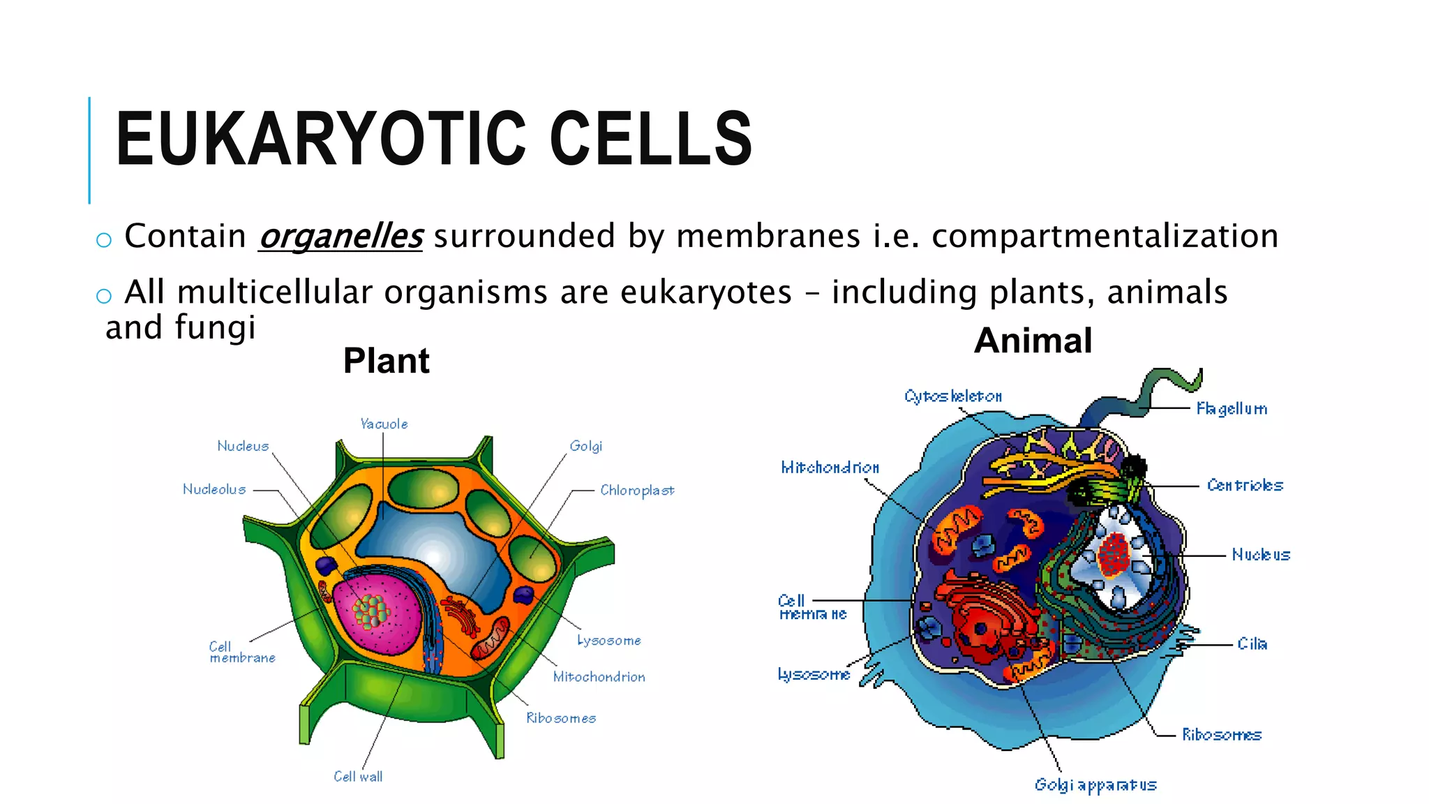
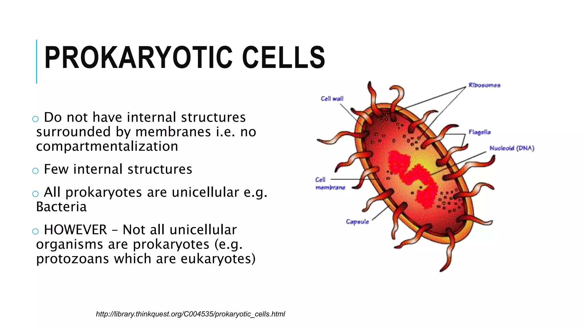
- Prokaryotic cells have a simple structure without internal compartments, while eukaryotic cells have organelles surrounded by membranes.
- The two main types of cells are prokaryotic and eukaryotic. Prokaryotic cells are smaller and simpler and were the first life forms on Earth. They lack internal membranes.
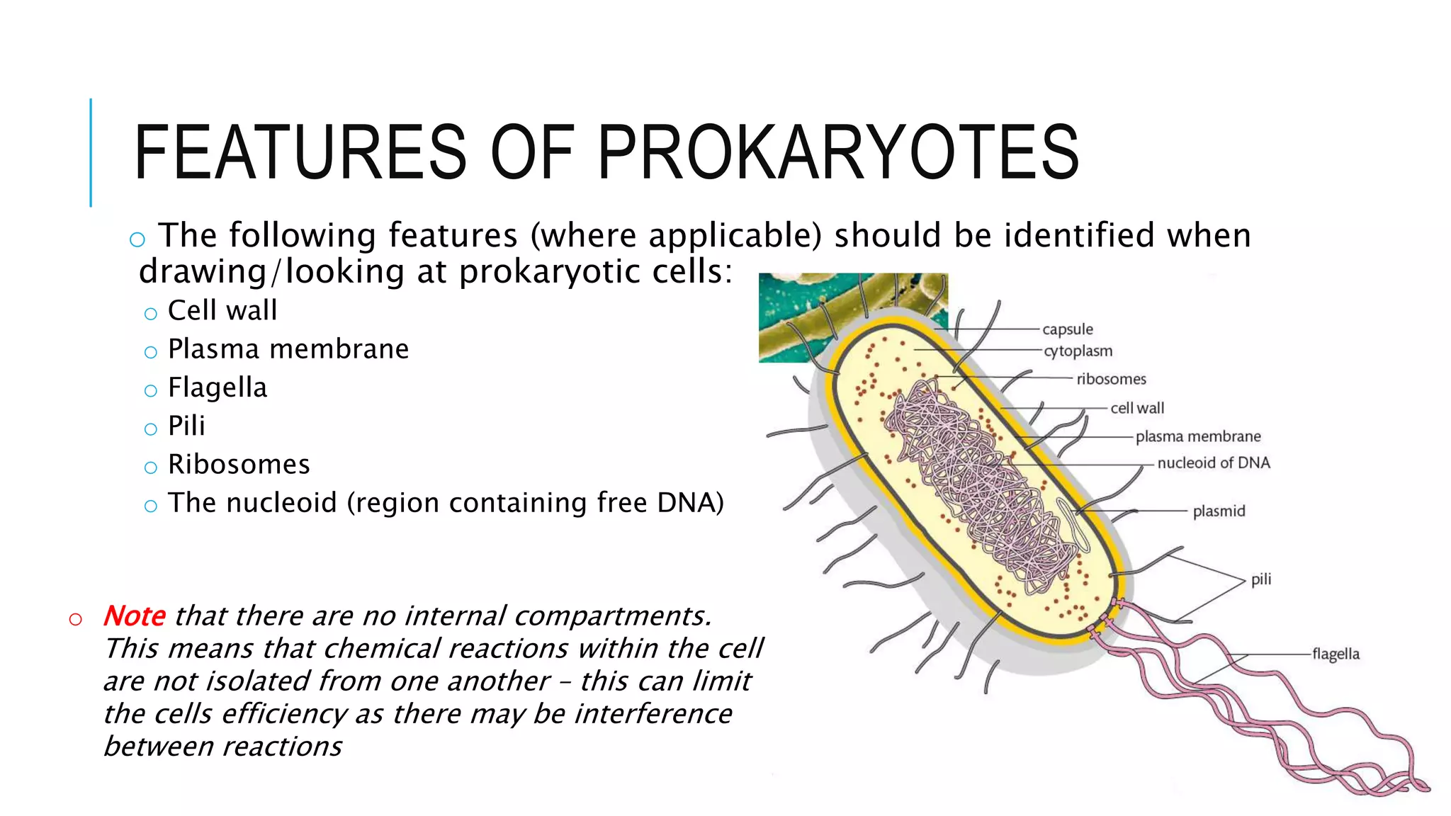
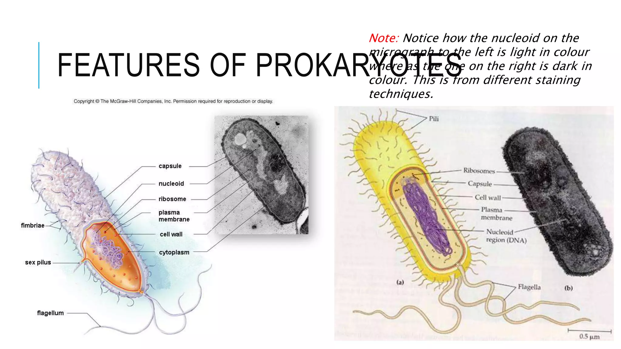
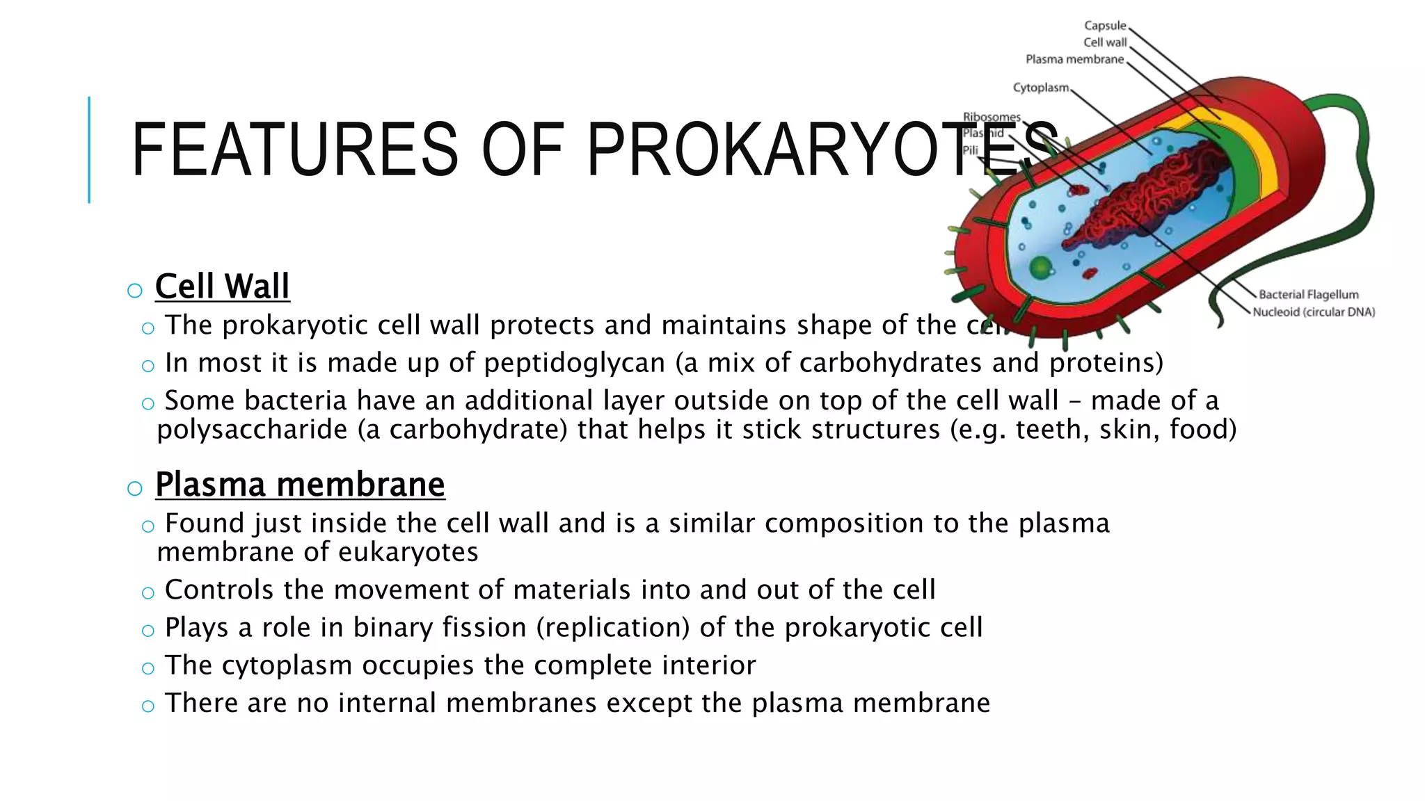
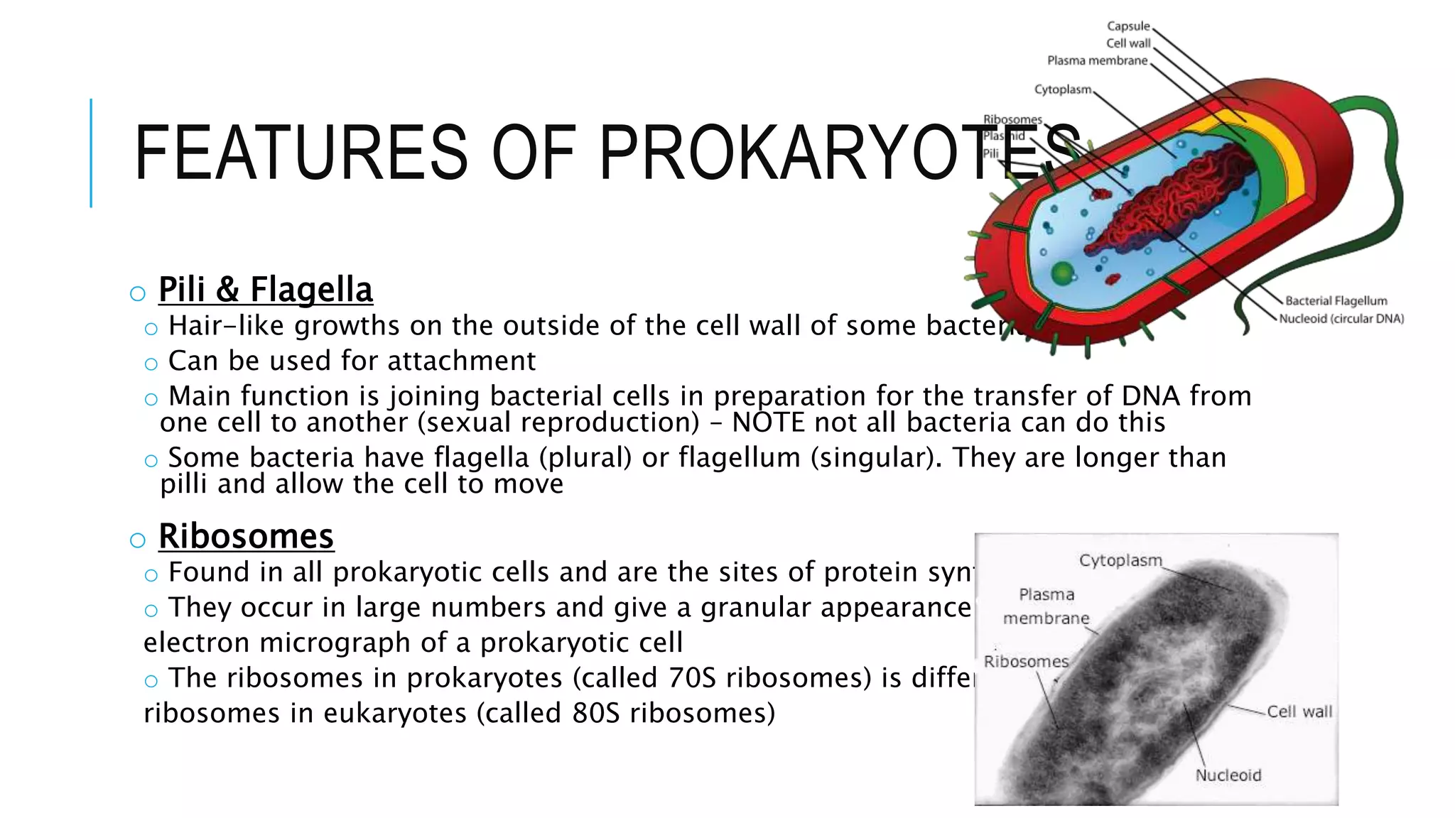
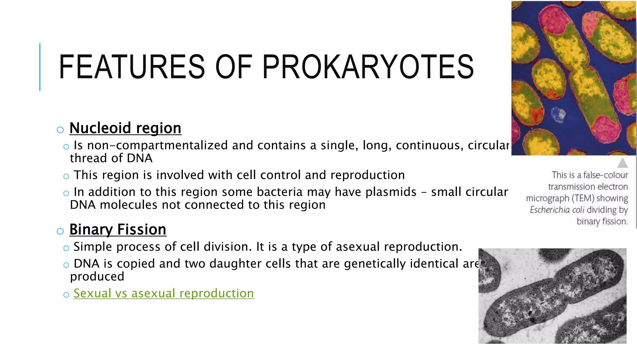
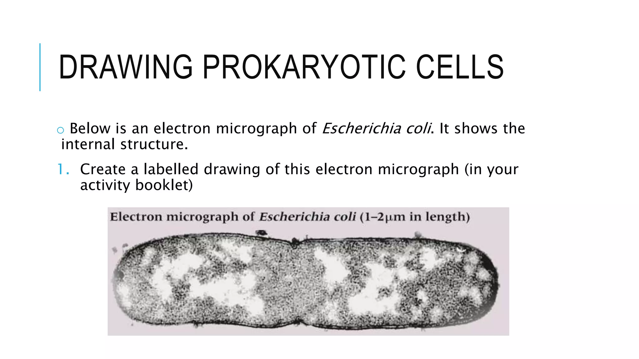
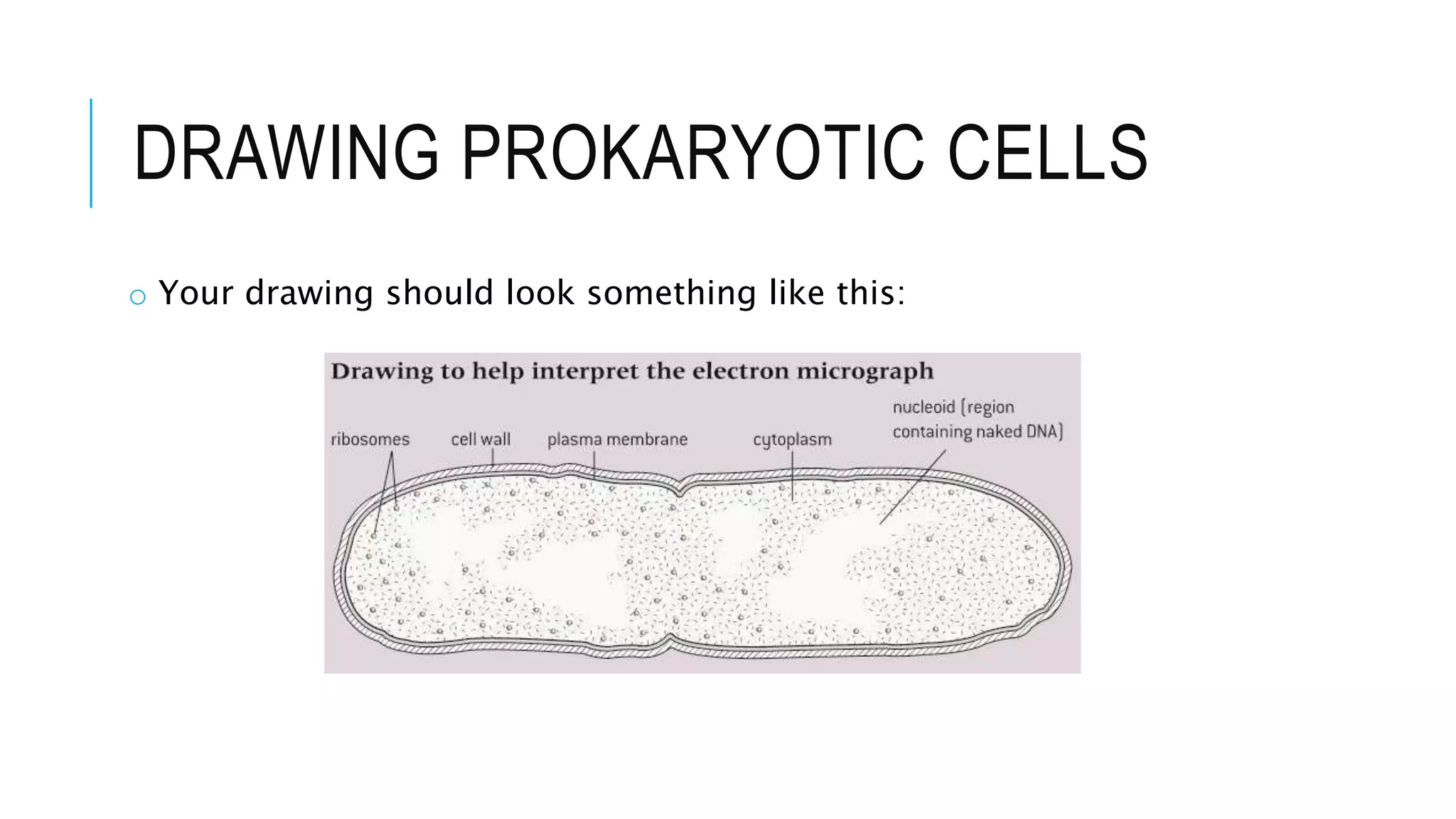
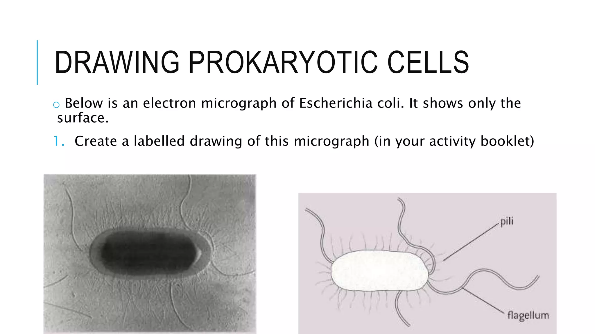
- Key structures of prokaryotic cells include the cell wall, plasma membrane, ribosomes, and nucleoid region containing DNA. Some bacteria also have flagella, pili, or plasmids. They divide through binary fission.