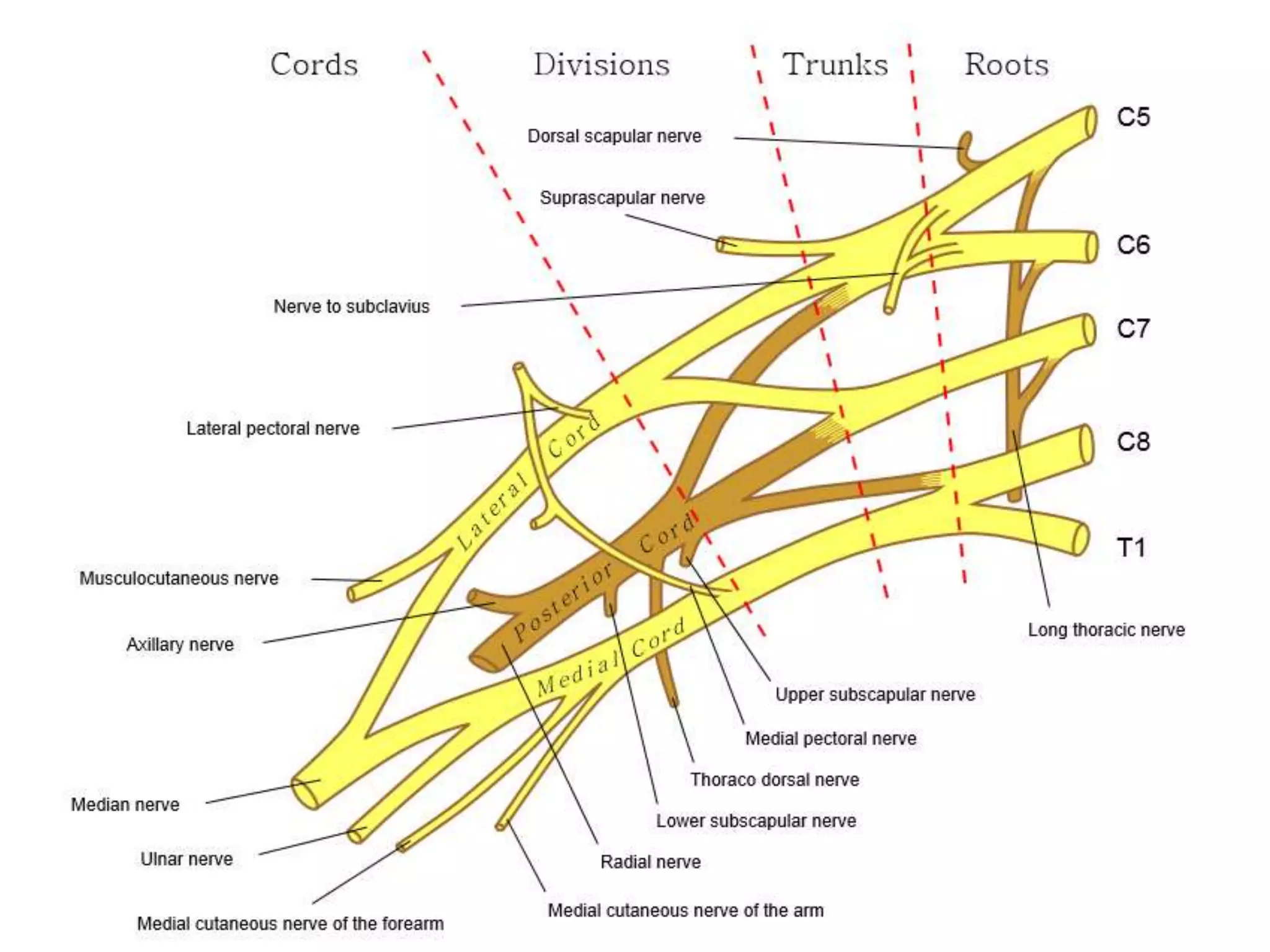
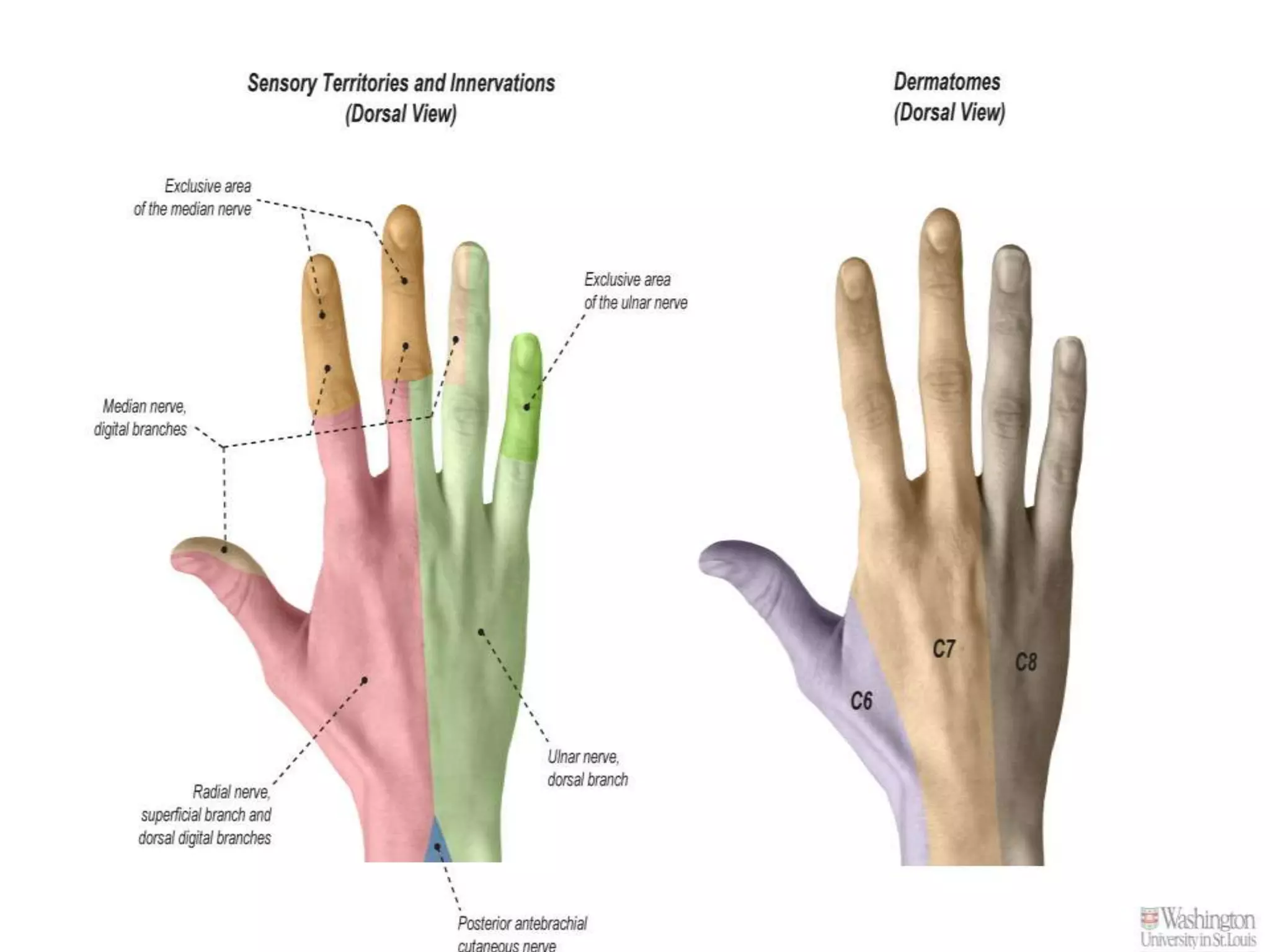
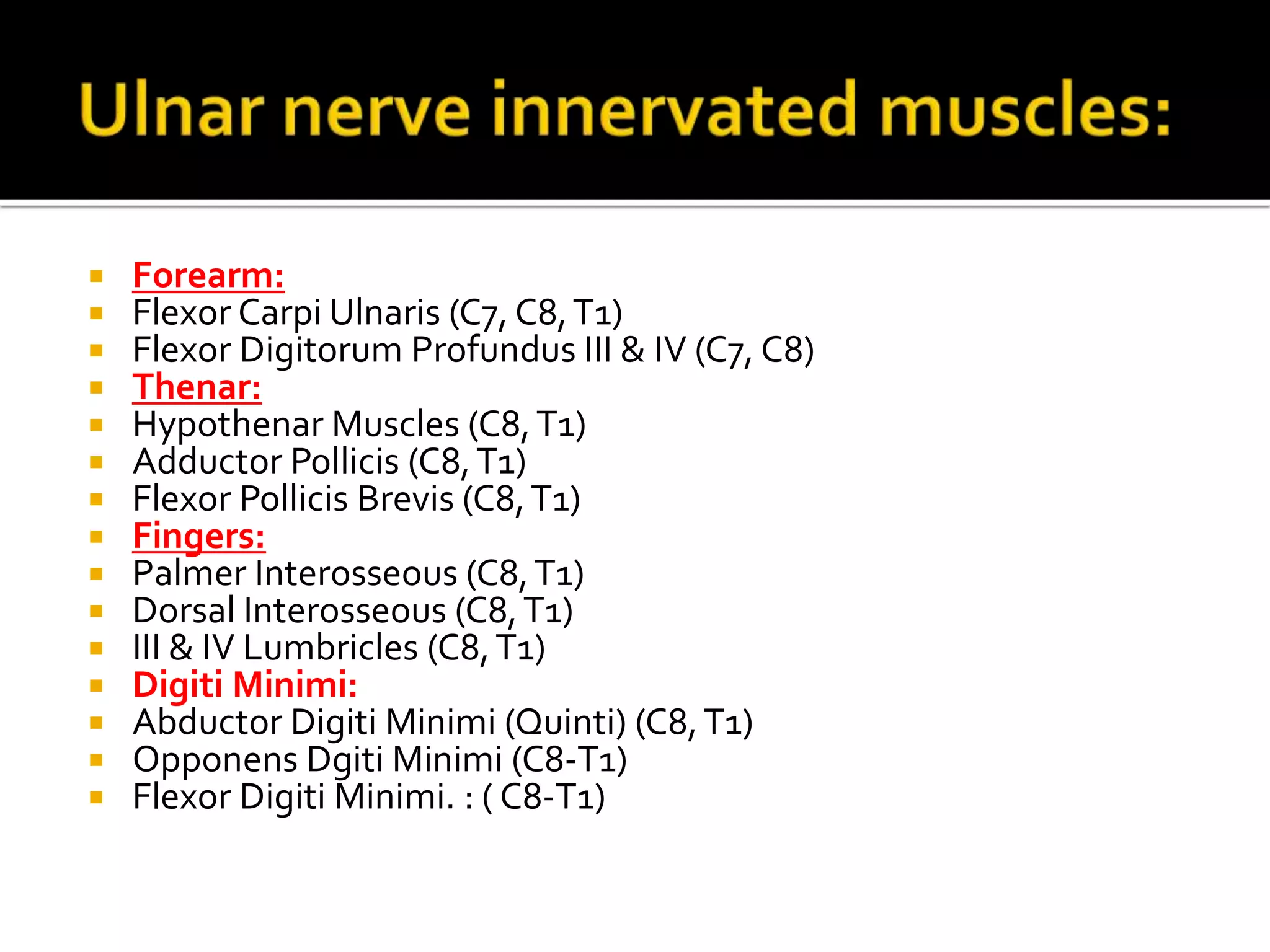
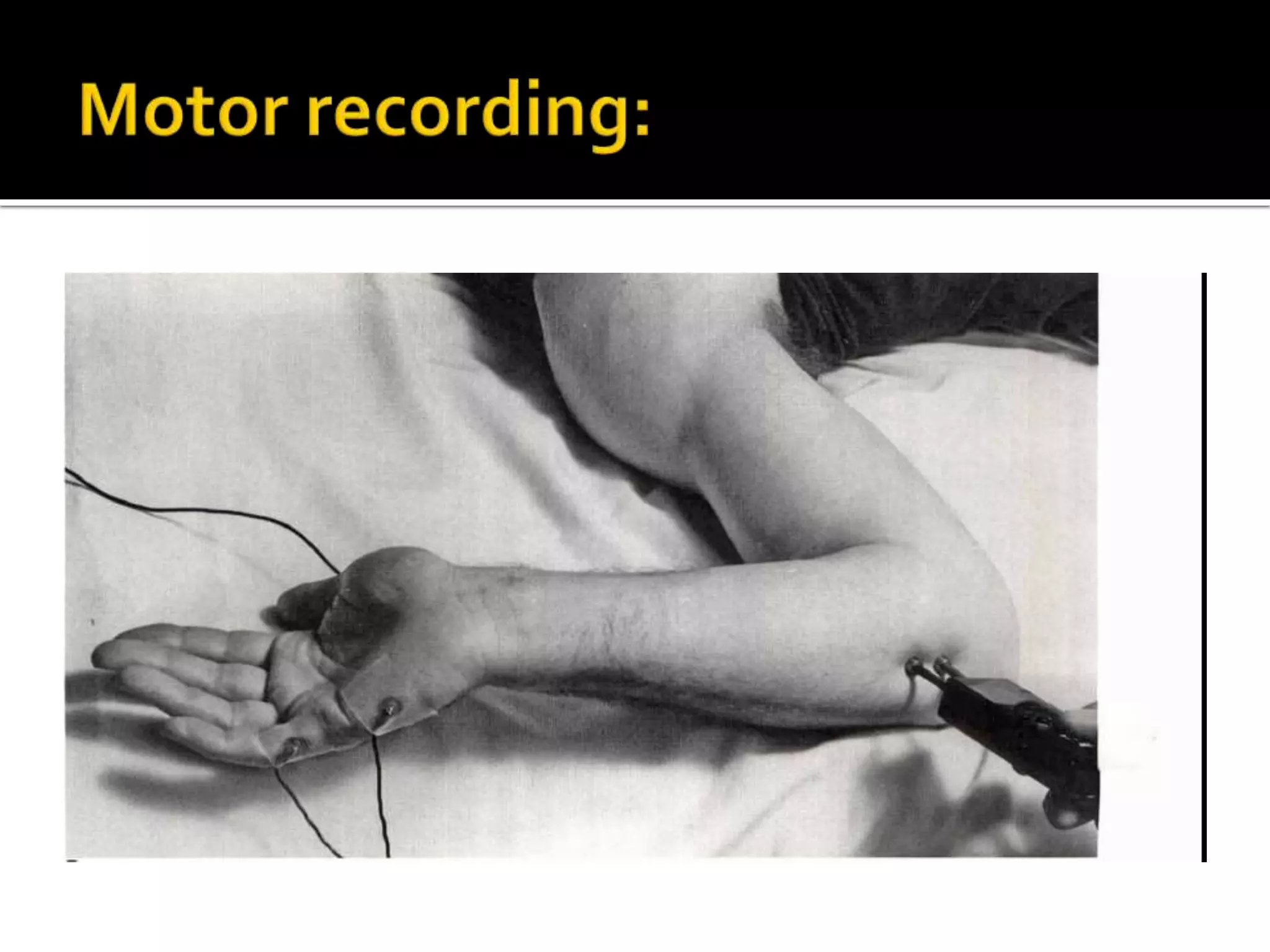
The ulnar nerve originates from the C8-T1 nerve roots and descends down the medial side of the upper arm. It enters the cubital tunnel at the elbow and gives sensation to the forearm. In the forearm, it innervates the flexor carpi ulnaris and part of the flexor digitorum profundus muscles. It passes through Guyon's canal at the wrist and innervates hand muscles like the hypothenar muscles and interossei, as well as providing sensation to the palmar side of the small and ring fingers. Recording and stimulation sites are described for sensory and motor nerve conduction studies of the ulnar nerve.