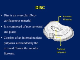
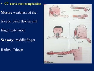
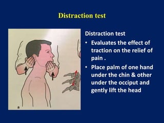
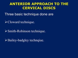
CERVICAL DISC PROLAPSE
DR.THOUSEEFA MAJEED
M S Orthopaedics PG
VMKVMCH Salem
Anatomy
Cervical vertebrae
2 TYPES
– Atypical
• Axis (C 1 )
• Atlas (C 2 )
• C 7
– Typical
• C 3-6
• 7 Vertebrae
• 6 Inter vertebral discs
• 8 Pairs of exiting nerve
roots
• Cervical spine gives a
normal lordosis
• Individual vertebrae are
connected to each other by
facet joints.
• The inter-body joints contain a
specialized structures called
intervertebral discs.
• The discs are found
throughout the vertebrae
except for the first and second
vertebrae.
DISC
• Disc is an avascular fibro-
cartilagenous material
• It is composed of two vertebral
end plates
• Consists of an internal nucleus
pulposus surrounded by the
external fibrous the annulus
fibrosus.
Annulus
Fibrosus
Nucleus
pulposus
 Vertebral end plates are 1mm thick sheets made of
fibrocartilage and hyaline cartilage.
 As age increases hyaline cartilage gets converted in to
fibrocartilage.
Nucleus pulposus
 Made up of type II-Collagen
 Contains 80 to 90% of water.
 Proteoglycan 65%
 Collagen 15 to 20 %0
Annulus fibrosus
• Made up of Type I Collagen
• 12 concentric layer of
lamellae with alternate
layer of collagen.
Annulus fibrous contains
• 60 to 70% of water.
• 50 to 60% of collagen
• 20% of proteoglycan.
Functions of disc
• Act as a shock absorber
• Ligaments by holding
together
• Allows compressive,
tensile, and rotational
motion
CERVICAL DISC PROLAPSE
History
 In 1838 - Herniation of the cervical disc was
identified by Key.
 In 1934 -Mixter and Barr reported disc herniation
Incidence and Etiology
 Common in men.
 Male :Female - 1.4:1
 Factors associated with injury are
 Frequent lifting of heavy objects on the job
 Cigarette Smoking
 Frequent diving from a board.
Pathopysiology
 Physiological annular degeneration.
 Frank extrusion of nuclear material
 As the degeneration proceeds there is hypermobility of the
segment resulting of the instability or degenerative arthritic
change.
 Internal disruption of the disc with annular tears results in
disc prolapse.
Types of prolapse
• Central
• Posterolateral
• Foraminal
• Extraforaminal
Common sites of cervical disc prolapse
 C5-C6
 More motion occurs between C5 and C6
 C6-C7 prolapse occurs in older patients
Cervical disc prolapse
 7 cervical vetebrae
 8 cervical nerve roots.
 First cervical root exists
between occiput and C1
 6th cervical root exists
between C5 and C6
 8th cervical root exists
between C7 and T1
A herniated disc impinges upon the nerve root
exiting above the disc and passing through the
near by foramen result in involvement of one
specific neurologic level
Each cervical root exists cephalad to the
pedicle of the vertebrae
This relationship changes at thoracic spine
because the C8 root exists between C7 and T1
pedicles ,requiring the T1 nerve root to exits
caudal or below the pedicle.
• Cervical disc Prolapse is
usually in the postero-lateral
direction, because the strong
posterior longitudinal
ligament prevents direct
posterior herniation.
• Unlike the lumbar region, the nerves pass directly laterally
from the cervical cord to their neural foramen, so that the
herniation compresses the nerve at that level.
Clinical Features
 Neck pain radiating to the arm or chest
 Numbness in the fingers
 Motor weakness of the arm.
 Mimics cardiac disease with chest and arm pain.
 Vertebral artery compression results in dizziness, tinnitus,
blurring of vision and retroocular pain.
Clinical examination
Restricted cervical spine movements.
The head is often moderately flexed, and tilted
towards the side of the pain in some patients but
occasionally away from it in others.
If the disc herniation is longstanding there may be
weakness & wasting in the appropriate muscle
group.
• C5 nerve root compression
Motor : Weakness of the deltoid
and biceps.
Sensory: upper lateral arm and
elbow.
Reflex-Biceps
• C6 nerve root compression
Motor: weakness of the biceps
and extensor carpi radialis brevis
and longus.
Sensory: lateral forearm, thumb
and index finger
Reflex – Biceps , Brachioradialis
• C7 nerve root compression
Motor: weakness of the
triceps, wrist flexion and
finger extension.
Sensory: middle finger
Reflex- Triceps
• C8 nerve root compression
Motor: weakness of the
interossei and finger flexors
and flexor carpi ulnaris.
Sensory: ulnar border of the
arm.
Reflex- no reflex changes
T-I Nerve root compression
Motor – weakness of
introssei muscles
Sensory – Medial aspect
of L –I
Reflex changes - None
SPECIAL TESTS
Sperlings Maneuver
• Extension and Ipsilateral rotation of the neck should
produce patients radicular pain
Lhermittes sign (Barber Chair Phenomenon)
• Flexion or extension produces electric shock like
sensations extend from spine and shoot to the limbs.
Compression test
Compression test
• Presses the head gently
down while the patient
is lying or sitting.
• Increase in radicular
pain
Distraction test
Distraction test
• Evaluates the effect of
traction on the relief of
pain .
• Place palm of one hand
under the chin & other
under the occiput and
gently lift the head
Confirmatory imaging
 X-Ray.
 Computerized Tomography (CT)
 Magnetic resonance imaging (MRI)
 Myelogram
X-RAY
MRI
Myelogram
Odom classification
 Type I : unilateral soft disc protrusion with nerve root
compression.
 Type II : foraminal spur or hard disc with nerve root
compression.
 Type III: medial soft disc protrusion with spinal root
compression.
 Type IV : transverse ridge or cervical spondylosis with
spinal cord compression.
TREATMENT
Non operative management:
 Rest
 Cervical Collar
 Ice
 Anti inflammatory agents with active mobilization.
 Muscle relaxents also can be given
 Cervical muscle strengthening excercises
 Cervical traction with weight less than 10 lbs.
OPERATIVE TREATMENT
Indications of surgery
 Failure of non-operative pain management
 Increasing and significant neurological deficit
 Cervical myelopathy
Surgical treatment:
• The most commonly performed surgeries for cervical
disc prolapse are:
– Cervical foraminotomy with excision of the disc
prolapse.
– Anterior cervical discectomy, with subsequent
fusion.
 Soft lateral discs are easily removed with the posterior
approach
 Soft central or hard discs are treated with anterior approach
 Spinal fusion are preceded with anterior approach with bone
grafting.
 Use of bone graft the collapse of the disc space and
maintains adequate foraminal disc.
Minimally invasive posterior approach
to the cervical spine
Indications:
 Radiculopathy
 Lateral disc herniation
 Foramnial stenosis
 Persistent or recurrent nerve root symptoms.
Contraindications:
 Pure axial neck pain without neurologic symptoms.
 Cervical instability
 Symptomatic central disc herniation
 Kyphotic deformity that would make posterior
decompression.
ANTERIOR APPROACH TO THE
CERVICAL DISCS
Three basic technique done are
Cloward technique.
Smith-Robinson technique.
Bailey-badgley technqiue.
Cloward technique
 Cloward technique involves making a round hole
centered at the disc space. A slightly larger, round
iliac crest into the disc space hole.
Smith robinson technique
It involves inserting a cortical strut of iliac
crest into the disc space after removing the
disc and cartilaginous endplate.
Bailey-badgley technique
 The technique involves the creation of slot in superior and
inferior vertebral bodies.
 This technique is most applicable to reconstruction when
one or more vertbrae are excised.
 Biomechanically the smith robinson technique provides the
greatest stability and least risk of extrusion.
ANTERIOR APPROACH
 Patient positioned supine, with bolster placed under the
shoulder.
 A 5-8 cms long transverse skin incision is made on skin
crease ipsilateral to the radiculopathy.
 Skin, subcutaneous tissue and platysma muscle are incised.
 Blunt dissection to access the anterior column of the
cervical spine.
 Carotid artery and sternocleidomastoid muscle are
retracted laterally and the muscle, trachea and
esophagus medial.
 Prevertebral fascia is split to expose the anterior
column of the cervical spine.
 longus colli muscle is identified and medial portion of
the muscle is excised to expose transverse process.
 using a high speed drill, a drill is made between
uncovertebral joint.
 Posterior longitudinal ligament is exposed and the lateral
remnant of the uncinate process are dissceted.
 The remaining uncinate process is removed and
decompression of the nerve root is done.
 A portion of the posterior longitudinal ligament is
removed to find hidden ruptured disc.
Posterior approach
 Midline incision is made on the spinous process.
 Ligamentum nuchae are divided, subperiosteal
division of the paravertebral muscles.
 With high speed burr minimal bone is removed.
 Ligamentum flavum is excised
 With the help of microscope the herniated nucleus
pulposus is removed.
CERVICAL DISC ARTHROPLASTY
 The primary argument favoring these device is that
adjacent segment degeneration is minimized.
 Maintenance of motion is better with arthroplasty.
 Complication includes implant migration,
heterotrophic ossification and recurrent radiculopathy.
THANK
YOU

Cervical disc prolapse

  • 1.
    CERVICAL DISC PROLAPSE DR.THOUSEEFAMAJEED M S Orthopaedics PG VMKVMCH Salem
  • 2.
  • 3.
    Cervical vertebrae 2 TYPES –Atypical • Axis (C 1 ) • Atlas (C 2 ) • C 7 – Typical • C 3-6
  • 5.
    • 7 Vertebrae •6 Inter vertebral discs • 8 Pairs of exiting nerve roots • Cervical spine gives a normal lordosis
  • 6.
    • Individual vertebraeare connected to each other by facet joints. • The inter-body joints contain a specialized structures called intervertebral discs. • The discs are found throughout the vertebrae except for the first and second vertebrae.
  • 7.
    DISC • Disc isan avascular fibro- cartilagenous material • It is composed of two vertebral end plates • Consists of an internal nucleus pulposus surrounded by the external fibrous the annulus fibrosus. Annulus Fibrosus Nucleus pulposus
  • 8.
     Vertebral endplates are 1mm thick sheets made of fibrocartilage and hyaline cartilage.  As age increases hyaline cartilage gets converted in to fibrocartilage.
  • 9.
    Nucleus pulposus  Madeup of type II-Collagen  Contains 80 to 90% of water.  Proteoglycan 65%  Collagen 15 to 20 %0
  • 10.
    Annulus fibrosus • Madeup of Type I Collagen • 12 concentric layer of lamellae with alternate layer of collagen. Annulus fibrous contains • 60 to 70% of water. • 50 to 60% of collagen • 20% of proteoglycan.
  • 11.
    Functions of disc •Act as a shock absorber • Ligaments by holding together • Allows compressive, tensile, and rotational motion
  • 12.
  • 13.
    History  In 1838- Herniation of the cervical disc was identified by Key.  In 1934 -Mixter and Barr reported disc herniation
  • 14.
    Incidence and Etiology Common in men.  Male :Female - 1.4:1  Factors associated with injury are  Frequent lifting of heavy objects on the job  Cigarette Smoking  Frequent diving from a board.
  • 15.
    Pathopysiology  Physiological annulardegeneration.  Frank extrusion of nuclear material  As the degeneration proceeds there is hypermobility of the segment resulting of the instability or degenerative arthritic change.  Internal disruption of the disc with annular tears results in disc prolapse.
  • 17.
    Types of prolapse •Central • Posterolateral • Foraminal • Extraforaminal
  • 18.
    Common sites ofcervical disc prolapse  C5-C6  More motion occurs between C5 and C6  C6-C7 prolapse occurs in older patients
  • 19.
    Cervical disc prolapse 7 cervical vetebrae  8 cervical nerve roots.  First cervical root exists between occiput and C1  6th cervical root exists between C5 and C6  8th cervical root exists between C7 and T1
  • 20.
    A herniated discimpinges upon the nerve root exiting above the disc and passing through the near by foramen result in involvement of one specific neurologic level
  • 21.
    Each cervical rootexists cephalad to the pedicle of the vertebrae This relationship changes at thoracic spine because the C8 root exists between C7 and T1 pedicles ,requiring the T1 nerve root to exits caudal or below the pedicle.
  • 22.
    • Cervical discProlapse is usually in the postero-lateral direction, because the strong posterior longitudinal ligament prevents direct posterior herniation.
  • 23.
    • Unlike thelumbar region, the nerves pass directly laterally from the cervical cord to their neural foramen, so that the herniation compresses the nerve at that level.
  • 24.
    Clinical Features  Neckpain radiating to the arm or chest  Numbness in the fingers  Motor weakness of the arm.  Mimics cardiac disease with chest and arm pain.  Vertebral artery compression results in dizziness, tinnitus, blurring of vision and retroocular pain.
  • 25.
    Clinical examination Restricted cervicalspine movements. The head is often moderately flexed, and tilted towards the side of the pain in some patients but occasionally away from it in others. If the disc herniation is longstanding there may be weakness & wasting in the appropriate muscle group.
  • 26.
    • C5 nerveroot compression Motor : Weakness of the deltoid and biceps. Sensory: upper lateral arm and elbow. Reflex-Biceps
  • 27.
    • C6 nerveroot compression Motor: weakness of the biceps and extensor carpi radialis brevis and longus. Sensory: lateral forearm, thumb and index finger Reflex – Biceps , Brachioradialis
  • 28.
    • C7 nerveroot compression Motor: weakness of the triceps, wrist flexion and finger extension. Sensory: middle finger Reflex- Triceps
  • 29.
    • C8 nerveroot compression Motor: weakness of the interossei and finger flexors and flexor carpi ulnaris. Sensory: ulnar border of the arm. Reflex- no reflex changes
  • 30.
    T-I Nerve rootcompression Motor – weakness of introssei muscles Sensory – Medial aspect of L –I Reflex changes - None
  • 31.
    SPECIAL TESTS Sperlings Maneuver •Extension and Ipsilateral rotation of the neck should produce patients radicular pain Lhermittes sign (Barber Chair Phenomenon) • Flexion or extension produces electric shock like sensations extend from spine and shoot to the limbs.
  • 32.
    Compression test Compression test •Presses the head gently down while the patient is lying or sitting. • Increase in radicular pain
  • 33.
    Distraction test Distraction test •Evaluates the effect of traction on the relief of pain . • Place palm of one hand under the chin & other under the occiput and gently lift the head
  • 34.
    Confirmatory imaging  X-Ray. Computerized Tomography (CT)  Magnetic resonance imaging (MRI)  Myelogram
  • 35.
  • 36.
  • 38.
  • 39.
    Odom classification  TypeI : unilateral soft disc protrusion with nerve root compression.  Type II : foraminal spur or hard disc with nerve root compression.  Type III: medial soft disc protrusion with spinal root compression.  Type IV : transverse ridge or cervical spondylosis with spinal cord compression.
  • 40.
    TREATMENT Non operative management: Rest  Cervical Collar  Ice  Anti inflammatory agents with active mobilization.  Muscle relaxents also can be given  Cervical muscle strengthening excercises  Cervical traction with weight less than 10 lbs.
  • 41.
    OPERATIVE TREATMENT Indications ofsurgery  Failure of non-operative pain management  Increasing and significant neurological deficit  Cervical myelopathy
  • 42.
    Surgical treatment: • Themost commonly performed surgeries for cervical disc prolapse are: – Cervical foraminotomy with excision of the disc prolapse. – Anterior cervical discectomy, with subsequent fusion.
  • 43.
     Soft lateraldiscs are easily removed with the posterior approach  Soft central or hard discs are treated with anterior approach  Spinal fusion are preceded with anterior approach with bone grafting.  Use of bone graft the collapse of the disc space and maintains adequate foraminal disc.
  • 44.
    Minimally invasive posteriorapproach to the cervical spine Indications:  Radiculopathy  Lateral disc herniation  Foramnial stenosis  Persistent or recurrent nerve root symptoms.
  • 45.
    Contraindications:  Pure axialneck pain without neurologic symptoms.  Cervical instability  Symptomatic central disc herniation  Kyphotic deformity that would make posterior decompression.
  • 46.
    ANTERIOR APPROACH TOTHE CERVICAL DISCS Three basic technique done are Cloward technique. Smith-Robinson technique. Bailey-badgley technqiue.
  • 47.
    Cloward technique  Clowardtechnique involves making a round hole centered at the disc space. A slightly larger, round iliac crest into the disc space hole.
  • 48.
    Smith robinson technique Itinvolves inserting a cortical strut of iliac crest into the disc space after removing the disc and cartilaginous endplate.
  • 49.
    Bailey-badgley technique  Thetechnique involves the creation of slot in superior and inferior vertebral bodies.  This technique is most applicable to reconstruction when one or more vertbrae are excised.  Biomechanically the smith robinson technique provides the greatest stability and least risk of extrusion.
  • 50.
    ANTERIOR APPROACH  Patientpositioned supine, with bolster placed under the shoulder.  A 5-8 cms long transverse skin incision is made on skin crease ipsilateral to the radiculopathy.  Skin, subcutaneous tissue and platysma muscle are incised.  Blunt dissection to access the anterior column of the cervical spine.
  • 51.
     Carotid arteryand sternocleidomastoid muscle are retracted laterally and the muscle, trachea and esophagus medial.  Prevertebral fascia is split to expose the anterior column of the cervical spine.  longus colli muscle is identified and medial portion of the muscle is excised to expose transverse process.
  • 53.
     using ahigh speed drill, a drill is made between uncovertebral joint.  Posterior longitudinal ligament is exposed and the lateral remnant of the uncinate process are dissceted.  The remaining uncinate process is removed and decompression of the nerve root is done.  A portion of the posterior longitudinal ligament is removed to find hidden ruptured disc.
  • 54.
    Posterior approach  Midlineincision is made on the spinous process.  Ligamentum nuchae are divided, subperiosteal division of the paravertebral muscles.  With high speed burr minimal bone is removed.  Ligamentum flavum is excised  With the help of microscope the herniated nucleus pulposus is removed.
  • 55.
    CERVICAL DISC ARTHROPLASTY The primary argument favoring these device is that adjacent segment degeneration is minimized.  Maintenance of motion is better with arthroplasty.  Complication includes implant migration, heterotrophic ossification and recurrent radiculopathy.
  • 56.