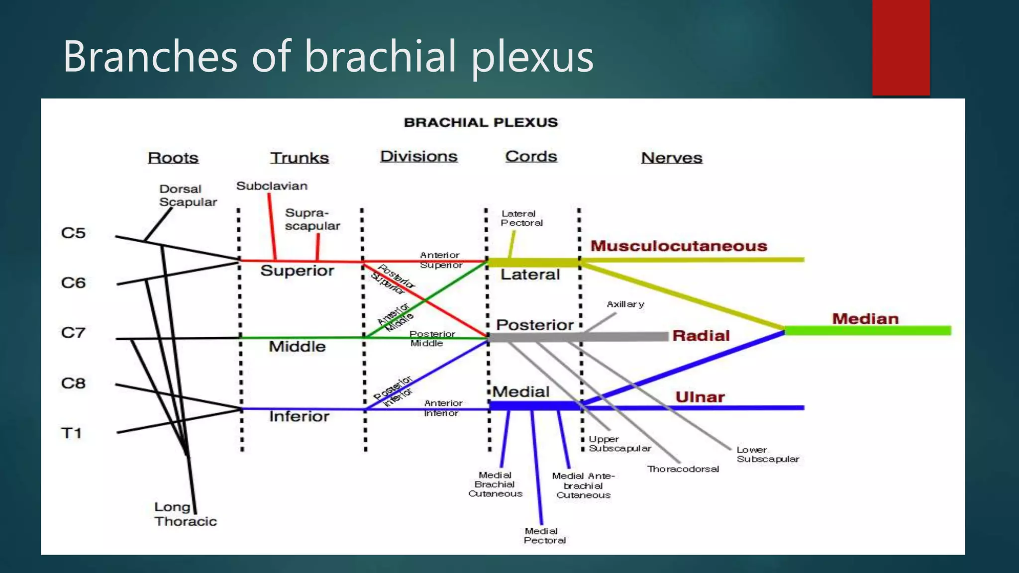
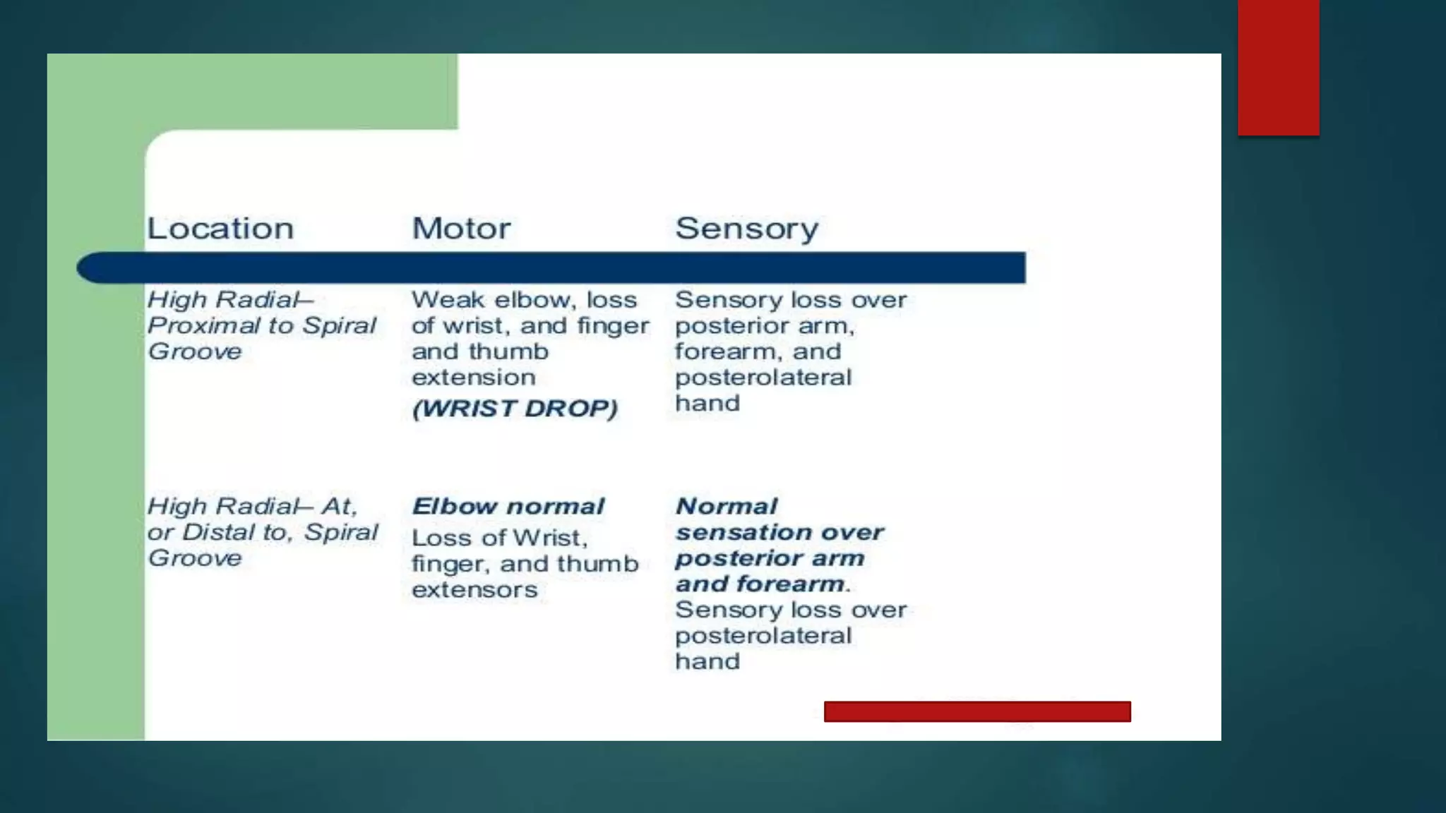
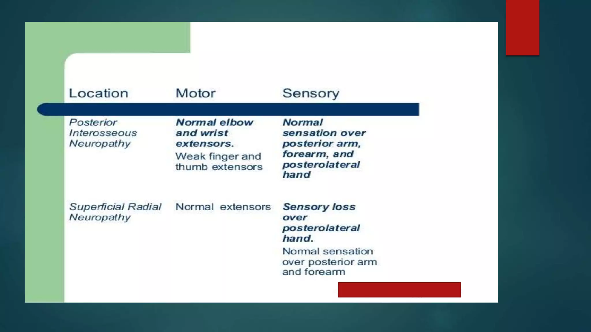
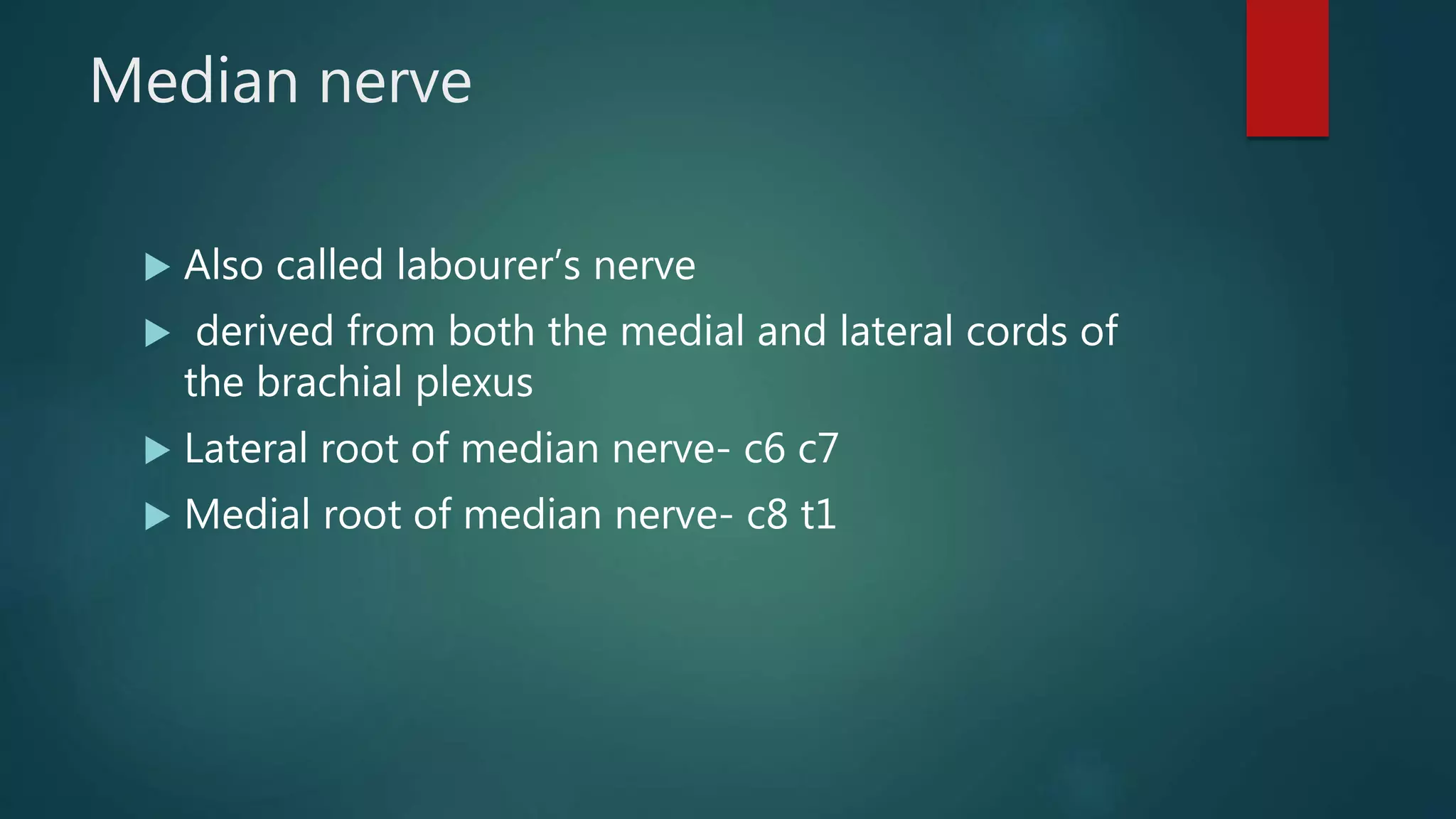
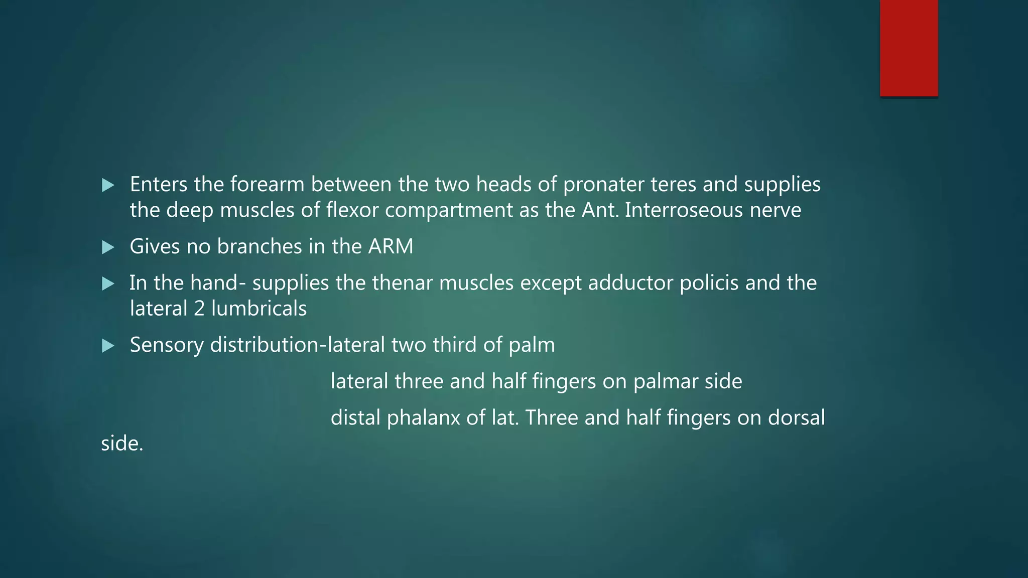
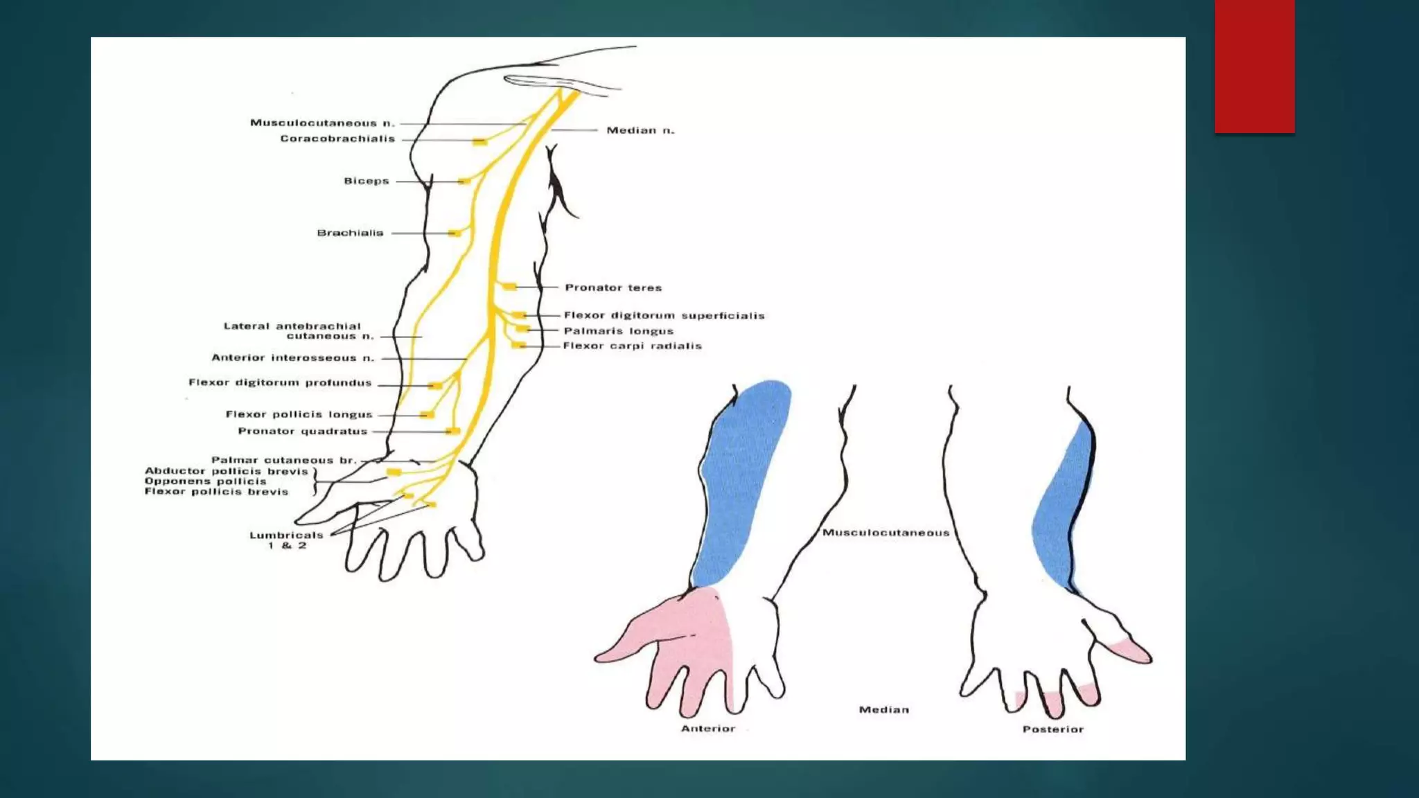
Upper limb nerve
examination
DR ADITYA APTE
VPMC & RC NASHIK
Brachial plexus
1.Formed by the ventral ramii of cervical spinal nerves
c5 to c8 and the thoracic spinal nerve T1
2.Prefix- includes the ramii of C4
3.Postfix- includes the ramii of T2
4.Consists of ROOTS, TRUNKS , DIVISIONS , CORDS and
BRANCHES
5.Responsible for sensory and motor innervation
of entire limb
 ROOTS- consists of ventral ramii of spinal nerves C5 to T1
 TRUNKS- roots c5 and c6 join to form the upper trunk
root c7 forms the middle trunk
roots c8 and T1 forms the lower trunk
CORDS- lateral cord- ventral division of upper and
middle trunk
medial cord-ventral division of lower trunk
posterior cord- dorsal division of all 3 trunks.
Branches of brachial plexus
Radial nerve
 Arises from posterior cord of brachial plexus
 Root value- c5 to T1
 Enters the arm behind the axillary artery in the post. Compartment of arm
 Enters the radial groove to emerge on the lateral aspect of arm in the
anterior compartment
 Reaches the distal part of humerus and passes anterior to the lateral
epicondyle to enter forearm
 Branches in arm-
 Before entering spiral groove- motor br. To long and medial head of
triceps and the posterior cutaneous nerve of arm
 In the spiral groove- motor br. To lateral and medial head of triceps and
the post cutaneous nerve of forearm and the lower lateral cut. Nerve of
arm
 Between the spiral groove and elbow-brachioradialis and ECRL
 After crossing elbow before piercing supinator-supinator and ECRB
 After piercing the supinator-continues as the post interroseous nerve and
supplies the post. Compartment of forearm
 ECRL- last muscle supplied by radial nerve
Radial nerve injury
Compression syndromes
Median nerve
 Also called labourer’s nerve
 derived from both the medial and lateral cords of
the brachial plexus
 Lateral root of median nerve- c6 c7
 Medial root of median nerve- c8 t1
 In the arm proximally the median nerve is lateral to the axillary artery
 Midway through the arm it crosses the artery from lateral to medial
anteriorly and lies medial to the brachial artery in the cubital fossa
 Gives an articular branch to the elbow and a branch to pronater teres just
before the joint
 In the cubital fossa gives branches to palmaris longus, FCR and FDS
 Enters the forearm between the two heads of pronater teres and supplies
the deep muscles of flexor compartment as the Ant. Interroseous nerve
 Gives no branches in the ARM
 In the hand- supplies the thenar muscles except adductor policis and the
lateral 2 lumbricals
 Sensory distribution-lateral two third of palm
lateral three and half fingers on palmar side
distal phalanx of lat. Three and half fingers on dorsal
side.
Aetiology of median nerve injury
 At the elbow- supracondylar humerus fractures; elbow dislocations
 At the wrist- sharp cuts ; lunate dislocation ; carpel tunnel syndrome
 Features-
 Pointing index-paralysis of FDS and FDP of index finger leads to failure of
its flexion
 Pen test- APB paralysis
 Ape thumb-thumb lies in the same plane as rest of the fingers in adducted
and laterally rotated position( due to overactivity of adductor policis)
 Wasting of thenar muscles
Ulnar nerve
 Originates from the medial cord ; c8 T1
 Descend medial to the brachial artery and enters the posterior
compartment of the arm
 Not a content of the cubital fossa as it passes behind the
medial epicondyle at elbow.
 Enters the anterior compartment of forearm between the two
heads of FCU.
 Does not give branch in arm
 In the forearm- FCU and medial third of FDP
 In the hand- superficial branch- hypothenar muscles
 deep branch- adductor pollicis, interrossie,
medial two lumbricals
 Called the muscians nerve as it controls the finer movements
of hand
 Claw hand- hyperextension at MCP and flexion at IP joints. (
due to paralysis of intrinsic musces)
 Ulnar paradox- clawing seen in low ulnar palsies and not in
high palsy
 In high palsy there is paralysis of FDP hence flexion at IP joint
does not occur.
THANK YOU.
Upper limb nerve examination

Upper limb nerve examination

  • 1.
    Upper limb nerve examination DRADITYA APTE VPMC & RC NASHIK
  • 2.
    Brachial plexus 1.Formed bythe ventral ramii of cervical spinal nerves c5 to c8 and the thoracic spinal nerve T1 2.Prefix- includes the ramii of C4 3.Postfix- includes the ramii of T2 4.Consists of ROOTS, TRUNKS , DIVISIONS , CORDS and BRANCHES 5.Responsible for sensory and motor innervation of entire limb
  • 3.
     ROOTS- consistsof ventral ramii of spinal nerves C5 to T1  TRUNKS- roots c5 and c6 join to form the upper trunk root c7 forms the middle trunk roots c8 and T1 forms the lower trunk CORDS- lateral cord- ventral division of upper and middle trunk medial cord-ventral division of lower trunk posterior cord- dorsal division of all 3 trunks.
  • 4.
  • 5.
    Radial nerve  Arisesfrom posterior cord of brachial plexus  Root value- c5 to T1  Enters the arm behind the axillary artery in the post. Compartment of arm  Enters the radial groove to emerge on the lateral aspect of arm in the anterior compartment  Reaches the distal part of humerus and passes anterior to the lateral epicondyle to enter forearm
  • 6.
     Branches inarm-  Before entering spiral groove- motor br. To long and medial head of triceps and the posterior cutaneous nerve of arm  In the spiral groove- motor br. To lateral and medial head of triceps and the post cutaneous nerve of forearm and the lower lateral cut. Nerve of arm  Between the spiral groove and elbow-brachioradialis and ECRL  After crossing elbow before piercing supinator-supinator and ECRB  After piercing the supinator-continues as the post interroseous nerve and supplies the post. Compartment of forearm  ECRL- last muscle supplied by radial nerve
  • 17.
  • 19.
  • 24.
    Median nerve  Alsocalled labourer’s nerve  derived from both the medial and lateral cords of the brachial plexus  Lateral root of median nerve- c6 c7  Medial root of median nerve- c8 t1
  • 26.
     In thearm proximally the median nerve is lateral to the axillary artery  Midway through the arm it crosses the artery from lateral to medial anteriorly and lies medial to the brachial artery in the cubital fossa  Gives an articular branch to the elbow and a branch to pronater teres just before the joint  In the cubital fossa gives branches to palmaris longus, FCR and FDS
  • 27.
     Enters theforearm between the two heads of pronater teres and supplies the deep muscles of flexor compartment as the Ant. Interroseous nerve  Gives no branches in the ARM  In the hand- supplies the thenar muscles except adductor policis and the lateral 2 lumbricals  Sensory distribution-lateral two third of palm lateral three and half fingers on palmar side distal phalanx of lat. Three and half fingers on dorsal side.
  • 39.
    Aetiology of mediannerve injury  At the elbow- supracondylar humerus fractures; elbow dislocations  At the wrist- sharp cuts ; lunate dislocation ; carpel tunnel syndrome  Features-  Pointing index-paralysis of FDS and FDP of index finger leads to failure of its flexion  Pen test- APB paralysis  Ape thumb-thumb lies in the same plane as rest of the fingers in adducted and laterally rotated position( due to overactivity of adductor policis)  Wasting of thenar muscles
  • 40.
    Ulnar nerve  Originatesfrom the medial cord ; c8 T1  Descend medial to the brachial artery and enters the posterior compartment of the arm  Not a content of the cubital fossa as it passes behind the medial epicondyle at elbow.  Enters the anterior compartment of forearm between the two heads of FCU.
  • 41.
     Does notgive branch in arm  In the forearm- FCU and medial third of FDP  In the hand- superficial branch- hypothenar muscles  deep branch- adductor pollicis, interrossie, medial two lumbricals
  • 52.
     Called themuscians nerve as it controls the finer movements of hand  Claw hand- hyperextension at MCP and flexion at IP joints. ( due to paralysis of intrinsic musces)  Ulnar paradox- clawing seen in low ulnar palsies and not in high palsy  In high palsy there is paralysis of FDP hence flexion at IP joint does not occur.
  • 53.