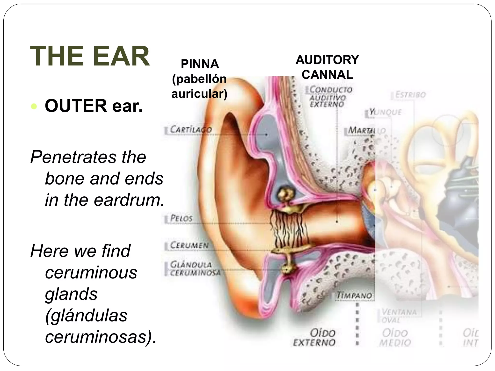
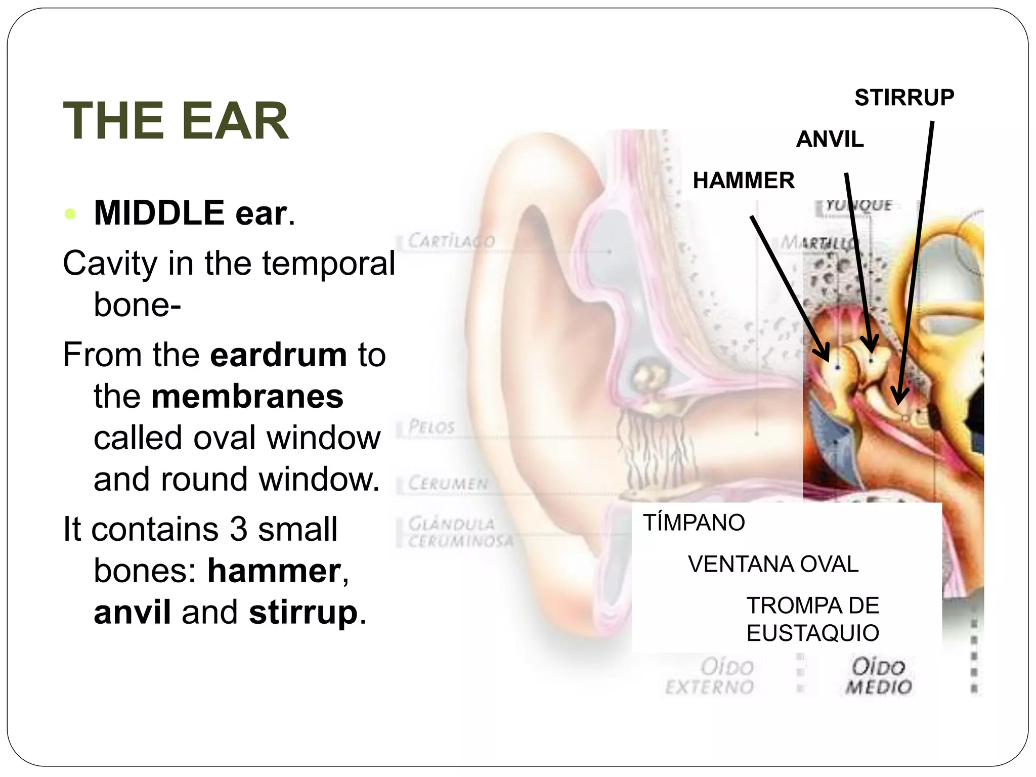
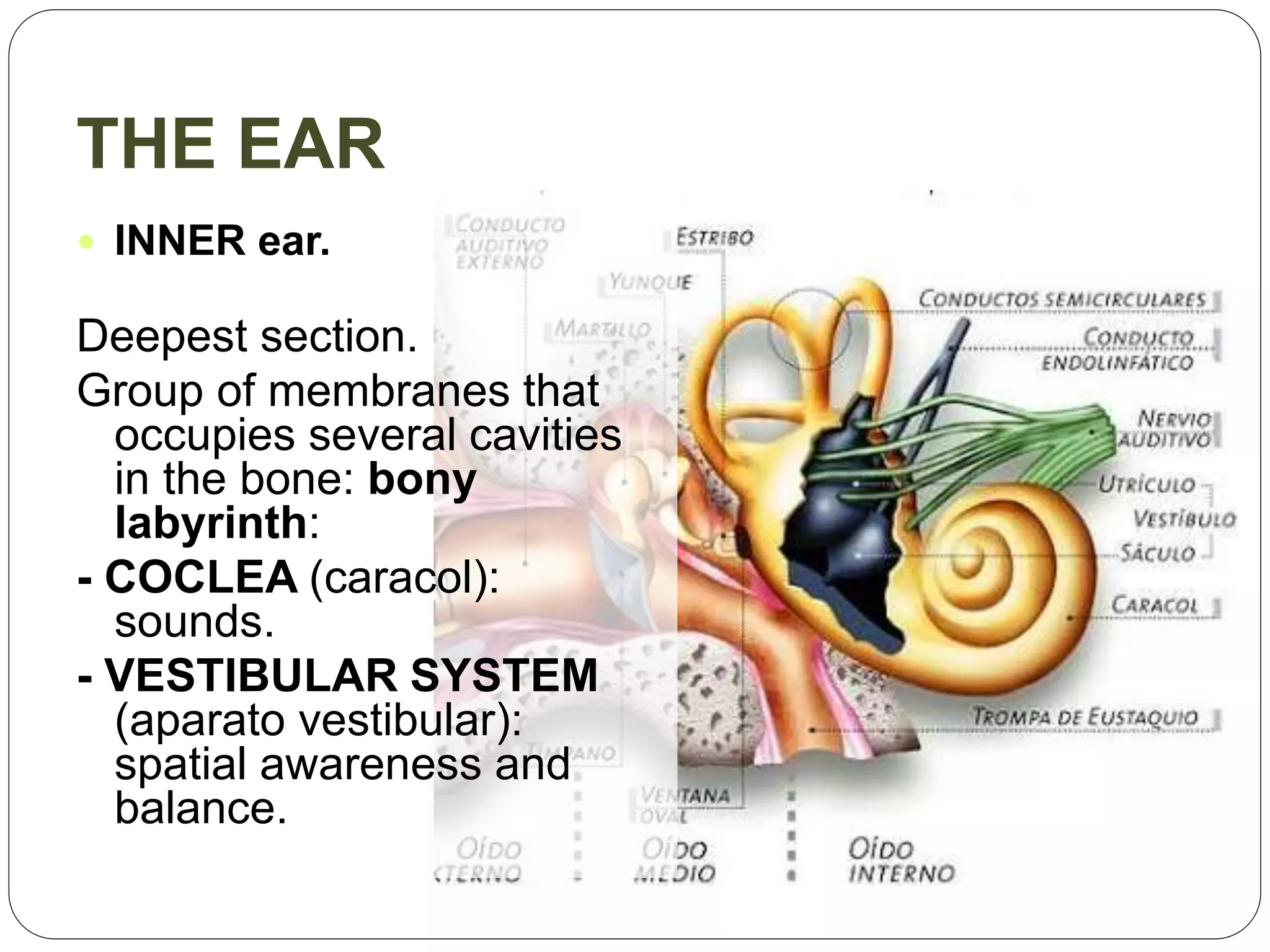
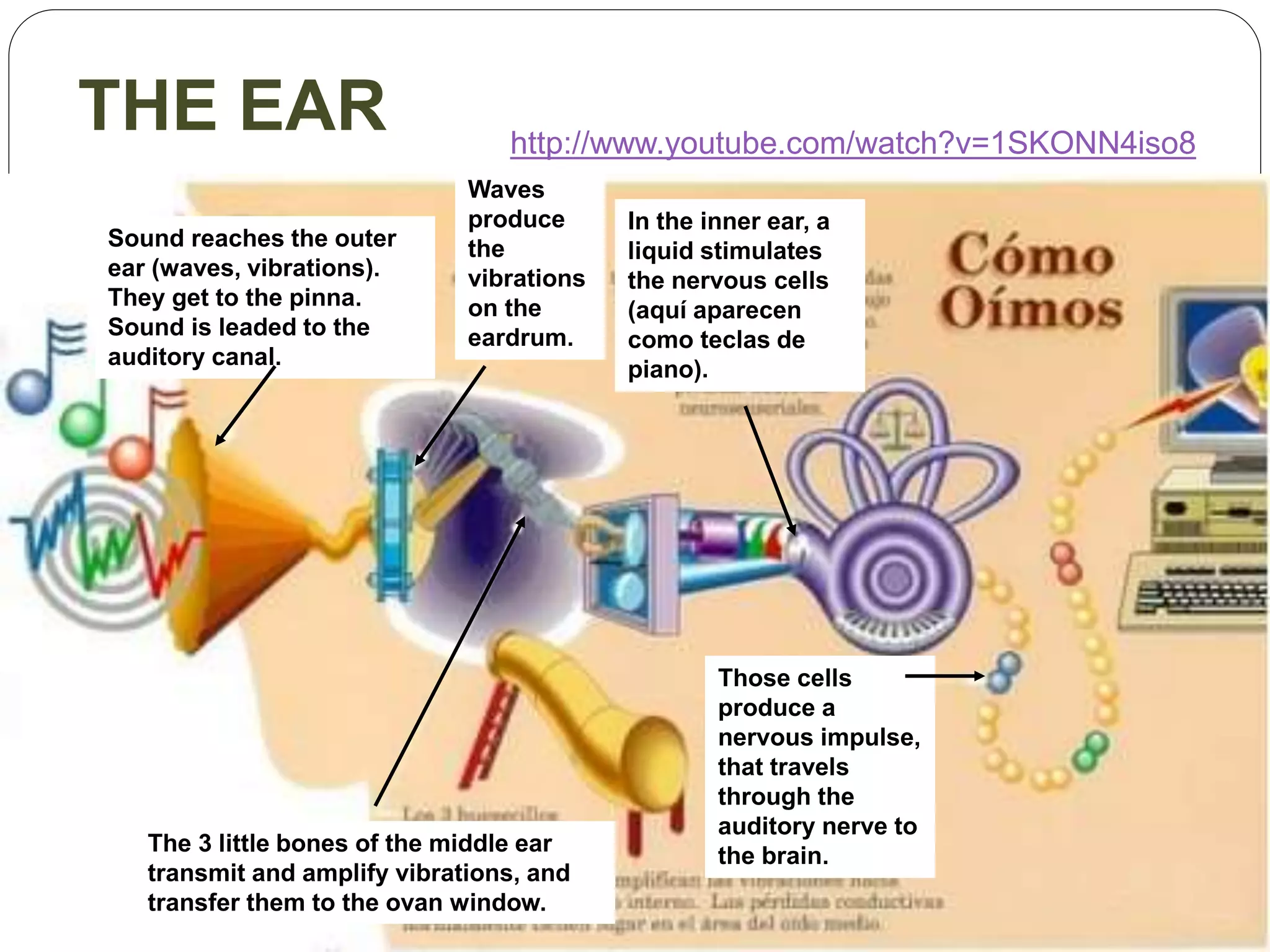
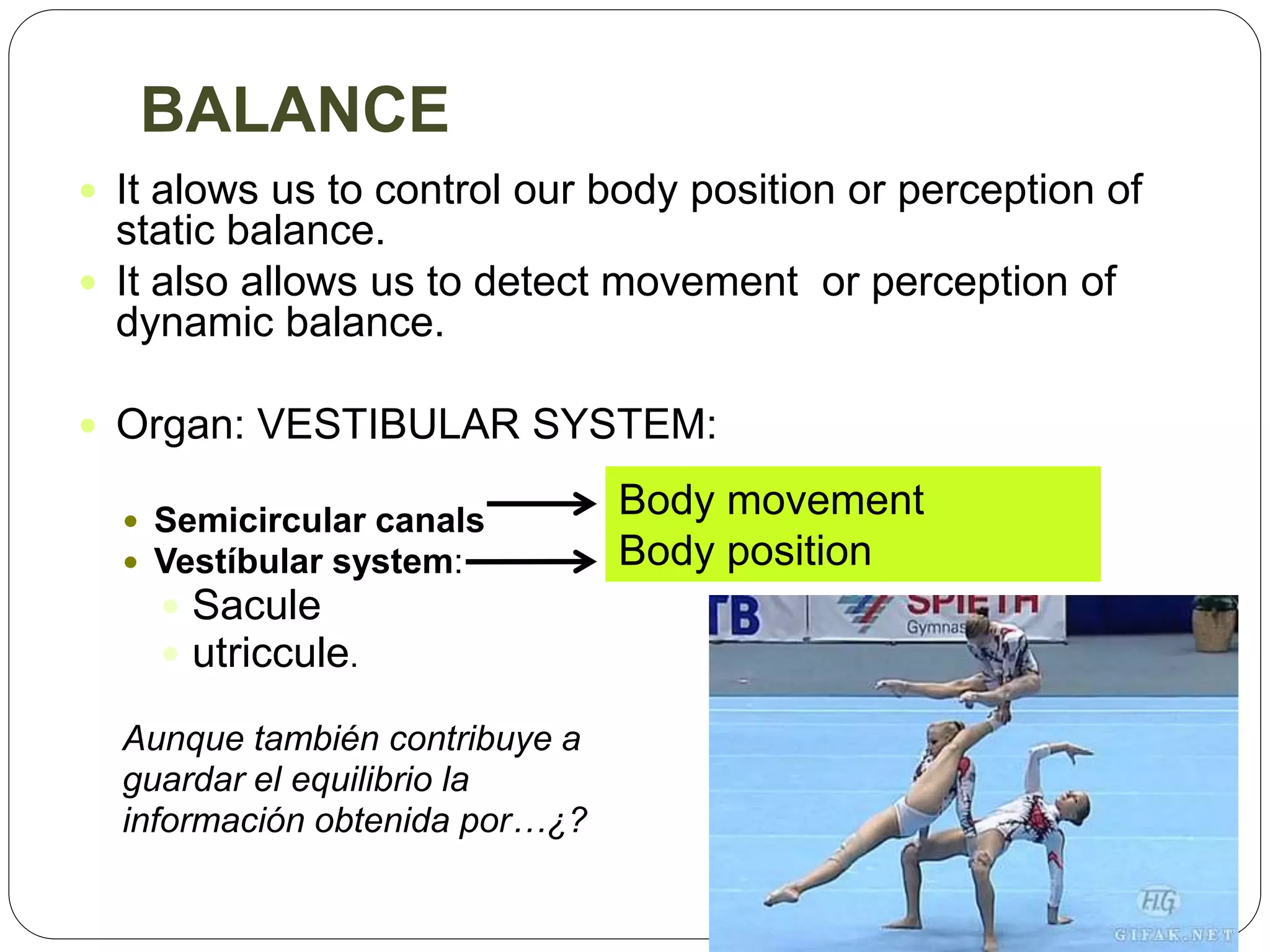
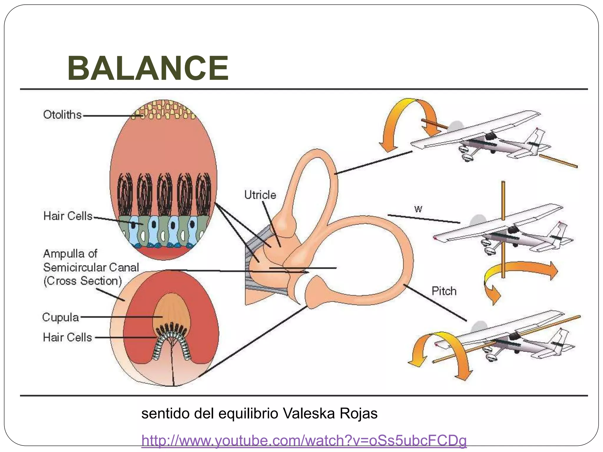
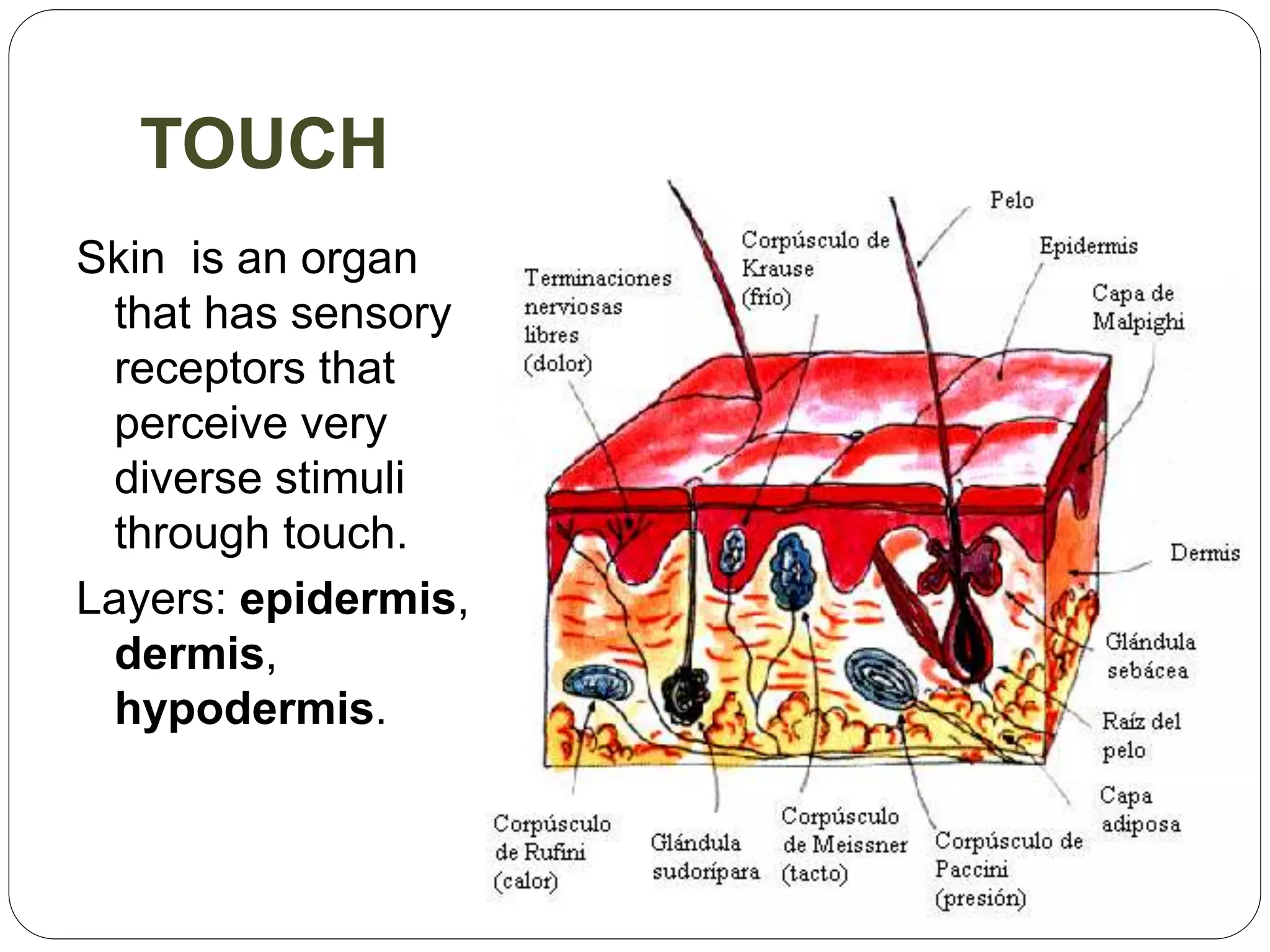
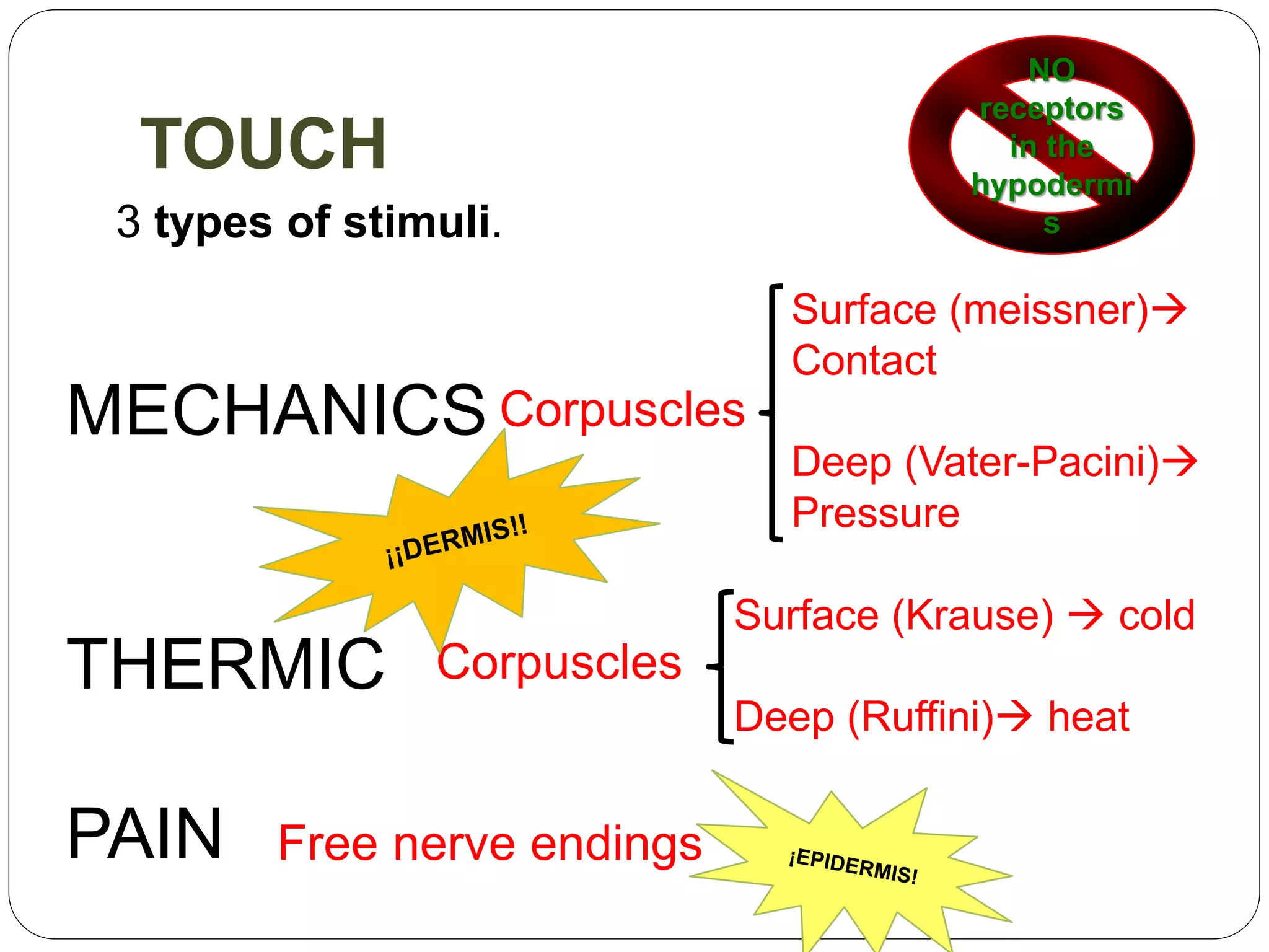
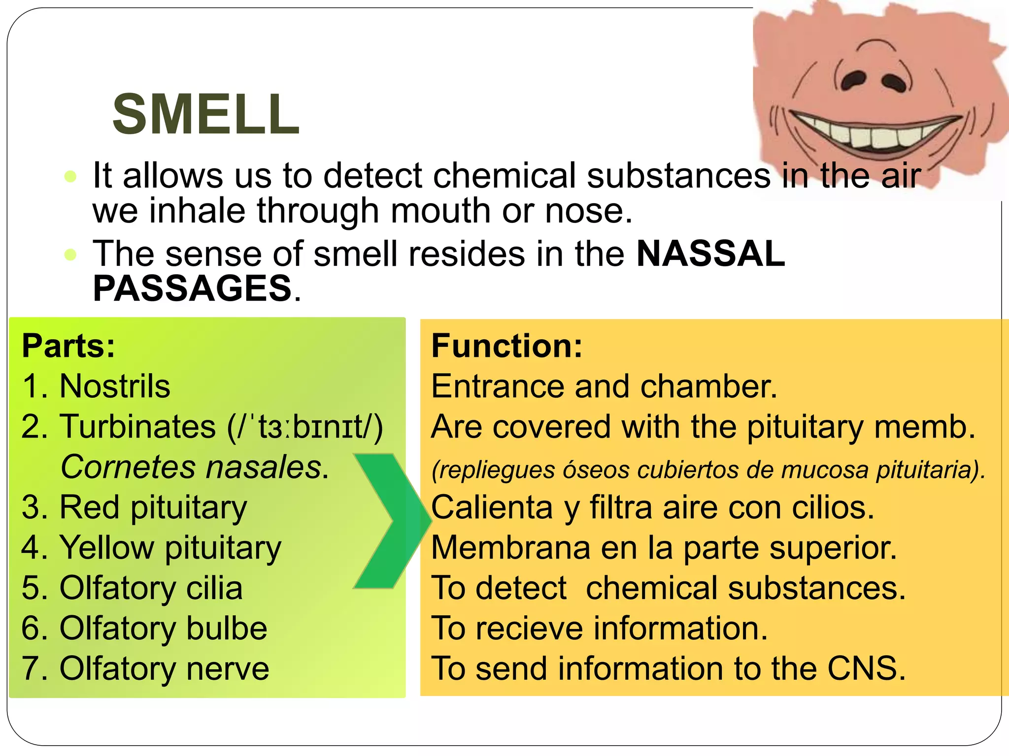
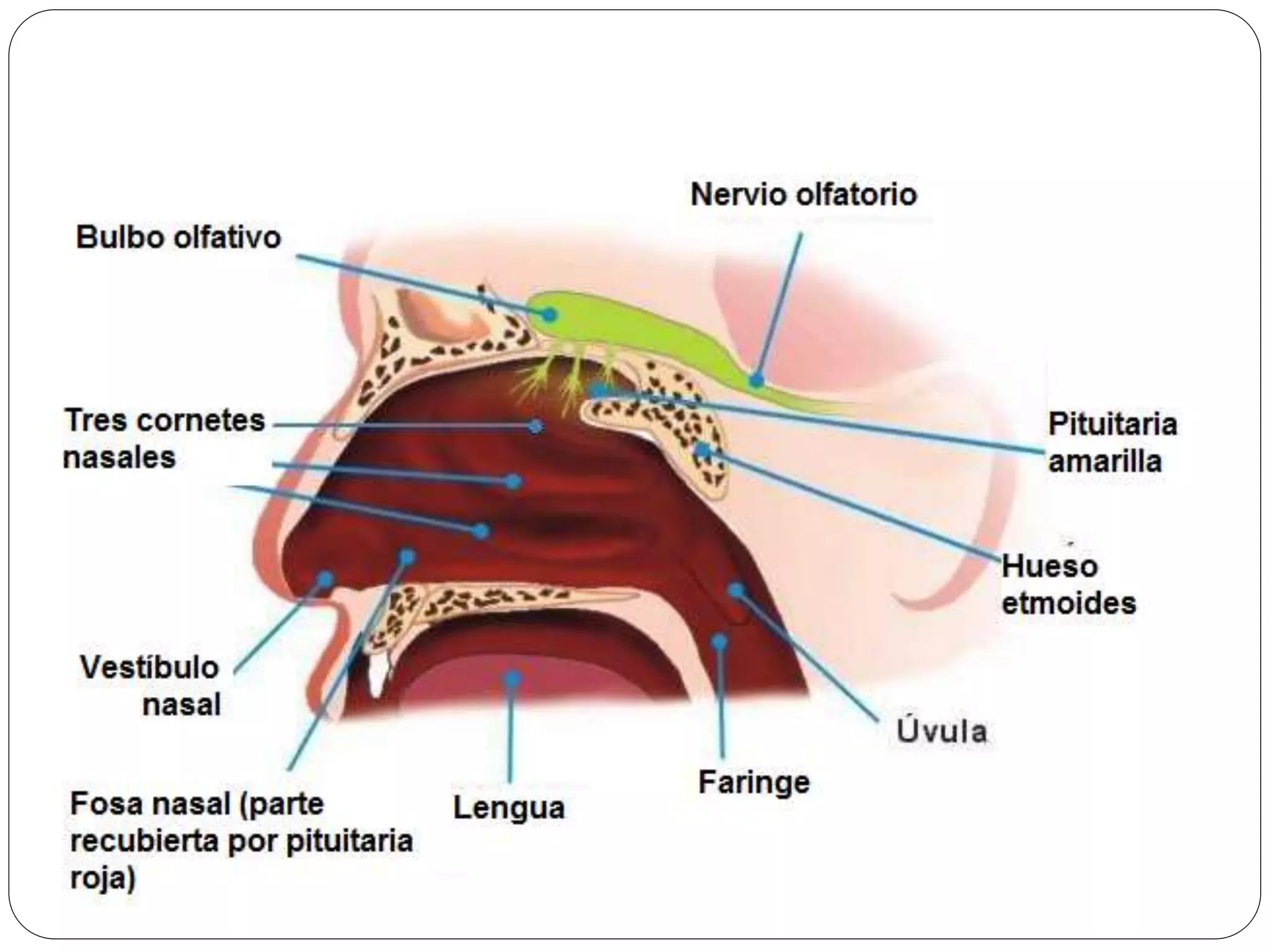
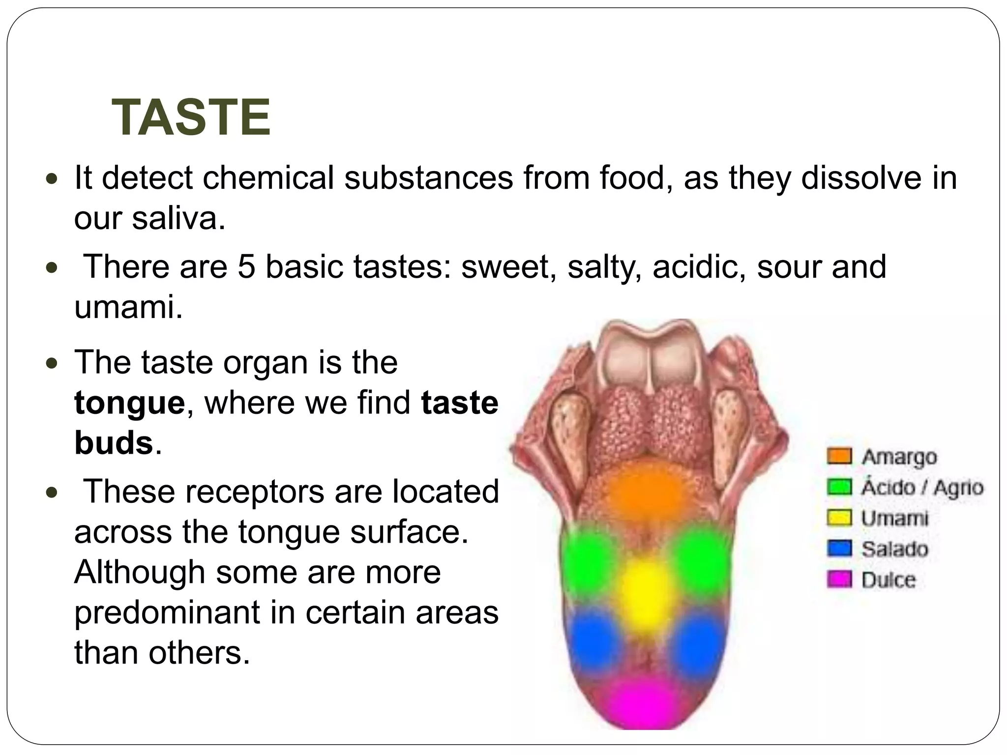
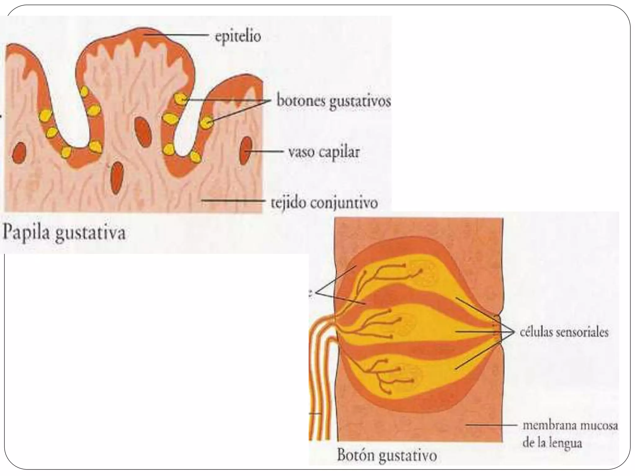
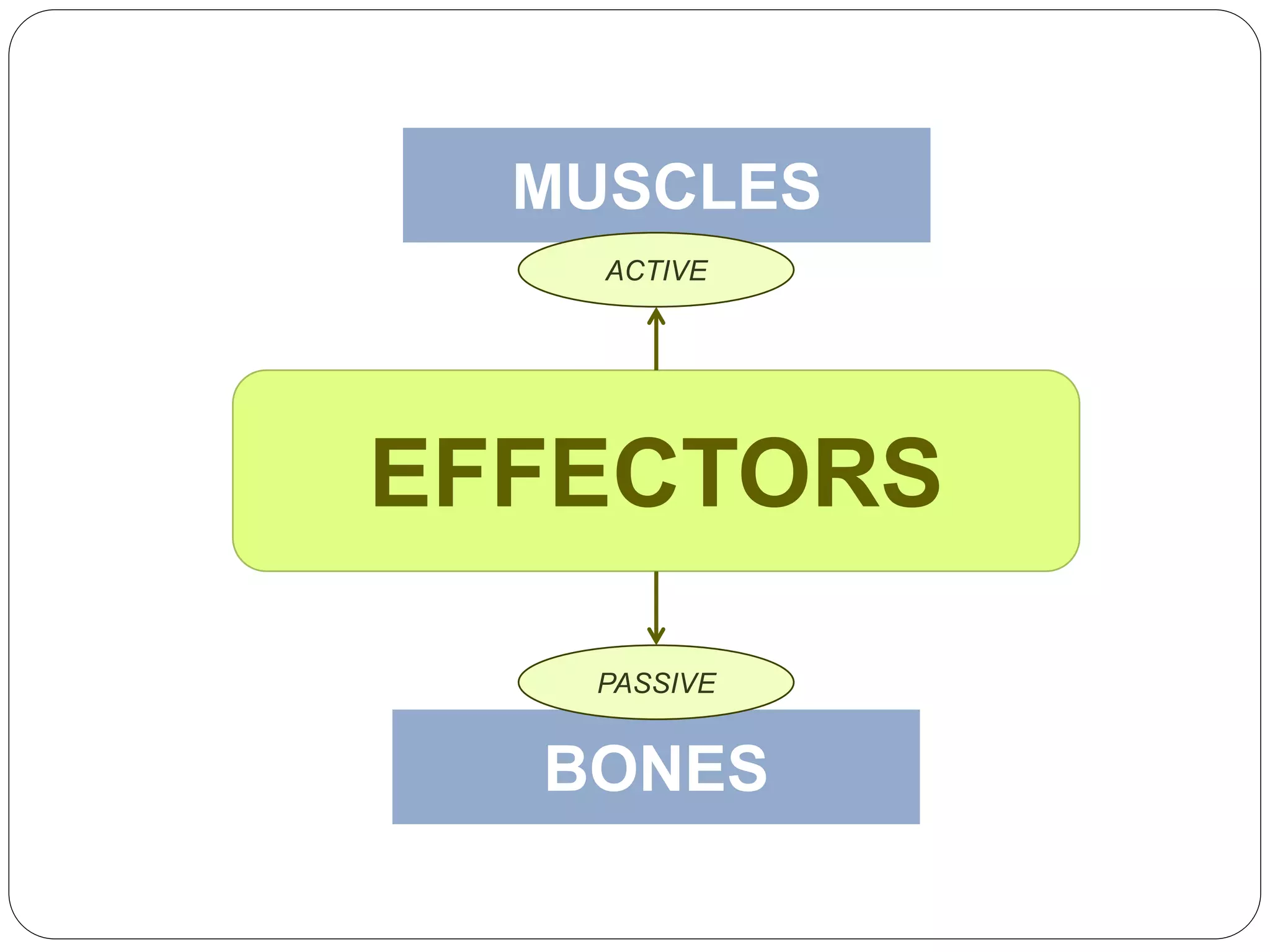
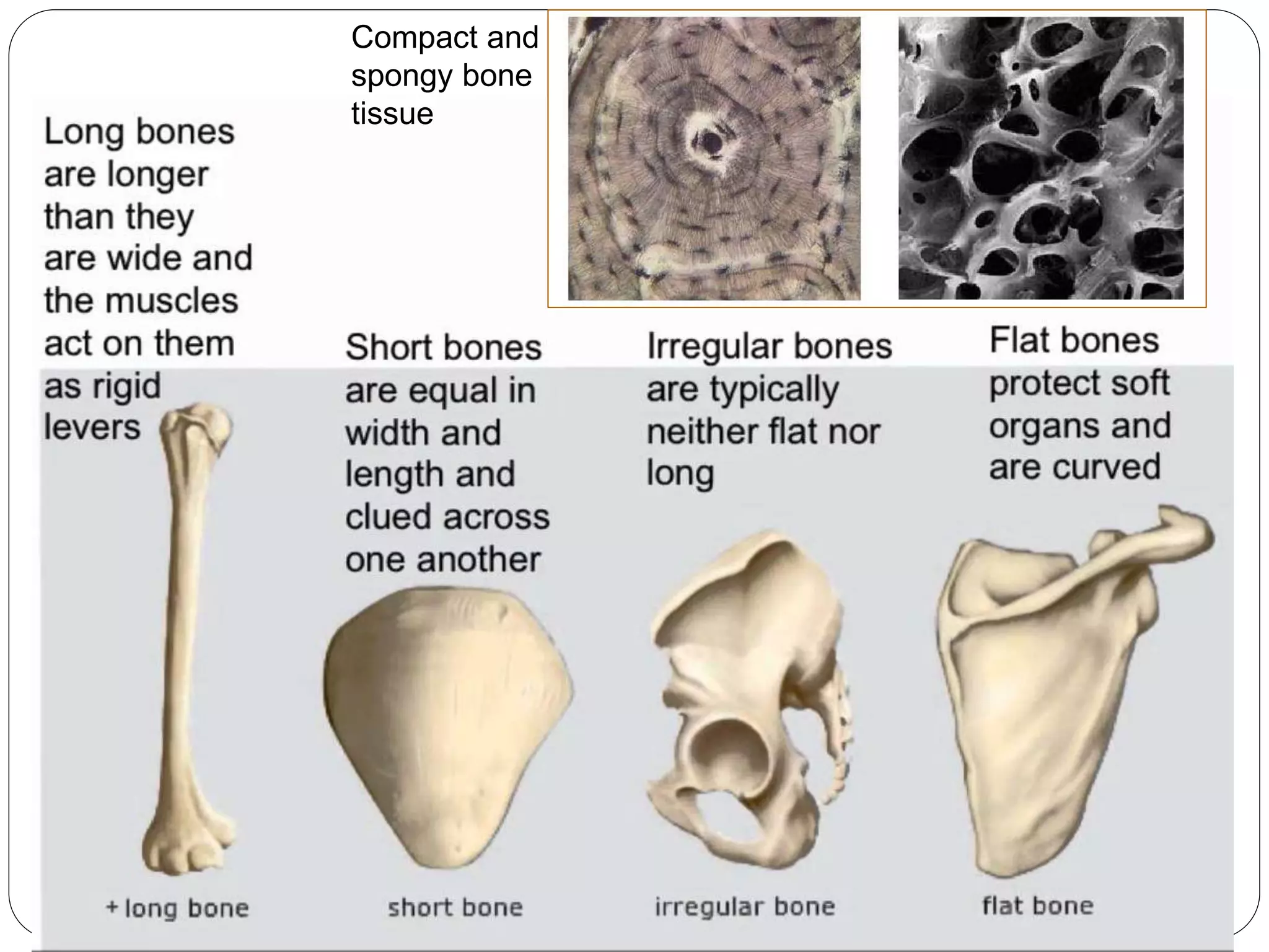
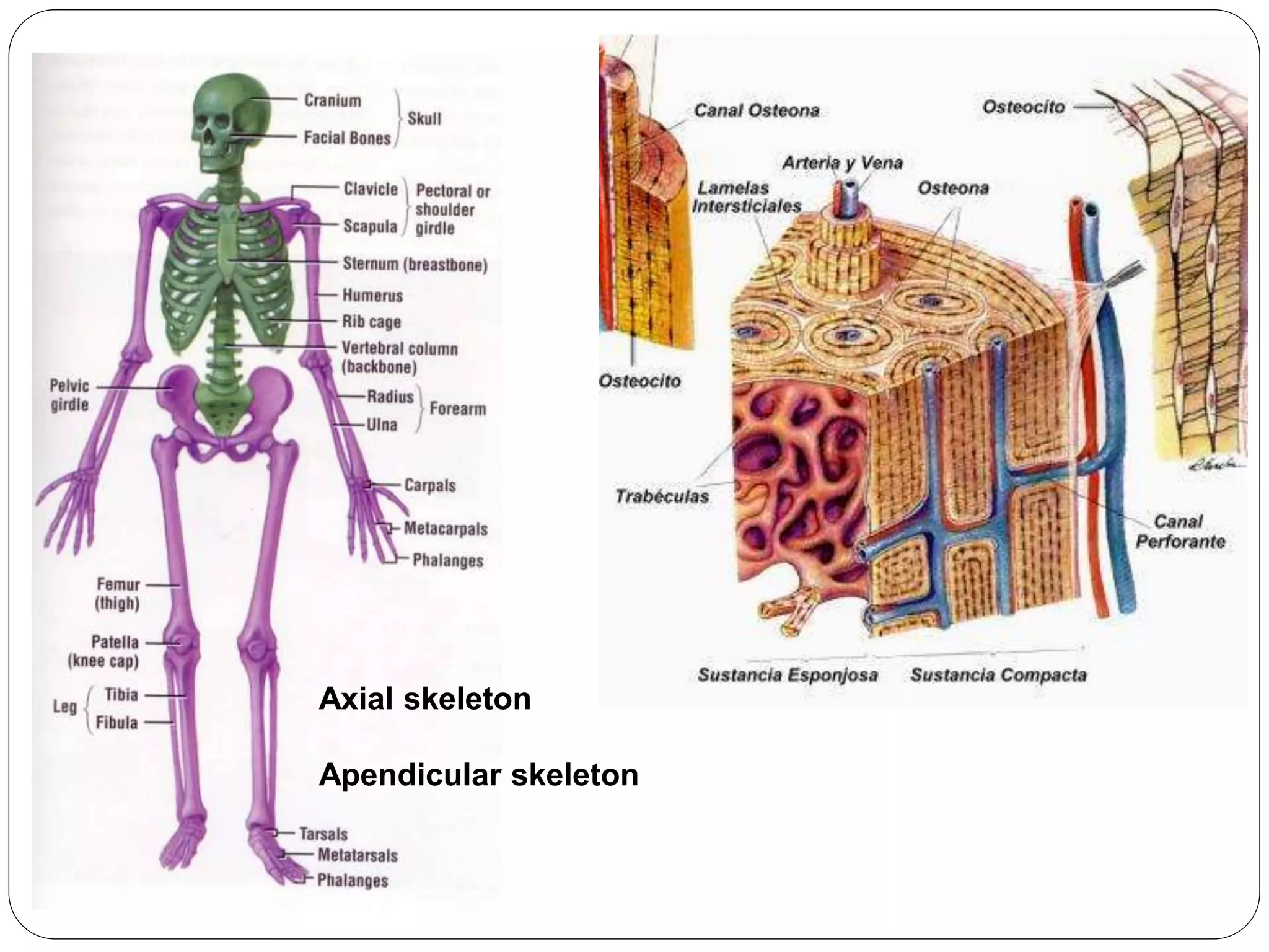
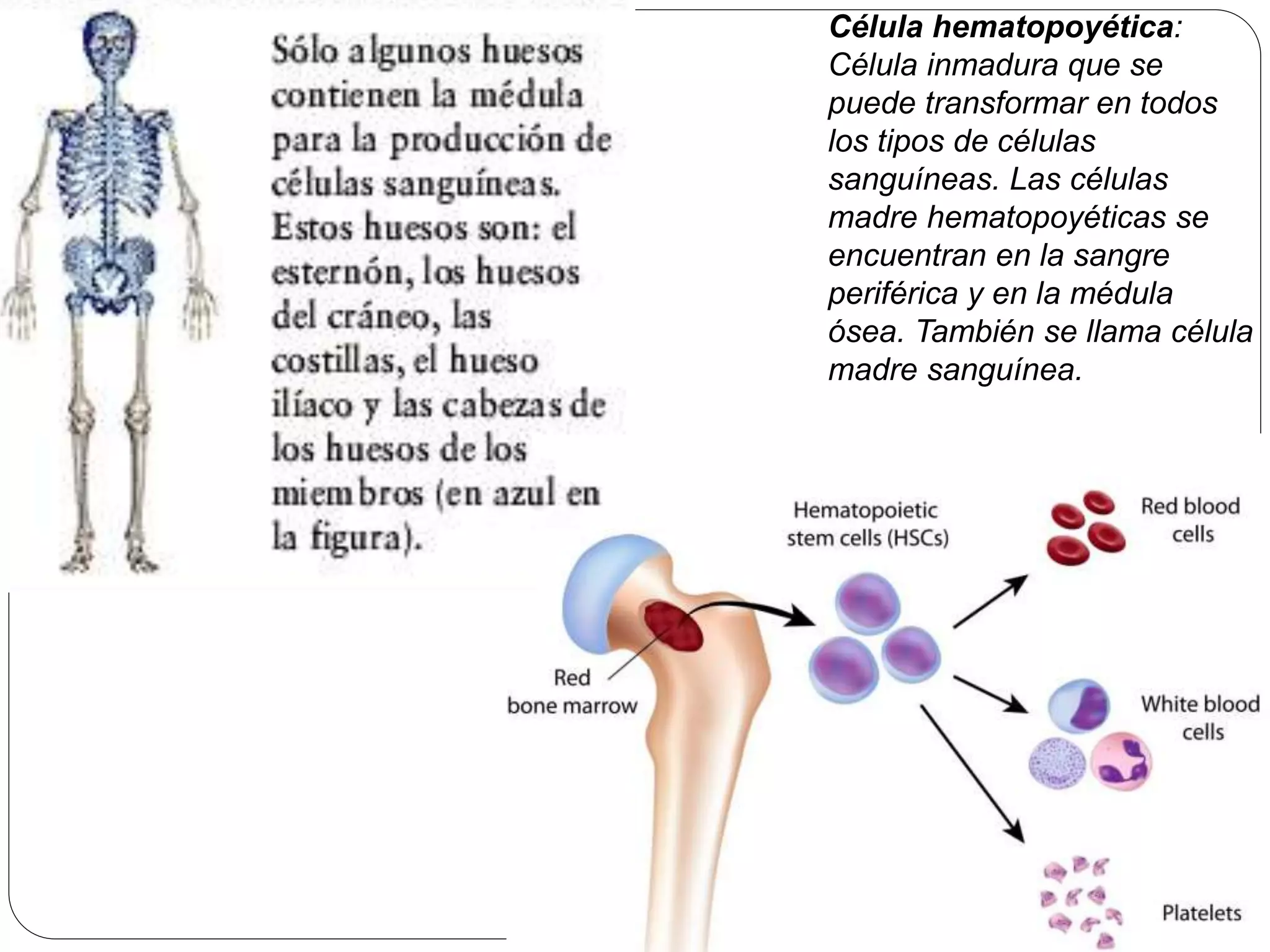
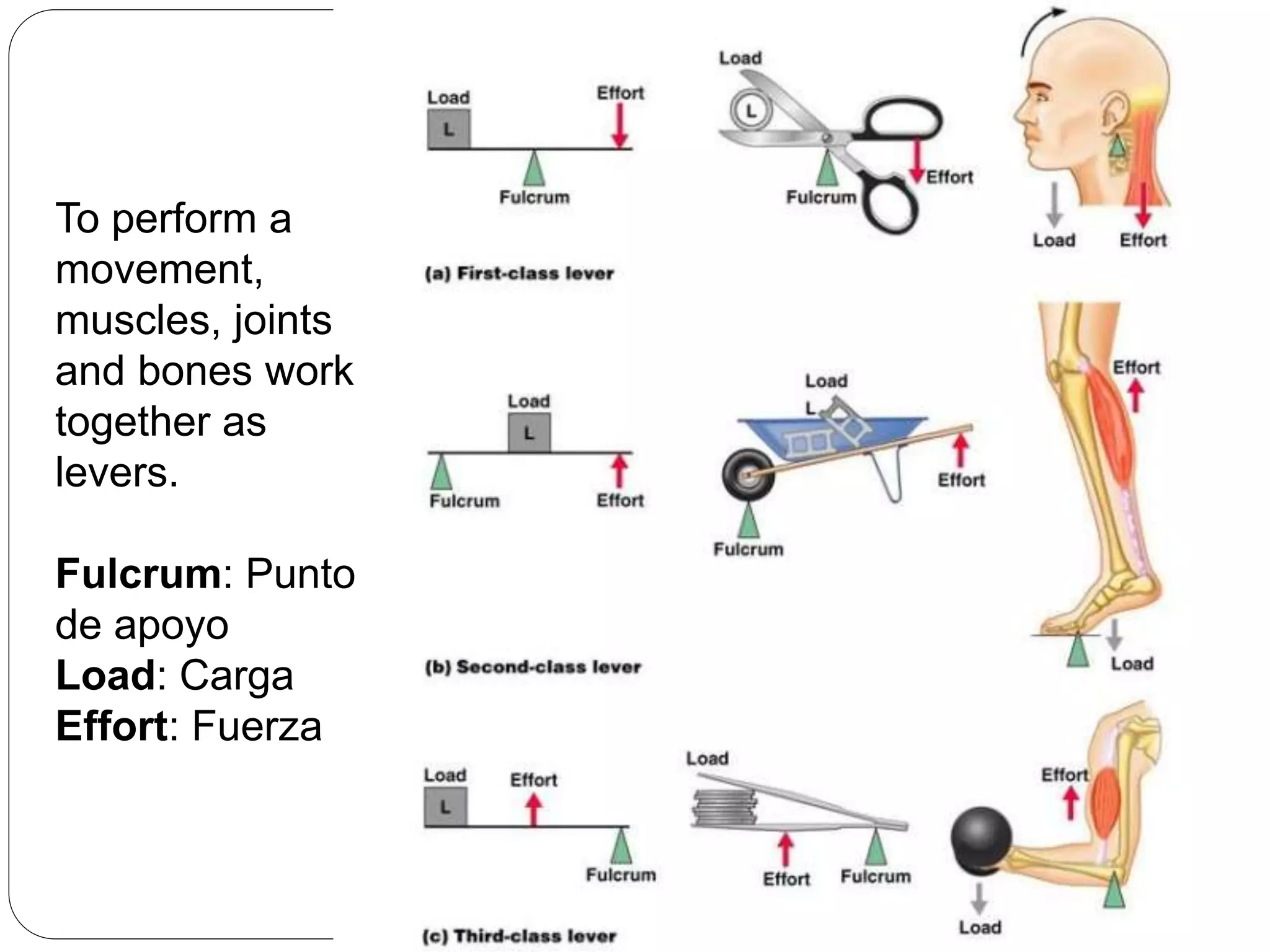
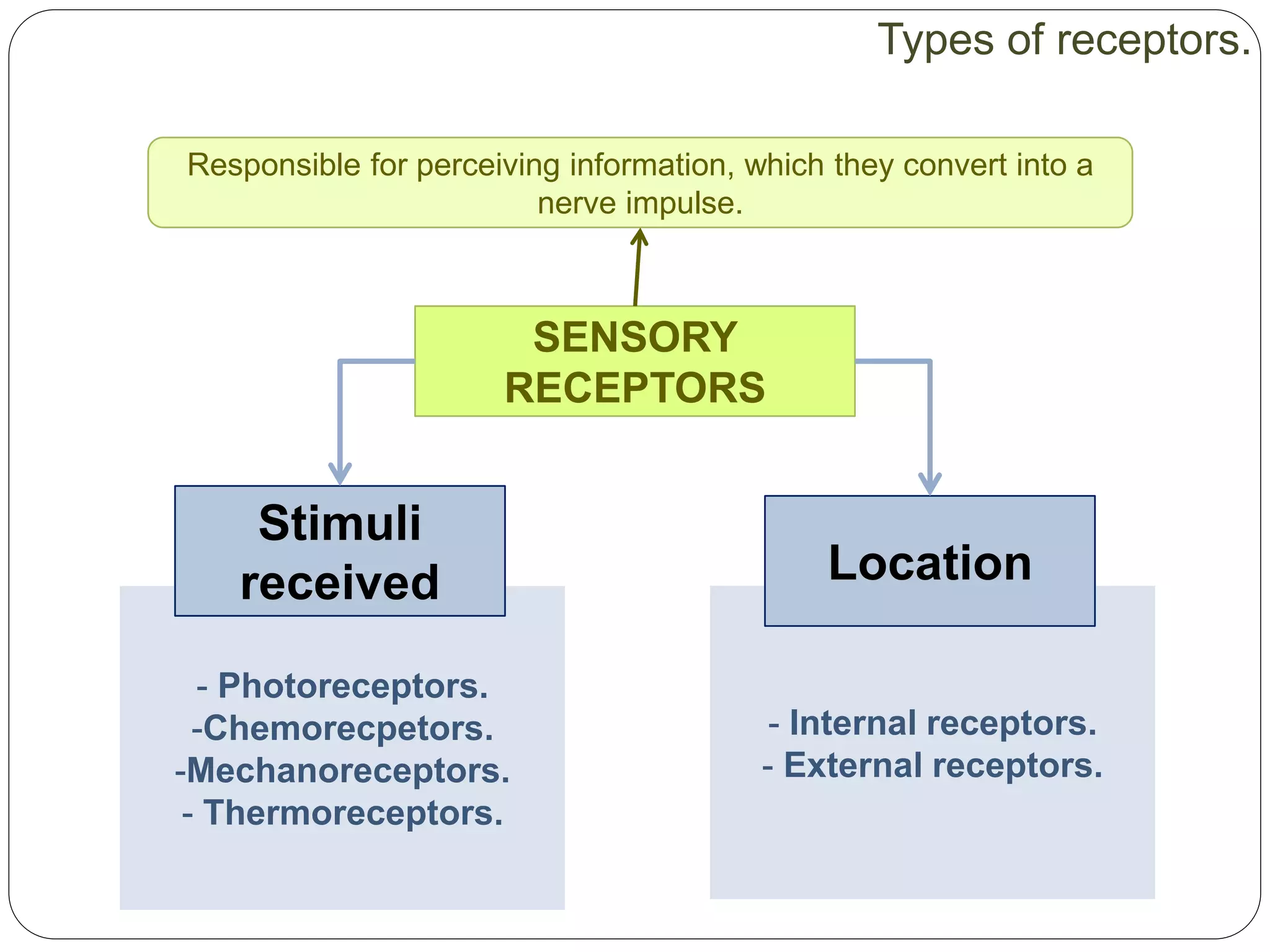
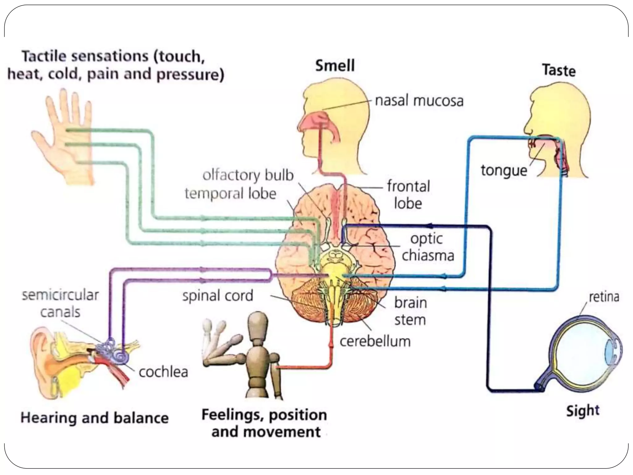
The document summarizes the main sensory and effector systems in the human body. It describes the key receptors for sight (eyes), hearing (ears), touch (skin), smell (nose), taste (tongue) and balance (inner ear). It also outlines the main components and functions of the skeletal system (bones and joints) and muscular system (striated, smooth and cardiac muscle).
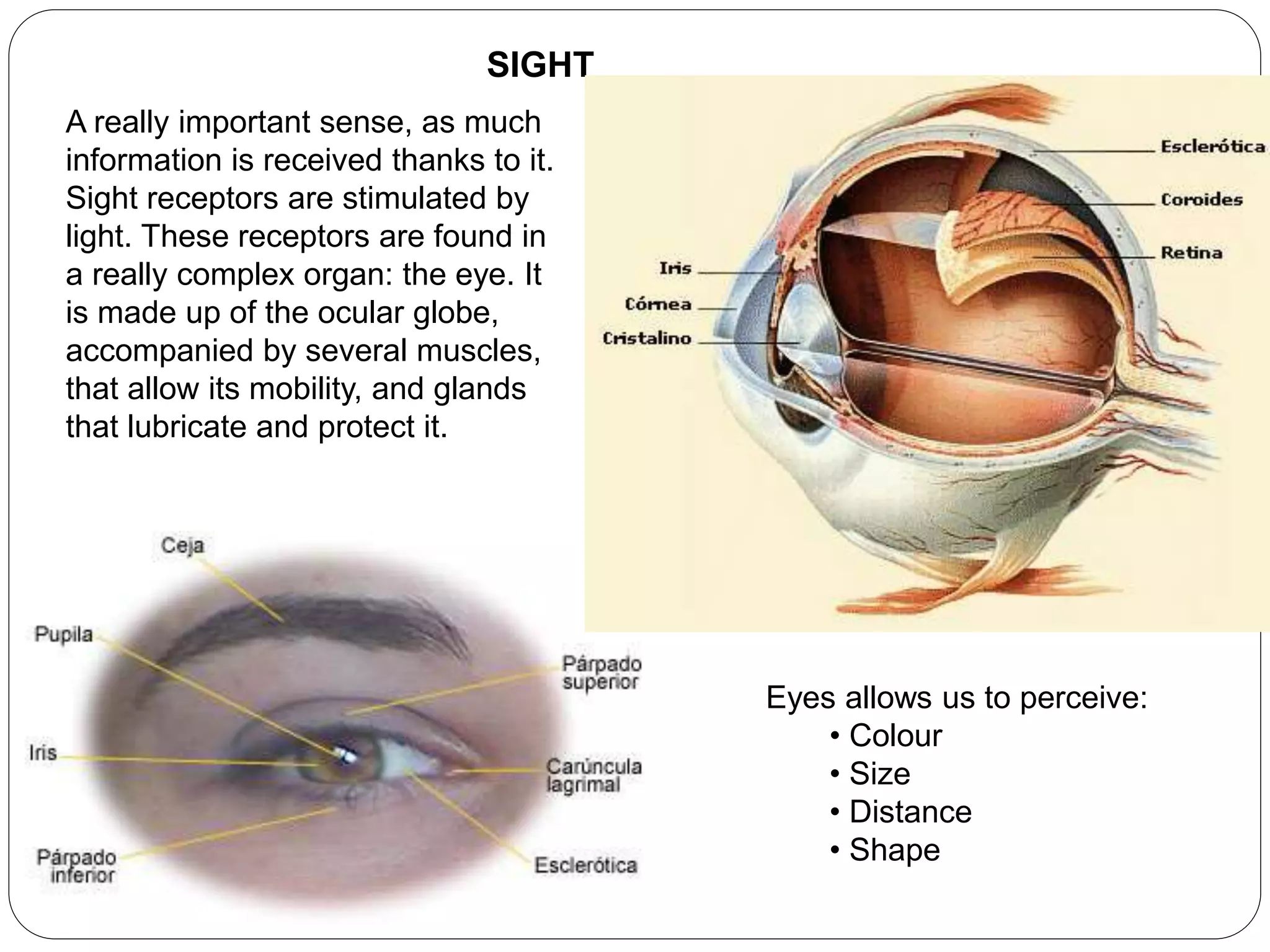
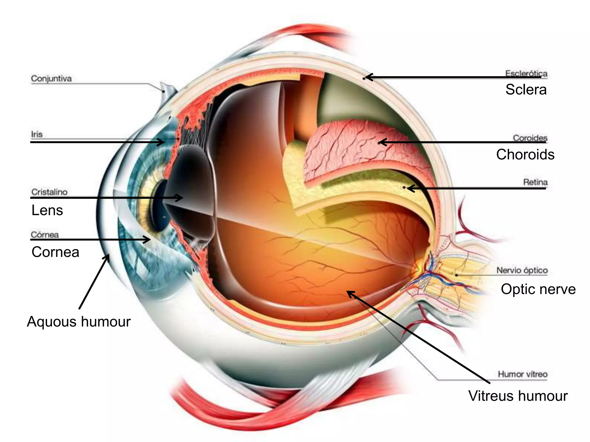
![Parts:
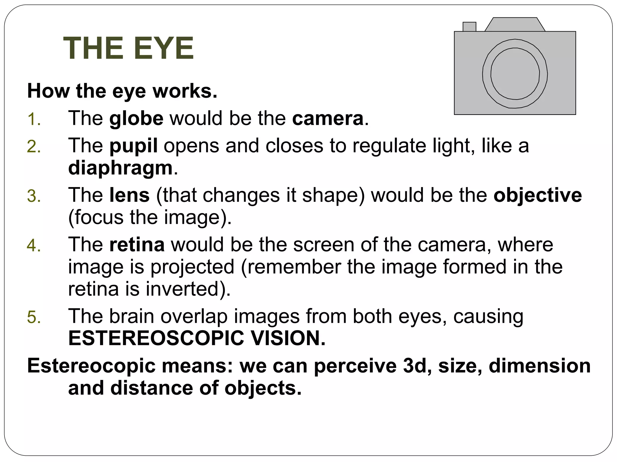
GLOBE:
Cornea
Iris
Sclera (esclerótica)
Choroid (coroides) [iris, pupil]
Aquous humour (humor acuoso)
Vitreous humour (humor vítreo)
Lens
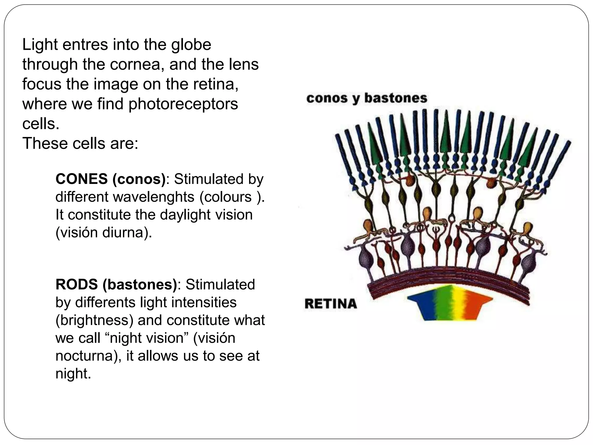
Retina [cells: rods, and cones]
ACCESORY ORGANS
Eyelids (párpados)
Eyelashes (pestañas)
Eyebrow (cejas)
Ocular muscles
Lacrimal glands
Description and functions in
your book.](https://image.slidesharecdn.com/receptorsandeffectors16-170307180108/75/Interaction-function-II-Receptors-and-effectors-7-2048.jpg)