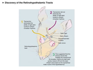
Circadian rhythms: sleep-waking cycle
Biological rhythms  (periodic physiological fluctuations) Types of rhythms Ultradian (Basic Rest-Activity Cycle) Circadian (sleep-wake cycle) Infradian (menstrual cycle) Circannual (annual breeding cycles) All rhythms allow us to time events and anticipate change!
Circadian Function Circadian (circa diem) rhythms   regular bodily rhythms that occur on a 24 hour cycle Internal biological clock(s)? Pacemaker cells; cryptochrome proteins; clock genes Fruit fly clock 4 regulatory proteins that interact to give the clock periodicity 2 proteins (CLOCK* and CYCLE) bind and increase production of PER (period) and TIM (timeless) – which accumulate over several hours When enough PER and TIM are made they inactivate CLOCK-CYCLE complex *   Circadian locomotor output cycles kaput
Human Clocks Cave Studies and free-running clocks (24 hrs 11 min) Why is this advantageous? Zeitgeber = External cue that helps to set the clock. Light/dark; temperature, social interactions, activity Entrainment/synchronization whether a cycle advances, is delayed, or remains unchanged differs depending on the phase in the cycle at which it is presented   REM and BRACS = Basic Resting Activity Cycles (approx. 90 minutes)
Measuring biological rhythms
FIGURE 1  Daily rhythms in rest–activity, body temperature, potassium excretion, computation speed (number of computations performed per minute), and time estimation (accuracy with which short intervals of time are assessed). From Wever (1974) with permission.
 
 
Suprachiasmatic nucleus is master pacemaker Activity in suprachiasmatic nucleus correlates with circadian rhythms Lesions of suprachiasmatic nucleus abolish free-running rhythms Isolated suprachiasmatic nucleus continues to cycle Transplanted suprachiasmatic nucleus imparts rhythm of the donor on the host
A Model proposed to explain circadian function
An anatomical route for regulating circadian function
Regulation of “The Clock”
A map of Activity Cycles in a Rat                                                                                                                                                      Pre SCN Ablation Post SCN Ablation
Subparaventricular zone is thought to reinforce or overlap function of the SCN.  Many of the projections are duplicate. Note: Consider effects across the listed categories
New Findings The existence of photoreceptors not specialized for visual functioning Regulate photoperiodism Entrainment of circadian rhythms Melanopsin-containing cells found in monkey retinal ganglion cell layer (Provencio et al., 2000)  Most likely comprise the retinohypothalamic tract Sensitive to wavelengths in the 484-500 nm (blue light)
SCN Rat SCN is divided into Dorsomedial (shell; nonvisual?) Vasopressin neurons ventrolateral (core; visual?) VIP neurons Geniculohypothalamic tract (GHT) Retinal afferents Overlapping  area containing Calretinin, Calbindin, gastrin-releasing peptide, substance P and enkephalin neurons
B: Retinohypothalamic Tract C: VIP in the ventrolateral or “core” of the SCN D: Vasopresin in the dorsomedial or “shell” of the SCN
Interconnectivity in SCN 40-70% of SCN neurons are GABAergic
IGL = Intergeniculate Leaflet, a subdivision of the LGN RHT raphe
SCN Herzog’s 2007 study:  VIP provides synchrony signal
Problems for SCN as a Pacemaker? SUPPORT Lesion SCN    loss of cycles Transplant SCN    resume cycles in animals PROBLEM? Connectivity of the transplant does not seem necessary for recovery. Other agents important: a peptide (prokineticin) appears important for transmitting info.  What is the spatial distribution of SCN neurons exhibiting clock-like behavior?  Are all SCN cells pacemakers? Do all pacemaker cells in the SCN oscillate at the same phase?
What is sleep? “ Natural periodic state of rest for the mind and body, in which the eyes usually close,  and consciousness is completely or partly lost , so that there is a decrease in bodily movement or external stimuli.” Not the absence of waking Not due to lack of sensory input An active process
Nathaniel Kleitman and the first sleep lab (1950s)
Single Cycle of Sleep
Characteristics of N-REM and REM
Getting the whole picture….
Sleep Stages Stage 1 (initial)- low voltage, fast wave Stage 2 - higher voltage, slower wave K complexes, sleep spindles Stage 3 - some delta waves Stage 4 - delta waves predominate Stage 1  emergent-  low muscle tone REM sleep
 
Typical Nightly Sleep Stages Hours of sleep Minutes of  Stage 4 and  REM 1 2 3 4 5 6 7 8 0 10 15 20 25 5 Decreasing  Stage 4 Increasing  REM
Night Terrors and Nightmares Night Terrors occur within 2 or 3 hours of falling asleep, usually during Stage 4 high arousal- appearance of being terrified Nightmares occur towards morning during REM sleep 0 1 2 3 4 5 6 7 4 3 2 1 Sleep stages Awake Hours of sleep REM
Sleep over the lifespan - early sleep patterns
Sleep changes over the lifespan Continuous REM in gestation Sleep quality changes with age:  Amount of time in slow wave and REM sleep decreases with age
Sleep architecture over the lifespan
Virtually all animals sleep Birds have short NREM and REM (9 seconds) waterfowl can sleep while  swimming transoceanic migrators can  sleep while flying Reptiles have no REM homeothermy? (but echidna * have no REM either) Smaller body size, more sleep     regulation of body temp? Longer life, less sleep * Spiny anteater—egg laying mammal Comparative Sleep Patterns
Half-sleep marine animals Either right or left side of the brain is in a sleep state Evidenced by EEG “ Half-asleep” for 8 hours a day Therefore, never fully unconscious/unaware Advantageous to prevent predation and drifting away
The functions and neural bases of sleep
Sleep deprivation I Sleep deprivation stunts Peter Tripp -- radio DJ sleep deprived self for 260 hours --> became psychotic Randy Gardner -- sleep deprived for 264 hours under supervision of sleep researcher Dement --> few reported ill-effects (played a mean game of  pinball)
Sleep deprivation II Rebound phenomena - following sleep deprivation, we recover much of our lost sleep but there is some segregation of recovery of different types of sleep. - following selective SWS or REM deprivation, there is selective recovery
Sleep deprivation III Sleep deprivation MAY cause death EXTREME sleep deprivation in animals will eventually cause death (thermoregulatory irregularities, loss of inflammatory responses, infection) Fatal familial insomnia leads to death but actual cause of death is unknown *  there’s a big stress confound here  *
Theories of Sleep I Sleep is adaptive ( Circadian Theory ) sleep forces us to be quiet at certain times of the day this allows us to share ecological niches with other  species allows us to conserve energy (species with high  metabolic demands sleep more, though metabolism is  high during REM) allows us to avoid predators (rough correlation  between predatory status and sleep properties, though  many animals are predator AND prey thermoregulation (sleep may help keep us cool -  alternating REM and SWS may prevent overcooling)
Theories of sleep function 2. Sleep is restorative ( Recuperation Theory ) -sleep helps us to get back something we lose during  waking -growth hormone is only secreted during sleep  (though not in kids under 4, not in adults over 60 and  not in all animals) -correlational studies not THAT convincing -small increase in SWS after ultramarathon -no decreases in sleep in quadraplegics
Theories of sleep function 3. Sleep promotes learning -sleep deprivation can have small effects on ability to  learn, but impossible to disentangle other effects of  deprivation - memory loss occurs when sleep is deprived on the  same night after material has been learned -some studies show a slight increase in REM after  difficult cognitive tasks -however, some people sleep little or not at all and  show no obvious deficits in ability to learn
Theories of sleep function No single theory of sleep function is completely satisfactory Perhaps sleep is multifactorial -- originally served to keep us quiet and still but now other functions (those that work best when we’re quiet and still?) piggy back onto the sleep state.
Reticular activating system integrates sensory input and regulates arousal Stimulating the system while the subject (usually a cat) is sleeping will awaken them . destruction results in somnolence Raphe nuclei lesions lead to insomnia  Serotonin source Normally promotes sleep destruction results in insomnia REM permanently inhibited Locus coeruleus:  dense nucleus of cells in brainstem NE source  promotes wakefulness The control of sleeping and waking is distributed in multiple areas of the brainstem to control the entire nervous system A balance and interaction between alert systems and rest systems Neural Mechanisms of Sleep
Raphe promotes sleep Locus coeruleus promotes wakefulness *so sleep control is distributed across centers
The reticular formation also promotes wakefulness
Narcolepsy Excessive daytime sleepiness  Abnormal REM sleep   Sleep paralysis Hypnagogic hallucinations   Cateplexy: sudden and transient paralysis triggered by high emotional arousal e.g hysteric laughing Hypothesis: Cholinergic hyperactivity and monaminergic hypoactivity in the pons Single autosomal recessive genetic disorder (in canines) Hypocretin…
Narcolepsy—Neurochemical Basis Narcolepsy has been studied since 1880 Hyocretin protein and receptor was discovered in 1998 and shown to be from the hypothalamus Hypocretin was attributed to narcolepsy in 1999 in canines, in 2000 for humans Greatly reduced levels of hypocretin peptides in CSF No or barely detectable hypocretin-containing neurons in their hypothalamus Mouse knockout for hypocretin made in 1999 and is an effective model for narcolepsy Modafinal drug treatment
What does hypocretin do and how? Increases wakefulness Suppresses REM sleep  Targets: Dorsal raphe Locus ceoruleus Pons Reticular formation Basal forbrain 2 receptor types Can have various effects Metabotrophic Narcolepsy—Neurochemical Basis SWS REM

Circadian Rhythms: Sleep-waking cycle

  • 1.
  • 2.
    Biological rhythms (periodic physiological fluctuations) Types of rhythms Ultradian (Basic Rest-Activity Cycle) Circadian (sleep-wake cycle) Infradian (menstrual cycle) Circannual (annual breeding cycles) All rhythms allow us to time events and anticipate change!
  • 3.
    Circadian Function Circadian(circa diem) rhythms regular bodily rhythms that occur on a 24 hour cycle Internal biological clock(s)? Pacemaker cells; cryptochrome proteins; clock genes Fruit fly clock 4 regulatory proteins that interact to give the clock periodicity 2 proteins (CLOCK* and CYCLE) bind and increase production of PER (period) and TIM (timeless) – which accumulate over several hours When enough PER and TIM are made they inactivate CLOCK-CYCLE complex * Circadian locomotor output cycles kaput
  • 4.
    Human Clocks CaveStudies and free-running clocks (24 hrs 11 min) Why is this advantageous? Zeitgeber = External cue that helps to set the clock. Light/dark; temperature, social interactions, activity Entrainment/synchronization whether a cycle advances, is delayed, or remains unchanged differs depending on the phase in the cycle at which it is presented REM and BRACS = Basic Resting Activity Cycles (approx. 90 minutes)
  • 5.
  • 6.
    FIGURE 1 Daily rhythms in rest–activity, body temperature, potassium excretion, computation speed (number of computations performed per minute), and time estimation (accuracy with which short intervals of time are assessed). From Wever (1974) with permission.
  • 7.
  • 8.
  • 9.
    Suprachiasmatic nucleus ismaster pacemaker Activity in suprachiasmatic nucleus correlates with circadian rhythms Lesions of suprachiasmatic nucleus abolish free-running rhythms Isolated suprachiasmatic nucleus continues to cycle Transplanted suprachiasmatic nucleus imparts rhythm of the donor on the host
  • 10.
    A Model proposedto explain circadian function
  • 11.
    An anatomical routefor regulating circadian function
  • 12.
  • 13.
    A map ofActivity Cycles in a Rat                                                                                                                                                      Pre SCN Ablation Post SCN Ablation
  • 14.
    Subparaventricular zone isthought to reinforce or overlap function of the SCN. Many of the projections are duplicate. Note: Consider effects across the listed categories
  • 15.
    New Findings Theexistence of photoreceptors not specialized for visual functioning Regulate photoperiodism Entrainment of circadian rhythms Melanopsin-containing cells found in monkey retinal ganglion cell layer (Provencio et al., 2000) Most likely comprise the retinohypothalamic tract Sensitive to wavelengths in the 484-500 nm (blue light)
  • 16.
    SCN Rat SCNis divided into Dorsomedial (shell; nonvisual?) Vasopressin neurons ventrolateral (core; visual?) VIP neurons Geniculohypothalamic tract (GHT) Retinal afferents Overlapping area containing Calretinin, Calbindin, gastrin-releasing peptide, substance P and enkephalin neurons
  • 17.
    B: Retinohypothalamic TractC: VIP in the ventrolateral or “core” of the SCN D: Vasopresin in the dorsomedial or “shell” of the SCN
  • 18.
    Interconnectivity in SCN40-70% of SCN neurons are GABAergic
  • 19.
    IGL = IntergeniculateLeaflet, a subdivision of the LGN RHT raphe
  • 20.
    SCN Herzog’s 2007study: VIP provides synchrony signal
  • 21.
    Problems for SCNas a Pacemaker? SUPPORT Lesion SCN  loss of cycles Transplant SCN  resume cycles in animals PROBLEM? Connectivity of the transplant does not seem necessary for recovery. Other agents important: a peptide (prokineticin) appears important for transmitting info. What is the spatial distribution of SCN neurons exhibiting clock-like behavior? Are all SCN cells pacemakers? Do all pacemaker cells in the SCN oscillate at the same phase?
  • 22.
    What is sleep?“ Natural periodic state of rest for the mind and body, in which the eyes usually close, and consciousness is completely or partly lost , so that there is a decrease in bodily movement or external stimuli.” Not the absence of waking Not due to lack of sensory input An active process
  • 23.
    Nathaniel Kleitman andthe first sleep lab (1950s)
  • 24.
  • 25.
  • 26.
    Getting the wholepicture….
  • 27.
    Sleep Stages Stage1 (initial)- low voltage, fast wave Stage 2 - higher voltage, slower wave K complexes, sleep spindles Stage 3 - some delta waves Stage 4 - delta waves predominate Stage 1 emergent- low muscle tone REM sleep
  • 28.
  • 29.
    Typical Nightly SleepStages Hours of sleep Minutes of Stage 4 and REM 1 2 3 4 5 6 7 8 0 10 15 20 25 5 Decreasing Stage 4 Increasing REM
  • 30.
    Night Terrors andNightmares Night Terrors occur within 2 or 3 hours of falling asleep, usually during Stage 4 high arousal- appearance of being terrified Nightmares occur towards morning during REM sleep 0 1 2 3 4 5 6 7 4 3 2 1 Sleep stages Awake Hours of sleep REM
  • 31.
    Sleep over thelifespan - early sleep patterns
  • 32.
    Sleep changes overthe lifespan Continuous REM in gestation Sleep quality changes with age: Amount of time in slow wave and REM sleep decreases with age
  • 33.
  • 34.
    Virtually all animalssleep Birds have short NREM and REM (9 seconds) waterfowl can sleep while swimming transoceanic migrators can sleep while flying Reptiles have no REM homeothermy? (but echidna * have no REM either) Smaller body size, more sleep  regulation of body temp? Longer life, less sleep * Spiny anteater—egg laying mammal Comparative Sleep Patterns
  • 35.
    Half-sleep marine animalsEither right or left side of the brain is in a sleep state Evidenced by EEG “ Half-asleep” for 8 hours a day Therefore, never fully unconscious/unaware Advantageous to prevent predation and drifting away
  • 36.
    The functions andneural bases of sleep
  • 37.
    Sleep deprivation ISleep deprivation stunts Peter Tripp -- radio DJ sleep deprived self for 260 hours --> became psychotic Randy Gardner -- sleep deprived for 264 hours under supervision of sleep researcher Dement --> few reported ill-effects (played a mean game of pinball)
  • 38.
    Sleep deprivation IIRebound phenomena - following sleep deprivation, we recover much of our lost sleep but there is some segregation of recovery of different types of sleep. - following selective SWS or REM deprivation, there is selective recovery
  • 39.
    Sleep deprivation IIISleep deprivation MAY cause death EXTREME sleep deprivation in animals will eventually cause death (thermoregulatory irregularities, loss of inflammatory responses, infection) Fatal familial insomnia leads to death but actual cause of death is unknown * there’s a big stress confound here *
  • 40.
    Theories of SleepI Sleep is adaptive ( Circadian Theory ) sleep forces us to be quiet at certain times of the day this allows us to share ecological niches with other species allows us to conserve energy (species with high metabolic demands sleep more, though metabolism is high during REM) allows us to avoid predators (rough correlation between predatory status and sleep properties, though many animals are predator AND prey thermoregulation (sleep may help keep us cool - alternating REM and SWS may prevent overcooling)
  • 41.
    Theories of sleepfunction 2. Sleep is restorative ( Recuperation Theory ) -sleep helps us to get back something we lose during waking -growth hormone is only secreted during sleep (though not in kids under 4, not in adults over 60 and not in all animals) -correlational studies not THAT convincing -small increase in SWS after ultramarathon -no decreases in sleep in quadraplegics
  • 42.
    Theories of sleepfunction 3. Sleep promotes learning -sleep deprivation can have small effects on ability to learn, but impossible to disentangle other effects of deprivation - memory loss occurs when sleep is deprived on the same night after material has been learned -some studies show a slight increase in REM after difficult cognitive tasks -however, some people sleep little or not at all and show no obvious deficits in ability to learn
  • 43.
    Theories of sleepfunction No single theory of sleep function is completely satisfactory Perhaps sleep is multifactorial -- originally served to keep us quiet and still but now other functions (those that work best when we’re quiet and still?) piggy back onto the sleep state.
  • 44.
    Reticular activating systemintegrates sensory input and regulates arousal Stimulating the system while the subject (usually a cat) is sleeping will awaken them . destruction results in somnolence Raphe nuclei lesions lead to insomnia Serotonin source Normally promotes sleep destruction results in insomnia REM permanently inhibited Locus coeruleus: dense nucleus of cells in brainstem NE source promotes wakefulness The control of sleeping and waking is distributed in multiple areas of the brainstem to control the entire nervous system A balance and interaction between alert systems and rest systems Neural Mechanisms of Sleep
  • 45.
    Raphe promotes sleepLocus coeruleus promotes wakefulness *so sleep control is distributed across centers
  • 46.
    The reticular formationalso promotes wakefulness
  • 47.
    Narcolepsy Excessive daytimesleepiness Abnormal REM sleep Sleep paralysis Hypnagogic hallucinations Cateplexy: sudden and transient paralysis triggered by high emotional arousal e.g hysteric laughing Hypothesis: Cholinergic hyperactivity and monaminergic hypoactivity in the pons Single autosomal recessive genetic disorder (in canines) Hypocretin…
  • 48.
    Narcolepsy—Neurochemical Basis Narcolepsyhas been studied since 1880 Hyocretin protein and receptor was discovered in 1998 and shown to be from the hypothalamus Hypocretin was attributed to narcolepsy in 1999 in canines, in 2000 for humans Greatly reduced levels of hypocretin peptides in CSF No or barely detectable hypocretin-containing neurons in their hypothalamus Mouse knockout for hypocretin made in 1999 and is an effective model for narcolepsy Modafinal drug treatment
  • 49.
    What does hypocretindo and how? Increases wakefulness Suppresses REM sleep Targets: Dorsal raphe Locus ceoruleus Pons Reticular formation Basal forbrain 2 receptor types Can have various effects Metabotrophic Narcolepsy—Neurochemical Basis SWS REM