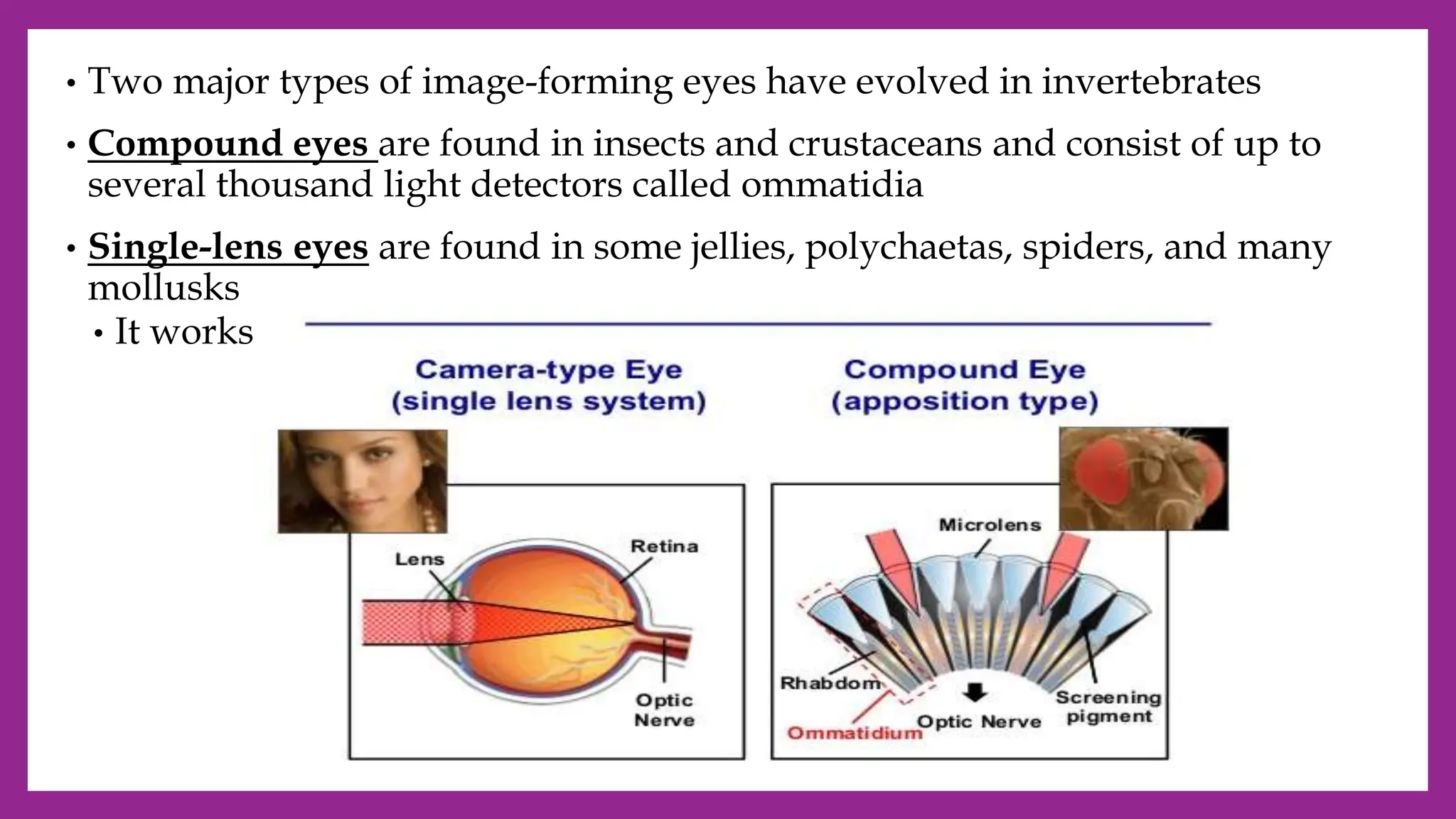
The document describes the major sensory systems and organs in animals. It discusses:
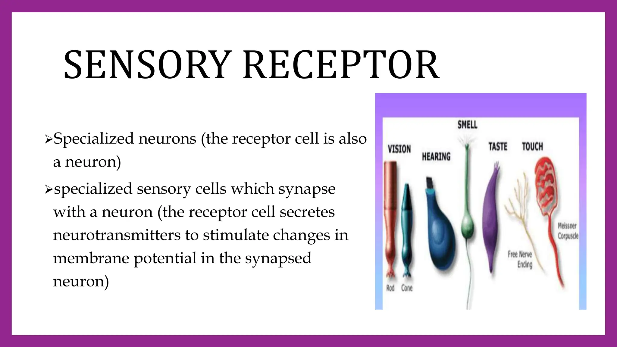
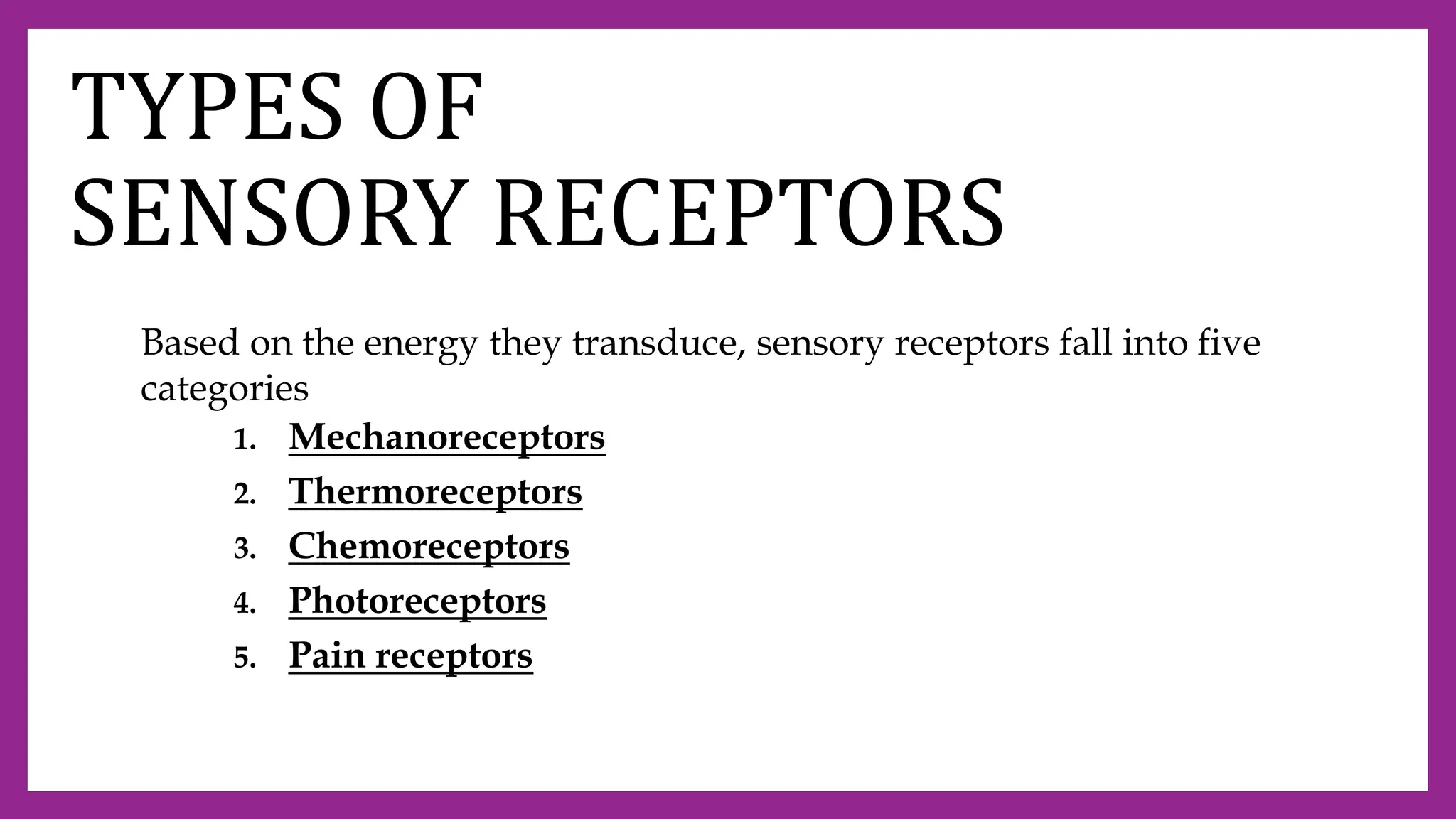
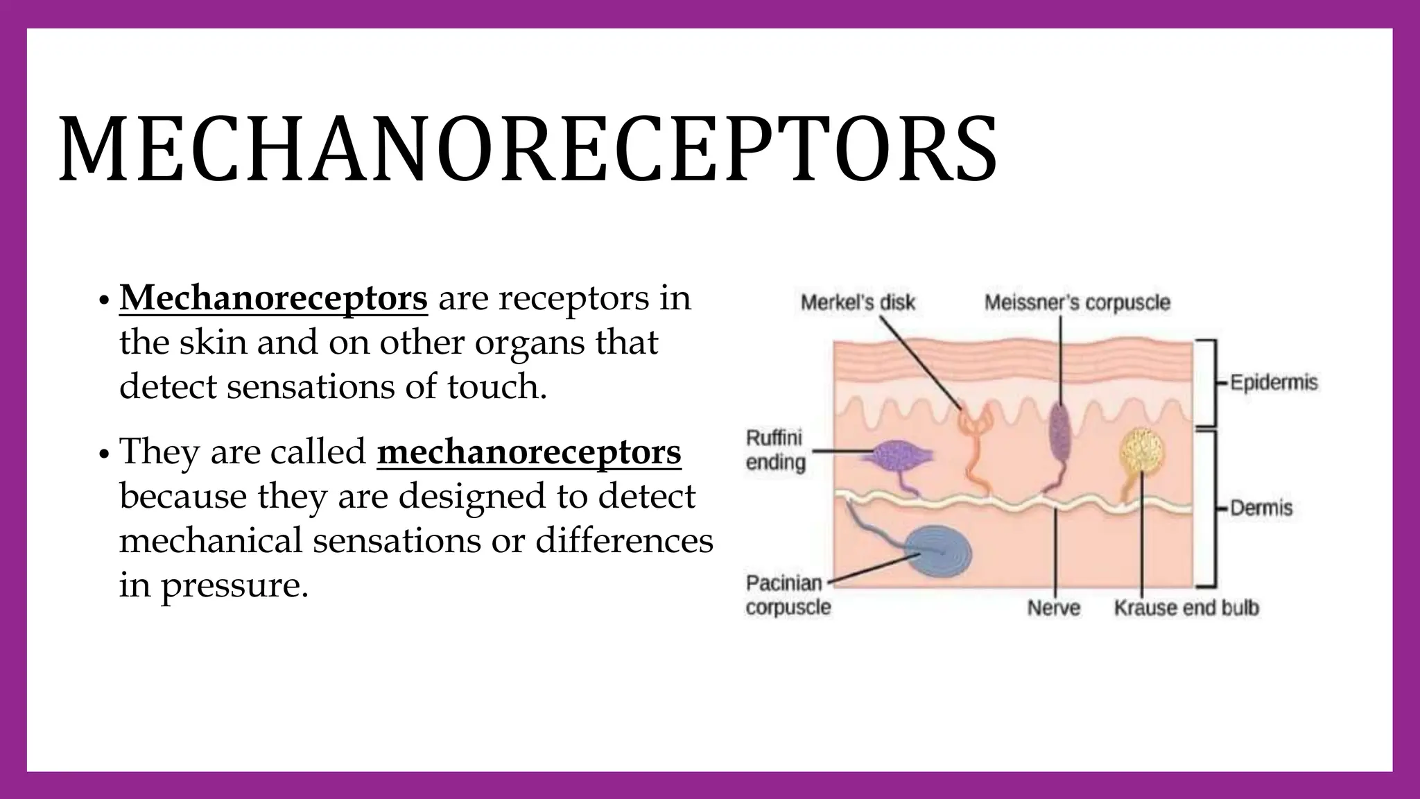
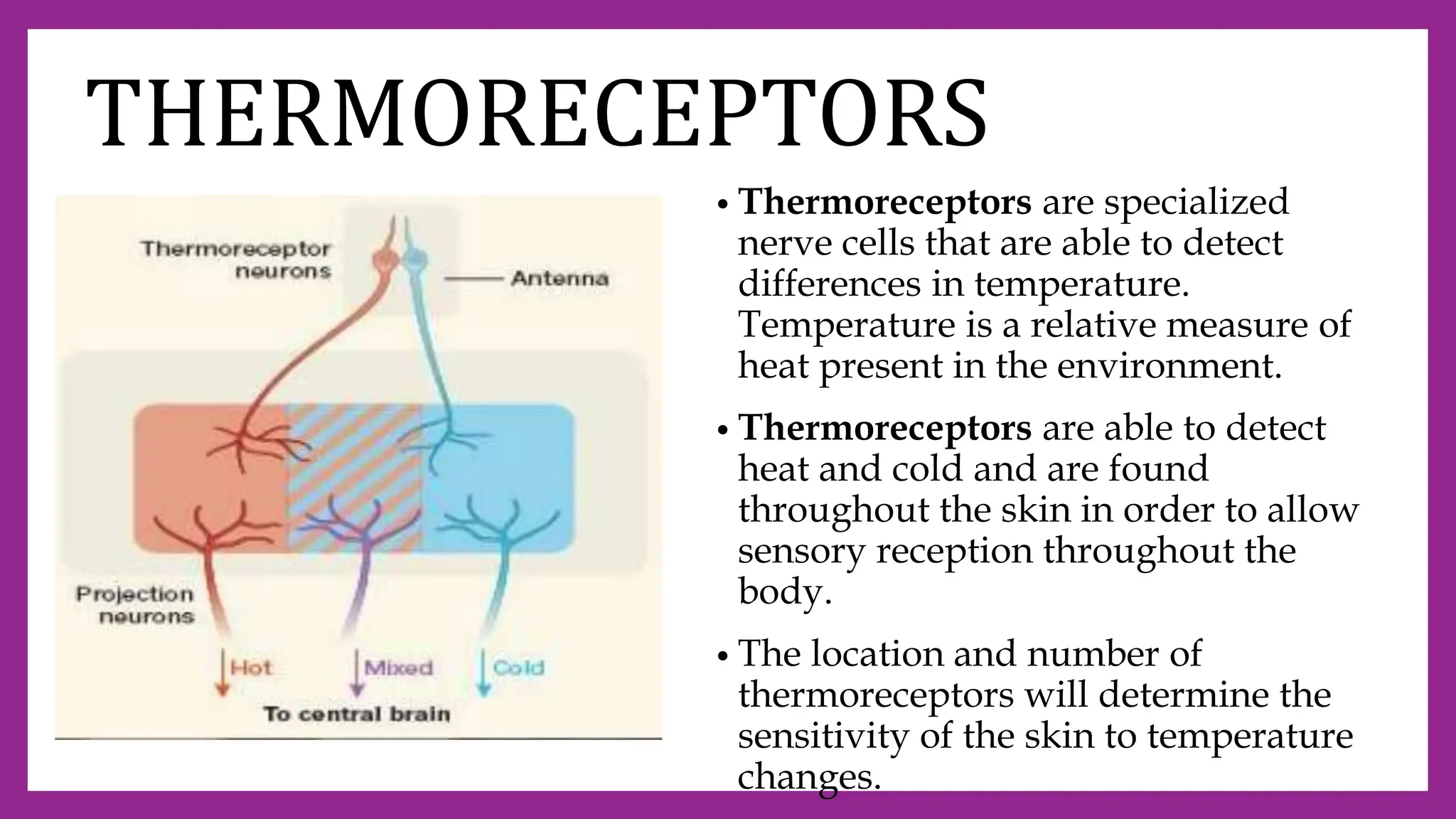
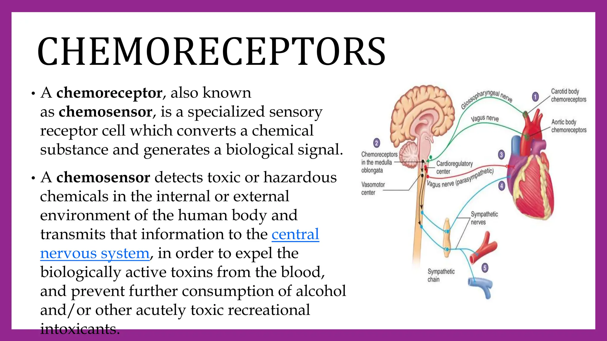
1. The five main categories of sensory receptors - mechanoreceptors, thermoreceptors, chemoreceptors, photoreceptors, and pain receptors - which detect different types of stimuli.
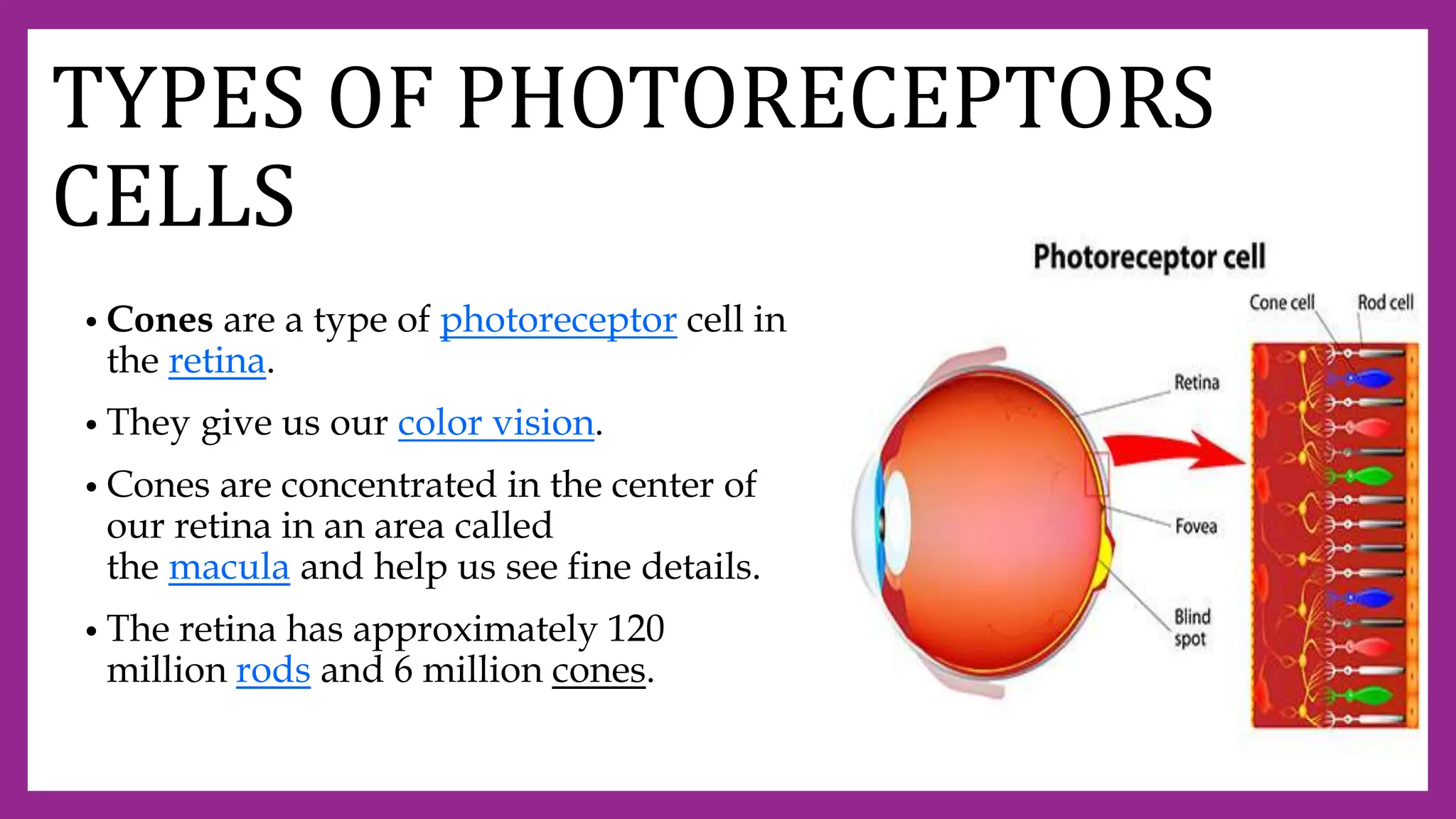
2. The structures and functions of sensory organs like the eye, which contains photoreceptors like rods and cones to detect light, and the ear, with outer, middle and inner sections that detect sound.
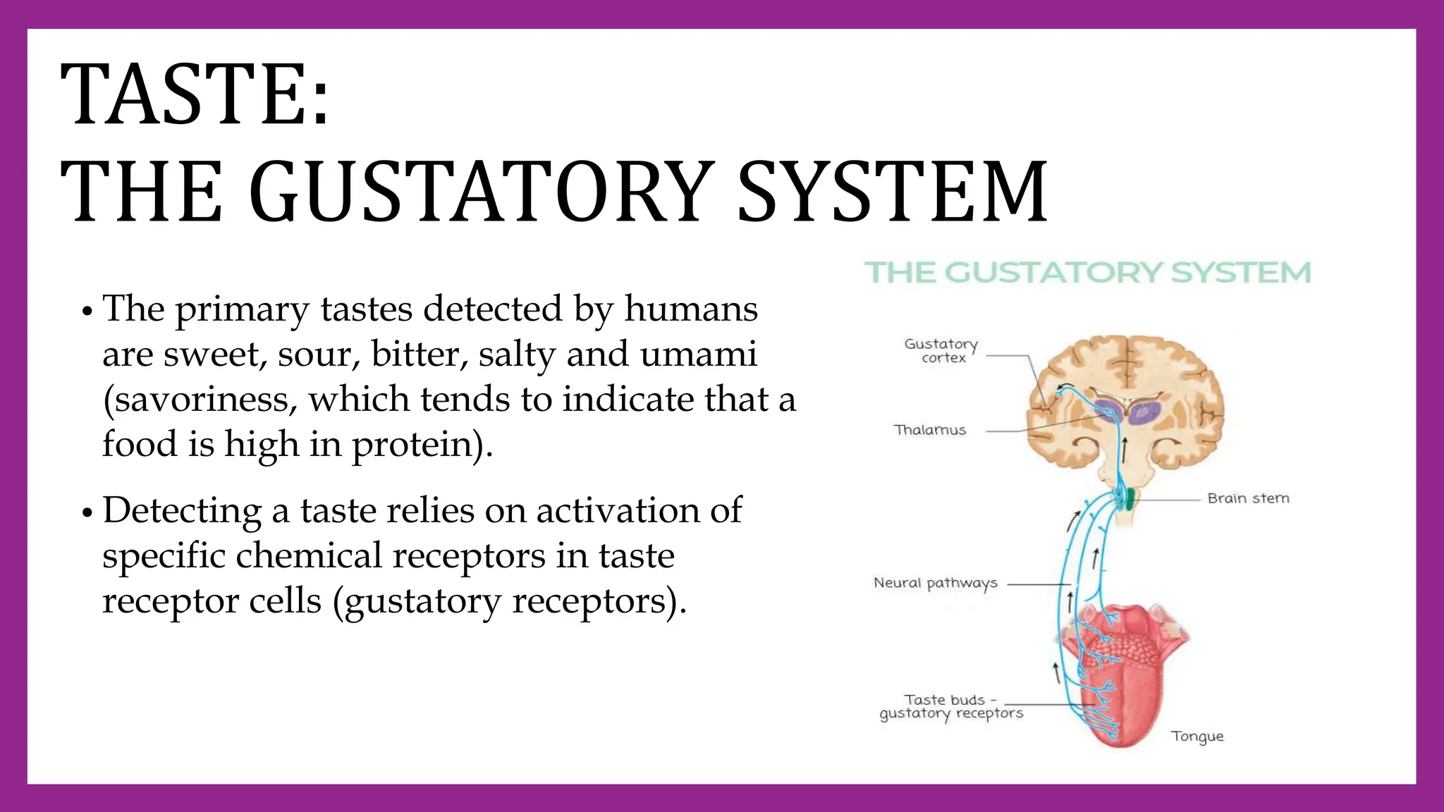
3. Other sensory systems like taste buds that contain gustatory receptors to detect the five basic tastes, and the olfactory system containing receptors that allow us to detect thousands of odors.


![CHAPTER 1_F3 [Autosaved].pptx](https://cdn.slidesharecdn.com/ss_thumbnails/chapter1f3autosaved-230327142030-7a281fc4-thumbnail.jpg?width=640&height=640&fit=bounds)


