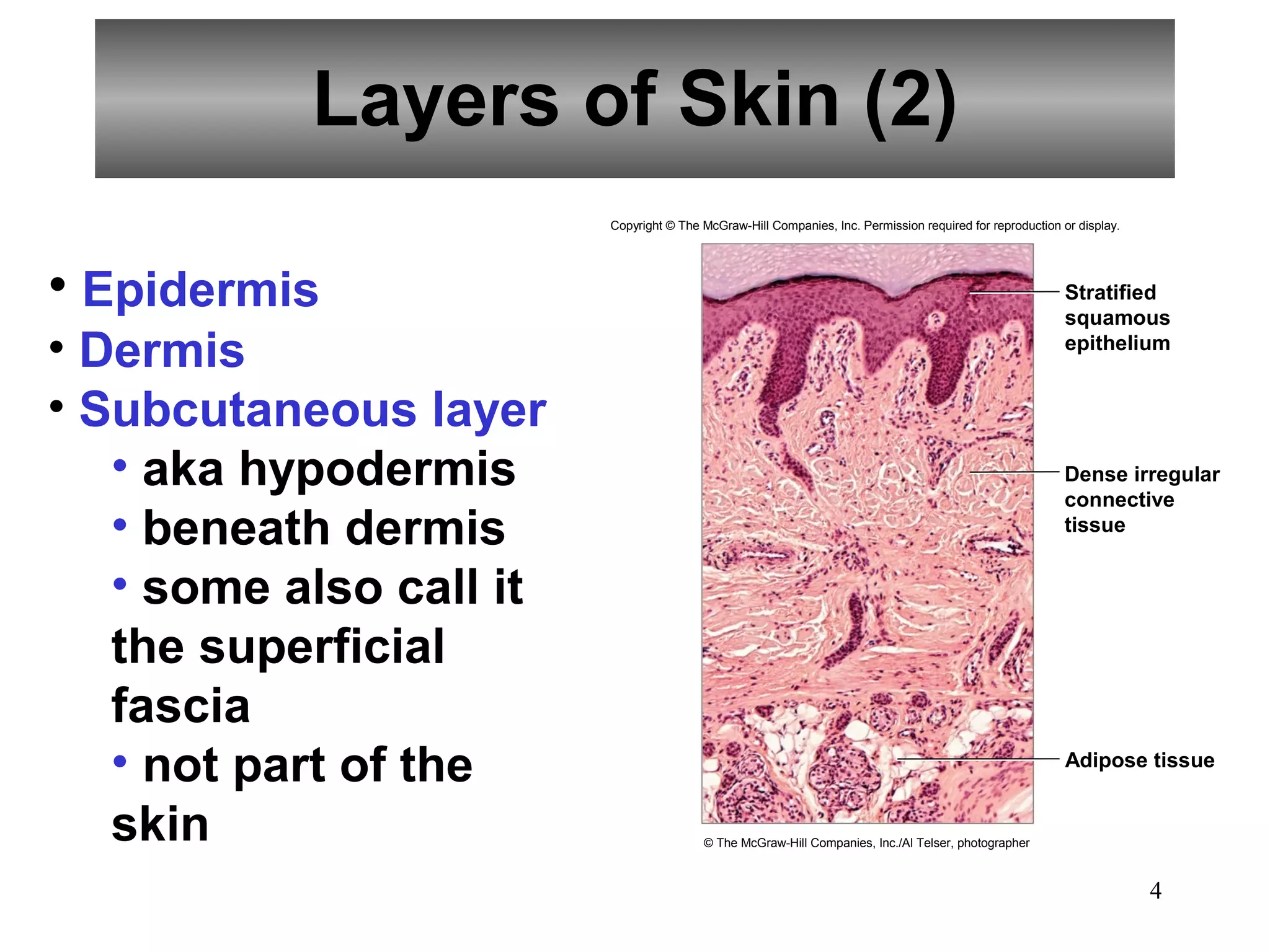
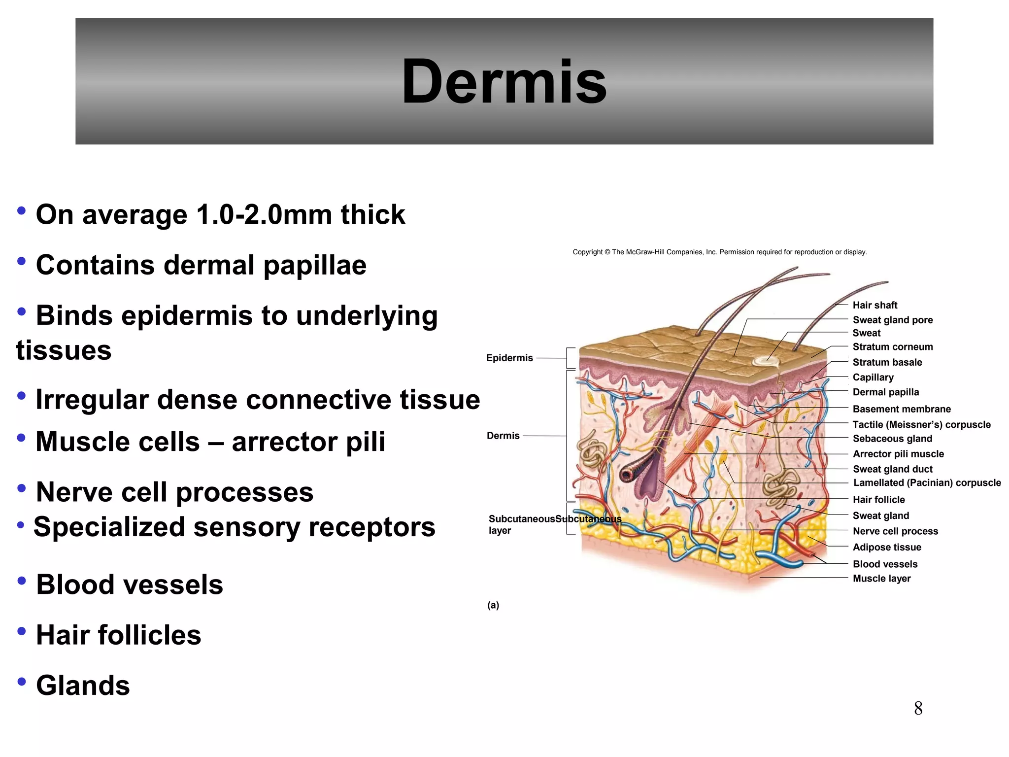
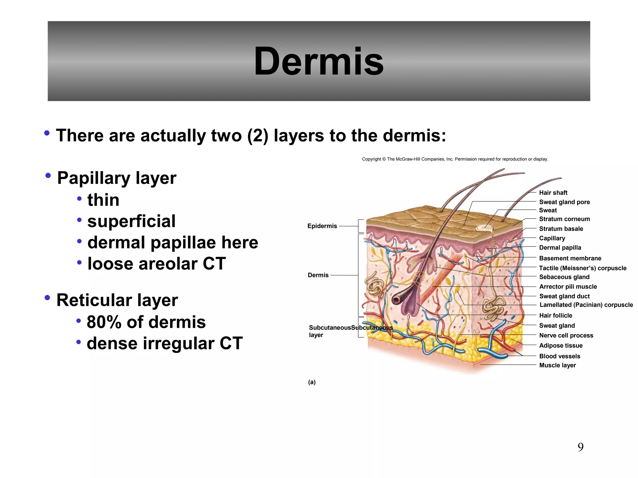
PowerPoint
                     Lecture

              Integumentary
                  System


Copyright © The McGraw-Hill Companies, Inc. Permission required for reproduction or display.
Introduction
• Two or more kinds of tissues grouped together
and performing specialized functions constitutes
an organ.

• The skin and its various structures make up the
integumentary system.


                                                    2
Skin and Its Tissues

• Composed of several tissue types
• Maintains homeostasis
• Protective covering
• Retards water loss
• Regulates body temperature
• Houses sensory receptors
• Contains immune system cells
• Synthesizes chemicals (such as vit D)
• Excretes small amounts of wastes


                                          3
Layers of Skin (2)
                         Copyright © The McGraw-Hill Companies, Inc. Permission required for reproduction or display.


• Epidermis                                                                                                Stratified
                                                                                                           squamous
• Dermis                                                                                                   epithelium


• Subcutaneous layer
   • aka hypodermis                                                                                       Dense irregular
                                                                                                          connective
   • beneath dermis                                                                                       tissue


   • some also call it
   the superficial
   fascia
   • not part of the                                                                                       Adipose tissue


   skin                                  © The McGraw-Hill Companies, Inc./Al Telser, photographer


                                                                                                                        4
Epidermis
                                             Copyright © The McGraw-Hill Companies, Inc. Permission required for reproduction or display.


• Lacks blood vessels                                                                                                               Hair shaft


• Keratinized
                                                                                                                                    Sweat gland pore
                                                                                                                                    Sweat
                                                                                                                                    Stratum corneum
                             Epidermis
                                                                                                                                    Stratum basale


• Thickest on palms and                                                                                                             Capillary
                                                                                                                                    Dermal papilla

                                                                                                                                    Basement membrane


  soles (0.8-1.4mm)          Dermis
                                                                                                                                    TTactile (Meissner’s) corpuscle
                                                                                                                                    Sebaceous gland
                                                                                                                                    Arrector pili muscle


• Melanocytes provide                                                                                                               Sweat gland duct
                                                                                                                                   Lamellated (Pacinian) corpuscle

                                                                                                                                    Hair follicle


melanin                     Subcutaneous
                            layer
                                                                                                                                    Sweat gland

                                                                                                                                    Nerve cell process


• Rests on basement membrane
                                                                                                                                    Adipose tissue

                                                                                                                                    Blood vessels
                                                                                                                                    Muscle layer


• Stratified squamous epithelia
                             (a)


                                                                                                                                            Hair shaft


                                                                                                                                            Epidermis


                           Hair follicle                                                                                                    Dermis


                           Sebaceous gland


                             (b)                                                                                                                     5
                                                                               b: © Victor Eroschenko
Epidermis
There are five (5) layers of the epidermis:
   • stratum corneum
   • stratum lucidum
   • stratum granulosum
   • stratum spinosum
   • stratum basale
                   Copyright © The McGraw-Hill Companies, Inc. Permission required for reproduction or display.


                                                  Stratum corneum


                                                   Stratum lucidum
                                                  Stratum granulosum
                                                  Stratum spinosum

                                                  Stratum basale


                                                   Basement
                                                   membrane
                                                   Dermal papilla
                                                   Dermis

    (a)                                                                                   (b)                     6
                                  b: © The McGraw-Hill Companies, Inc./Al Telser, photographer
Epidermis
• Heredity and environment determine skin color
• Genetic Factors              • Physiological Factors
   • Varying amounts of melanin    • Dilation of dermal blood vessels
   • Varying size of melanin       • Constriction of dermal blood
   granules                        vessels
   • Albinos lack melanin          • Accumulation of carotene
                                   • Jaundice
• Environmental Factors            • Cyanosis
   • Sunlight
   • UV light from sunlamps
   • X-rays
   • Darkens melanin


                                                              7
Dermis
• On average 1.0-2.0mm thick
• Contains dermal papillae
                                                     Copyright © The McGraw-Hill Companies, Inc. Permission required for reproduction or display.


• Binds epidermis to underlying                                                                                                             Hair shaft
                                                                                                                                            Sweat gland pore
                                                                                                                                            Sweat

tissues                               Epidermis
                                                                                                                                            Stratum corneum
                                                                                                                                            Stratum basale
                                                                                                                                            Capillary

• Irregular dense connective tissue                                                                                                         Dermal papilla
                                                                                                                                            Basement membrane


• Muscle cells – arrector pili
                                                                                                                                            Tactile (Meissner’s) corpuscle
                                      Dermis                                                                                                Sebaceous gland
                                                                                                                                            Arrector pili muscle
                                                                                                                                            Sweat gland duct

• Nerve cell processes                                                                                                                      Lamellated (Pacinian) corpuscle
                                                                                                                                            Hair follicle


• Specialized sensory receptors       SubcutaneousSubcutaneous                                                                              Sweat gland
                                      layer                                                                                                 Nerve cell process
                                                                                                                                            Adipose tissue
                                                                                                                                            Blood vessels

• Blood vessels                       (a)
                                                                                                                                            Muscle layer


• Hair follicles
• Glands
                                                                                                                                                            8
Dermis
• There are actually two (2) layers to the dermis:
                                               Copyright © The McGraw-Hill Companies, Inc. Permission required for reproduction or display.


• Papillary layer
    • thin
                                                                                                                                      Hair shaft
                                                                                                                                      Sweat gland pore
                                                                                                                                      Sweat

    • superficial               Epidermis
                                                                                                                                      Stratum corneum
                                                                                                                                      Stratum basale

    • dermal papillae here                                                                                                            Capillary
                                                                                                                                      Dermal papilla


    • loose areolar CT          Dermis
                                                                                                                                      Basement membrane
                                                                                                                                      Tactile (Meissner’s) corpuscle
                                                                                                                                      Sebaceous gland
                                                                                                                                      Arrector pili muscle

• Reticular layer                                                                                                                     Sweat gland duct
                                                                                                                                      Lamellated (Pacinian) corpuscle


    • 80% of dermis
                                                                                                                                      Hair follicle
                                SubcutaneousSubcutaneous                                                                              Sweat gland
                                layer                                                                                                 Nerve cell process
    • dense irregular CT                                                                                                              Adipose tissue
                                                                                                                                      Blood vessels
                                                                                                                                      Muscle layer

                                (a)


                                                                                                                                                      9
Subcutaneous Layer
• Aka hypodermis                    Copyright © The McGraw-Hill Companies, Inc. Permission required for reproduction or display.


• Loose connective                                                                                                         Hair shaft
                                                                                                                           Sweat gland pore
                                                                                                                           Sweat

tissue and …         Epidermis
                                                                                                                           Stratum corneum
                                                                                                                           Stratum basale
                                                                                                                           Capillary
                                                                                                                           Dermal papilla

• Adipose tissue     Dermis
                                                                                                                           Basement membrane
                                                                                                                           Tactile (Meissner’s) corpuscle
                                                                                                                           Sebaceous gland

are present                                                                                                                Arrector pili muscle
                                                                                                                           Sweat gland duct
                                                                                                                           Lamellated (Pacinian) corpuscle


• Insulates          Subcutaneous
                                                                                                                           Hair follicle
                                                                                                                           Sweat gland
                     layer                                                                                                 Nerve cell process
                                                                                                                           Adipose tissue


• Major blood                                                                                                              Blood vessels
                                                                                                                           Muscle layer


vessels present
                     (a)


                                                                                                                                       10
Accessory Structures
            of the Skin
• Accessory structures of the skin originate from the
epidermis and include:
   • Hair follicles
   • Nails
   • Skin glands


                                                        11
Hair Follicles
                         Copyright © The McGraw-Hill Companies, Inc. Permission required for reproduction or display.


• Epidermal cells
                                                                                                          Hair shaft
• Tube-like depression                                                                                    Pore


• Extends into dermis
• Three (3) parts:                                                                                        Sebaceous
                                                                                                          gland

   • Hair root
    • Hair shaft                                                                                          Arrector pili
                                                                                                          muscle
                                                                                                          Hair root

   • Hair papilla                                                                                         (keratinized
                                                                                                          cells)
                                                                                                          Hair follicle

• Melanin                                                                                                 Eccrine
                                                                                                          sweat gland
                                                                                                          Region of

• Arrector pili muscle                                                                                    cell division
                                                                                                          Hair papilla


                                                                                                          DermalDermal
                                                                                                          blood
                                                                                                          vessels
                           (a)
                                                                                                                          12
Nails
                           Copyright © The McGraw-Hill Companies, Inc. Permission required for reproduction or display.


• Protective coverings                                     Lunula Nail bed Nail plate


• Three (3) parts:
   • Nail plate
    • Nail bed
    • Lunula


                                                                                                                          13
Sebaceous Glands
                                 Copyright © The McGraw-Hill Companies, Inc. Permission required for reproduction or display.


• Usually associated with hair                                                                                              Sebaceous
                                                                                                                            gland
follicles
                                                                                                                            Hair follicle


• Holocrine glands

• Secrete sebum (oil)                                                                                                      Hair


• Absent on palms and soles


                                                                      © Per H. Kjeldsen
                                                                                                                                  14
Sweat Glands
                            Copyright © The McGraw-Hill Companies, Inc. Permission required for reproduction or display.


• Aka sudoriferous glands
                       Hair shaft
• Widespread in skin                                                                                                Pore


• Originates in deeper dermis                                                                                       Dermal
                                                                                                                    papilla
or hypodermis
                                                                                                                    Sebaceous
                                                                                                                    gland
• Eccrine glands                                                                                                    Duct

                                                                                                                    Hair

• Apocrine glands                                                                                                   follicle

                                                                                                                    Eccrine
                                                                                                                    sweat

• Ceruminous glands                                                                                                 gland

                                                                                                                    Apocrine
                                                                                                                    sweat
• Mammary glands                                                                                                    gland
                                                                                                                           15
Regulation of
           Body Temperature
• Regulation of body temperature is vitally important
because even slight shifts can disrupt metabolic
reactions.


                                                        16
Regulation of Body
  Temperature
       Copyright © The McGraw-Hill Companies, Inc. Permission required for reproduction or display.


                           Control center
                           Hypothalamus
                           detects the deviation
                           from the set point and
                           signals effector organs.


      Receptors                                      Effectors
      Thermoreceptors                                Dermal blood vessels
      send signals to the                            dilate and sweat glands
      control center.                                secrete.


    Stimulus                                       Response
    Body temperature rises                         Body heat is
    above normal.                                  lost to surroundings,
                                                   temperature drops toward
                                                   normal.

                        too high


                      Normal body
                      temperature
                      37°C (98.6°F)


                         too low


    Stimulus                                        Response
    Body temperature                                Body heat is conserved,
    drops below normal.                             temperature rises toward normal.


       Receptors                                    Effectors                   Effectors
       Thermoreceptors                              Dermal blood                Dermal blood
       send signals to the                          vessels constrict           vessels constrict
       control center.                              and sweat glands            and sweat glands
                                                    remain inactive.            remain inactive.


                           Control center
                           Hypothalamus
                           detects the deviation
                           from the set point and
                           signals effector organs.                 If body temperature
                                                                    continues to drop,
                                                                    control center signals            17
                                                                    muscles to contract
                                                                    involuntarily.
Heat Production and Loss
• Heat is a product of cellular metabolism
   • The most active body cells are the heat producers
   and include:
       • Skeletal muscle
       • Cardiac muscle
       • Cells of certain glands such as the liver
• The primary means of heat loss is radiation
   • Also there is conduction, convection and
   evaporation


                                                     18
Problems in Temperature
       Regulation
• Hyperthermia – abnormally high body temperature


• Hypothermia – abnormally low body temperature


                                                  19
Healing of Wounds and Burns

• Inflammation is a normal response to injury or stress.
• Blood vessels in affected tissues dilate and become
more permeable, allowing fluids to leak into the
damaged tissues.
• Inflammed skin may become:
    • Reddened
    • Swollen
    • Warm
    • Painful


                                                           20
Healing of Cuts
                          Copyright © The McGraw-Hill Companies, Inc. Permission required for reproduction or display.


                                                                                                                         Site of injury


                                                                                                                         Blood cells


                    (a)                                                           (b)


                                                                                                                                          Scab


                                                                                           Blood
                                                                                           clot


(c)                                      (d)                                                              (e)


                                                                                                                         Scab


                                                                                                                         Scar
                                                                                                                         tissue

      Scar
      tissue
      Fibroblasts


                                                                                                                                                 21
                    (f)                                                           (g)
Types of Burns
• First degree burn – superficial, partial-thickness

• Second degree burn – deep, partial-thickness

• Third degree burn – full-thickness


                                                       22
Rule of Nines for Adults
                       Copyright © The McGraw-Hill Companies, Inc. Permission required for reproduction or display.


Anterior head                       41/2%        Anterior and posterior head and neck                   41/ 2%                 Posterior head
and neck 41/2%                                                    9%                                                           and neck 41/2%


                                  Anterior                     Anterior and                            Posterior
                                  trunk                       posterior upper                          trunk
                                  18%                           extremities                            18%
                                                                    18%
Anterior upper                                                 Anterior and                                                    Posterior upper
extremities 9%                                                posterior trunk                                                  extremities 9%
                                                                   36%
                  41/ 2%                            41/2%                             41/2%                           41/ 2%


                                                                  Perineum 1%
                               9%           9%                                                     9%            9%
Anterior lower                                                 Anterior and                                                    Posterior lower
extremities 18%                                               posterior lower                                                  extremities 18%
                                                                extremities
                                                                    36%


                                                                                                                                                 23
                                                                    100%
Lifespan Changes
• Skin becomes scaly   • Melanin production slows
• Age spots appear     • Hair thins
• Epidermis thins      • Number of hair follicles
• Dermis becomes       decreases
reduced                • Nail growth becomes
• Loss of fat          impaired
• Wrinkling            • Sensory receptors decline
• Sagging              • Body temperature unable to
• Sebaceous glands     be controlled
secrete less oil       • Diminished ability to
                       activate Vitamin D

                                               24

Skin ap

  • 1.
    PowerPoint Lecture Integumentary System Copyright © The McGraw-Hill Companies, Inc. Permission required for reproduction or display.
  • 2.
    Introduction • Two ormore kinds of tissues grouped together and performing specialized functions constitutes an organ. • The skin and its various structures make up the integumentary system. 2
  • 3.
    Skin and ItsTissues • Composed of several tissue types • Maintains homeostasis • Protective covering • Retards water loss • Regulates body temperature • Houses sensory receptors • Contains immune system cells • Synthesizes chemicals (such as vit D) • Excretes small amounts of wastes 3
  • 4.
    Layers of Skin(2) Copyright © The McGraw-Hill Companies, Inc. Permission required for reproduction or display. • Epidermis Stratified squamous • Dermis epithelium • Subcutaneous layer • aka hypodermis Dense irregular connective • beneath dermis tissue • some also call it the superficial fascia • not part of the Adipose tissue skin © The McGraw-Hill Companies, Inc./Al Telser, photographer 4
  • 5.
    Epidermis Copyright © The McGraw-Hill Companies, Inc. Permission required for reproduction or display. • Lacks blood vessels Hair shaft • Keratinized Sweat gland pore Sweat Stratum corneum Epidermis Stratum basale • Thickest on palms and Capillary Dermal papilla Basement membrane soles (0.8-1.4mm) Dermis TTactile (Meissner’s) corpuscle Sebaceous gland Arrector pili muscle • Melanocytes provide Sweat gland duct Lamellated (Pacinian) corpuscle Hair follicle melanin Subcutaneous layer Sweat gland Nerve cell process • Rests on basement membrane Adipose tissue Blood vessels Muscle layer • Stratified squamous epithelia (a) Hair shaft Epidermis Hair follicle Dermis Sebaceous gland (b) 5 b: © Victor Eroschenko
  • 6.
    Epidermis There are five(5) layers of the epidermis: • stratum corneum • stratum lucidum • stratum granulosum • stratum spinosum • stratum basale Copyright © The McGraw-Hill Companies, Inc. Permission required for reproduction or display. Stratum corneum Stratum lucidum Stratum granulosum Stratum spinosum Stratum basale Basement membrane Dermal papilla Dermis (a) (b) 6 b: © The McGraw-Hill Companies, Inc./Al Telser, photographer
  • 7.
    Epidermis • Heredity andenvironment determine skin color • Genetic Factors • Physiological Factors • Varying amounts of melanin • Dilation of dermal blood vessels • Varying size of melanin • Constriction of dermal blood granules vessels • Albinos lack melanin • Accumulation of carotene • Jaundice • Environmental Factors • Cyanosis • Sunlight • UV light from sunlamps • X-rays • Darkens melanin 7
  • 8.
    Dermis • On average1.0-2.0mm thick • Contains dermal papillae Copyright © The McGraw-Hill Companies, Inc. Permission required for reproduction or display. • Binds epidermis to underlying Hair shaft Sweat gland pore Sweat tissues Epidermis Stratum corneum Stratum basale Capillary • Irregular dense connective tissue Dermal papilla Basement membrane • Muscle cells – arrector pili Tactile (Meissner’s) corpuscle Dermis Sebaceous gland Arrector pili muscle Sweat gland duct • Nerve cell processes Lamellated (Pacinian) corpuscle Hair follicle • Specialized sensory receptors SubcutaneousSubcutaneous Sweat gland layer Nerve cell process Adipose tissue Blood vessels • Blood vessels (a) Muscle layer • Hair follicles • Glands 8
  • 9.
    Dermis • There areactually two (2) layers to the dermis: Copyright © The McGraw-Hill Companies, Inc. Permission required for reproduction or display. • Papillary layer • thin Hair shaft Sweat gland pore Sweat • superficial Epidermis Stratum corneum Stratum basale • dermal papillae here Capillary Dermal papilla • loose areolar CT Dermis Basement membrane Tactile (Meissner’s) corpuscle Sebaceous gland Arrector pili muscle • Reticular layer Sweat gland duct Lamellated (Pacinian) corpuscle • 80% of dermis Hair follicle SubcutaneousSubcutaneous Sweat gland layer Nerve cell process • dense irregular CT Adipose tissue Blood vessels Muscle layer (a) 9
  • 10.
    Subcutaneous Layer • Akahypodermis Copyright © The McGraw-Hill Companies, Inc. Permission required for reproduction or display. • Loose connective Hair shaft Sweat gland pore Sweat tissue and … Epidermis Stratum corneum Stratum basale Capillary Dermal papilla • Adipose tissue Dermis Basement membrane Tactile (Meissner’s) corpuscle Sebaceous gland are present Arrector pili muscle Sweat gland duct Lamellated (Pacinian) corpuscle • Insulates Subcutaneous Hair follicle Sweat gland layer Nerve cell process Adipose tissue • Major blood Blood vessels Muscle layer vessels present (a) 10
  • 11.
    Accessory Structures of the Skin • Accessory structures of the skin originate from the epidermis and include: • Hair follicles • Nails • Skin glands 11
  • 12.
    Hair Follicles Copyright © The McGraw-Hill Companies, Inc. Permission required for reproduction or display. • Epidermal cells Hair shaft • Tube-like depression Pore • Extends into dermis • Three (3) parts: Sebaceous gland • Hair root • Hair shaft Arrector pili muscle Hair root • Hair papilla (keratinized cells) Hair follicle • Melanin Eccrine sweat gland Region of • Arrector pili muscle cell division Hair papilla DermalDermal blood vessels (a) 12
  • 13.
    Nails Copyright © The McGraw-Hill Companies, Inc. Permission required for reproduction or display. • Protective coverings Lunula Nail bed Nail plate • Three (3) parts: • Nail plate • Nail bed • Lunula 13
  • 14.
    Sebaceous Glands Copyright © The McGraw-Hill Companies, Inc. Permission required for reproduction or display. • Usually associated with hair Sebaceous gland follicles Hair follicle • Holocrine glands • Secrete sebum (oil) Hair • Absent on palms and soles © Per H. Kjeldsen 14
  • 15.
    Sweat Glands Copyright © The McGraw-Hill Companies, Inc. Permission required for reproduction or display. • Aka sudoriferous glands Hair shaft • Widespread in skin Pore • Originates in deeper dermis Dermal papilla or hypodermis Sebaceous gland • Eccrine glands Duct Hair • Apocrine glands follicle Eccrine sweat • Ceruminous glands gland Apocrine sweat • Mammary glands gland 15
  • 16.
    Regulation of Body Temperature • Regulation of body temperature is vitally important because even slight shifts can disrupt metabolic reactions. 16
  • 17.
    Regulation of Body Temperature Copyright © The McGraw-Hill Companies, Inc. Permission required for reproduction or display. Control center Hypothalamus detects the deviation from the set point and signals effector organs. Receptors Effectors Thermoreceptors Dermal blood vessels send signals to the dilate and sweat glands control center. secrete. Stimulus Response Body temperature rises Body heat is above normal. lost to surroundings, temperature drops toward normal. too high Normal body temperature 37°C (98.6°F) too low Stimulus Response Body temperature Body heat is conserved, drops below normal. temperature rises toward normal. Receptors Effectors Effectors Thermoreceptors Dermal blood Dermal blood send signals to the vessels constrict vessels constrict control center. and sweat glands and sweat glands remain inactive. remain inactive. Control center Hypothalamus detects the deviation from the set point and signals effector organs. If body temperature continues to drop, control center signals 17 muscles to contract involuntarily.
  • 18.
    Heat Production andLoss • Heat is a product of cellular metabolism • The most active body cells are the heat producers and include: • Skeletal muscle • Cardiac muscle • Cells of certain glands such as the liver • The primary means of heat loss is radiation • Also there is conduction, convection and evaporation 18
  • 19.
    Problems in Temperature Regulation • Hyperthermia – abnormally high body temperature • Hypothermia – abnormally low body temperature 19
  • 20.
    Healing of Woundsand Burns • Inflammation is a normal response to injury or stress. • Blood vessels in affected tissues dilate and become more permeable, allowing fluids to leak into the damaged tissues. • Inflammed skin may become: • Reddened • Swollen • Warm • Painful 20
  • 21.
    Healing of Cuts Copyright © The McGraw-Hill Companies, Inc. Permission required for reproduction or display. Site of injury Blood cells (a) (b) Scab Blood clot (c) (d) (e) Scab Scar tissue Scar tissue Fibroblasts 21 (f) (g)
  • 22.
    Types of Burns •First degree burn – superficial, partial-thickness • Second degree burn – deep, partial-thickness • Third degree burn – full-thickness 22
  • 23.
    Rule of Ninesfor Adults Copyright © The McGraw-Hill Companies, Inc. Permission required for reproduction or display. Anterior head 41/2% Anterior and posterior head and neck 41/ 2% Posterior head and neck 41/2% 9% and neck 41/2% Anterior Anterior and Posterior trunk posterior upper trunk 18% extremities 18% 18% Anterior upper Anterior and Posterior upper extremities 9% posterior trunk extremities 9% 36% 41/ 2% 41/2% 41/2% 41/ 2% Perineum 1% 9% 9% 9% 9% Anterior lower Anterior and Posterior lower extremities 18% posterior lower extremities 18% extremities 36% 23 100%
  • 24.
    Lifespan Changes • Skinbecomes scaly • Melanin production slows • Age spots appear • Hair thins • Epidermis thins • Number of hair follicles • Dermis becomes decreases reduced • Nail growth becomes • Loss of fat impaired • Wrinkling • Sensory receptors decline • Sagging • Body temperature unable to • Sebaceous glands be controlled secrete less oil • Diminished ability to activate Vitamin D 24