SlideShare a Scribd company logo
Radiological Imaging of Gastrointestinal stromal tumor.
Dr/ ABD ALLAH NAZEER. MD.
Gastrointestinal stromal tumours (GIST) are the most common
mesenchymal tumours of the gastrointestinal tract. They account for ~5%
of all sarcomas. They respond remarkably well to chemotherapy.
Terminology
Previously these tumours have been variably referred to
as leiomyomas, leiomyosarcomas, leiomyoblastomas.
Epidemiology
GISTs usually occurs after the age of 40, with most seen in older
patients except when associated with tumour syndromes when they may
present earlier and are often multiple. It is equal in both male and female
with possible slight male predilection.
Clinical presentation
Clinical presentation is variable and reflects the variability of radiographic
appearances, location and biological behavior. Many tumours are incidentally
identified on imaging for other indications but mostly present with dysphagia.
Some tumours, on the other hand, are aggressive and present with
metastases or symptoms relating to local disease. Ulceration is seen in 50% of
tumours although it is commoner in large tumours. Hemorrhage may occur,
with presentation relating to gastrointestinal bleeding.
Pathology
GISTs are believed to arise from the interstitial cells of Cajal, with 95% staining
positive for CD117 (c-KIT) and 70% for CD34. The former is a tyrosine kinase
growth factor receptor and the target of ST-571 (imatinib/Gleevec/GlivecTM) .
As the tumours are intramural but submucosal, the overlying mucosa often
appears intact on pathological and imaging assessment.
Grading GISTs requires assessment of both tumour size and mitotic index. Smaller
lesions have less aggressive biological behavior, as do stomach GISTs when
compared to tumours elsewhere along the gastrointestinal tract.
Macroscopic appearance
They are rounded with frequent hemorrhage. Larger tumours may also
demonstrate necrosis and cystic change. Size is variable, ranging from 1 to 30 cm.
Histology
Histology demonstrates a relatively cellular tumour composed of spindle cells (70-
80%) and plump epithelioid cells (20-30%). They appear to arise from
the muscularis propria layer.
Associations
The vast majority of GISTs are sporadic. However, they occasionally occur in the
setting of a recognized syndrome, including:
Carney triad
neurofibromatosis type 1 (NF1).
Location and classification
Common sites of involvement include
stomach: 70%
small intestine: 20-25%
anorectum: 7%
colon
esophagus
GISTs occur not only anywhere along the gastrointestinal tract, but also in
the mesentery, omentum and retroperitoneum, these are then called
extra-gastrointestinal GISTs.
Metastatic lesions may also be seen in cases of malignant extra-
gastrointestinal GISTs.
Radiographic features
Specific appearances will vary according to location, but in general, these
tumours appear as rounded soft tissue masses, arising from the wall of a
hollow viscus (most commonly the stomach) and projecting into the
lumen, or less commonly outwards from the serosa. Mucosal ulceration is
present in 50% of cases with large necrotic cavities communicating with
the lumen also seen.
Plain radiograph
When large, soft tissue density displacing bowel loops may be seen, which is, of
course, non-specific.
Fluoroscopy
On upper abdominal studies, filling defect projecting from the wall of the stomach
may be seen, with overlying ulceration or cavitation. The tumour margins are
normally seen as smooth and may form right or obtuse angles with the adjacent
mucosa due to its intramural origin.
CT
Appearances vary with size and location. Typically the mass is of soft tissue
density with central areas of lower density when necrosis is present (usually in
larger tumours) that occasionally appear as fluid-fluid levels.
As the tumours are often exophytic, it can be difficult to delineate them on CT if
the stomach is distended with barium, though the non-enhancing central necrotic
area may be helpful. A deep crescent-shaped ulceration demonstrating an internal
air-fluid level may be referred to as the Torricelli-Bernoulli sign.
Enhancement is typically peripheral (due to central necrosis). Calcification is
uncommon (3%).
Metastases (distant, peritoneal, omental) or direct invasion into adjacent organs
may be seen in more aggressive lesions. Lymph node enlargement is not a feature
US:
Typically hypoechoic, Variable vascularity on color Doppler and usually difficult to identify
organ of origin.
MRI
Presence of necrosis, hemorrhagic and cystic change make appearances variable:
T1:
low signal intensity solid component
enhancement is usually present, and predominantly peripheral in larger
lesions
T2: high signal intensity solid component.
T1 post-contrast: Enhancement of solid areas and non-enhancing regions
consistent with necrosis or hemorrhage.
Angiography:
SMA angiogram show hypervascular GIST and the tumour blush on arteriogram.
18 FDG-PET, PET/CT:
Metabolic responses to imatinib treatment can be followed using positron
emission tomography(PET) using fluorine-18-flourodeoxyglucose(18 FDG).
Significant reduction in tumour 18FDG uptake in imatinib –responsive tumors as
early as 24 hours later and by 1 month after initiating imatinib therapy.
Lack of metabolic response on 18FD-PET indicates primary or secondary resistance
to chromo therapeutic agent.
Ilial perivesical GIST, appearing on ultrasound as an hypoechoic
mass, markedly heterogeneous and slightly lobulated.
Endoscopic Ultrasonography (EU) of a duodenal bulb GIST, with calcifications on its deeper segment.
EU of a large gastric body GIST, with areas of necrosis and hemorrhage.
Cystic GIST. Longitudinal USG scan of the abdomen (A) in a 41-year-old male shows a predominantly
cystic mass with septae (arrows) in the left hypochondrium. Axial contrast-enhanced CT scan (B) shows
a peripherally enhancing cystic mass (arrow) in the lesser sac region along the tail of the pancreas.
Gastric gastrointestinal stromal tumor (GIST) en face. Upper GI image obtained
during the single contrast enhancement portion shows an incidentally found mass.
The smooth borders suggest a submucosal process. At surgery, a GIST was found.
Multifocal gastrointestinal stromal tumors. Barium examination and CT Scan reveals a
smooth mass that causes narrowing in the second portion of the duodenum (arrows).
Malignant GIST. Barium meal study (A) shows an irregular filling defect along the fundus and greater curvature of the
stomach, suggestive of ulceration (arrow). Axial contrast-enhanced CT scan (B) shows an exophytic heterogeneously
enhancing mass (arrowhead) arising from the stomach (small arrow) with areas of ulceration (long arrow)
Small-bowel gastrointestinal stromal tumor with a diffusely thickened bowel wall.
Gastric gastrointestinal stromal tumor (GIST) en face. Upper GI image obtained during the single contrast enhancement
portion shows an incidentally found mass. The smooth borders suggest a submucosal process. At surgery, a GIST was found.
Gastric gastrointestinal stromal tumor with huge exophytic component, which has become ulcerated. Barium
collects in the exophytic ulcer crater (arrows). CT demonstrates the GIST with large exophytic ulceration (arrows).
Proximal jejunal gastrointestinal stromal tumor that is completely exophytic and not visible at small-
bowel barium examination. Contrast-enhanced CT obtained in the same patient as in the previous image
demonstrates a peripherally enhancing mass immediately adjacent to the pancreas that was thought to
be a pancreatic neoplasm. However, at surgery, this proved to be a proximal exophytic jejunal GIST.
Gastrointestinal stromal tumor. Enteroclysis
results were surprisingly normal. No
abnormalities are seen, and no significant
mass effect suggests the presence of the mass.
Gastrointestinal stromal tumor (GIST). CT
demonstrates a complex mass originating from
the small bowel with characteristics of a GIST.
Massive ileal GIST, with invasion of adjacent bowel loops. Note
the small calcification at the bottom portion of the mass.
Large gastric GIST on a T2-weighted MRI, with areas of hemorrhage and necrosis.
T2-weighted MRI: large rectal GIST with mild hyperintensity relative to skeletal muscle.
Gastrointestinal stromal tumor (GIST). Patient in the previous image then underwent angiography, which
demonstrates a mass (white arrows) in the same location as the small-bowel bleed. The blood supply is from the
superior mesenteric artery (red arrow). Minimal mass effect on the bladder is noted (yellow arrows). Findings are
consistent with a small bowel mass, which stained positive for CD117; this result is consistent with a GIST.
GIST of the jejunum in a 53-year-old woman with acute GI bleeding. CT show prominent feeding and draining veins (c) Digital
subtraction angiography shows a hypervascular mass (arrows) fed by branches of the superior mesenteric artery (arrowhead).
Larger and multiple small bowel gastrointestinal stromal tumors. A 52-year-old woman with 2 gastrointestinal stromal tumors in the
middle jejunum. The tumors (white arrows) were 5.5 cm and 1.5 cm in diameter, respectively, and were also hypervascular and
homogeneous in the early arterial phrase. The feeding artery of the large tumor (A: white arrowhead) was slightly thickened. The draining
veins of the tumors (B: black arrowheads) were also clear. The pathological risk classification was high (large) and very low (small).
Small bowel gastrointestinal stromal tumor. A 70-year-old man with a 1.2 cm gastrointestinal stromal tumor of very
low risk classification in the upper jejunum. The tumor (white arrows) was round, well-defined and homogeneous in the
early arterial phase (A, B). The feeding arteries were not significantly enlarged, and the draining veins (B: black
arrowheads) developed clearly and early, merging into the portal vein system (B, asterisk).
Small bowel gastrointestinal stromal tumor case with active bleeding. A 35-year-old woman with a 3.2 cm gastrointestinal stromal tumor in the middle
jejunum. The hemoglobin was 34 g/L. Digital subtraction angiography showed many draining veins (arrows) on the tumor merging into the portal vein
system (arrowheads) without signs of active bleeding. The tumor was slightly lobular and was low risk based on the pathological classification.
Gastrointestinal stromal tumor. A 62-year-old women presented with 2 episodes of massive GI bleeding. Nuclear medicine GI bleed
scanning demonstrates increased tracer activity in the mid abdomen (arrows) consistent with a small bowel active bleed.
This correlates with computed tomography (CT) scans, on this side shrinking. On this side, you see the PET scan
is very hot, there is lots of uptake, then it goes cold. The speed of this is what has been amazing to all of us.
A 52-year-old man with suspected recurrence of gastrointestinal stromal tumors (GISTs). (A) An upper abdominal image reveals a 10- x 7-cm mass in
the right liver lobe (arrow). (B) Mid abdominal image shows a 12- x 6-cm subhepatic mass (arrow). (C) A necrotic pelvic lymph node (arrow) measuring
3 x 3 cm was also noted. (D) A positron emission tomography scan with 18F-fluorodeoxyglucose (FDG) shows markedly increased uptake in the liver
(short arrow) and the subhepatic masses (long arrow). Lack of FDG uptake in the pelvis excludes this mass as a GIST. Positron emission tomography
helped in accurate restaging by revealing the malignant nature of the hepatic and subhepatic masses and excluding spread to the pelvis.
A 42-year-old woman with a history of surgical removal of gastrointestinal stromal tumor had a PET scan with 18F-fluorodeoxyglucose
for restaging. (A) The positron emission tomography (PET) scan showed a small focus of markedly increased FDG uptake in the right lower
neck (black arrow). (B) An ultrasound image revealed a hypoechoic lesion (white arrow) in the right lobe of the thyroid gland correlating
with the abnormal uptake noted on the PET scan. Biopsy revealed an unsuspected incidental papillary carcinoma.
41-year-old woman following partial resection of gastrointestinal stromal tumors. (A) Before therapy with Gleevec
(Novartis Pharmaceuticals Corp., East Hanover, NJ), positron emission tomography (PET) shows markedly increased
uptake in the left abdomen and 3 liver lesions (arrows). (B) A PET scan performed 5 weeks after the initiation of
therapy was normal, with no abnormal uptake in the abdomen or liver, indicating an excellent tumor response to
therapy. The uptake of 18F-fluorodeoxyglucose in the heart, renal collecting systems, and bladder is physiologic.
Gastrointestinal Stromal Tumors.
Thank You.

More Related Content

What's hot

Presentation1.pptx, radiological imaging of spinal cord tumour.
Presentation1.pptx, radiological imaging of spinal cord tumour.Presentation1.pptx, radiological imaging of spinal cord tumour.
Presentation1.pptx, radiological imaging of spinal cord tumour.
Abdellah Nazeer
 
Retroperitoneal masses
 Retroperitoneal masses Retroperitoneal masses
Retroperitoneal masses
Vamshi Medico
 
Radiodiagnosis of salivary gland tumours
Radiodiagnosis of salivary gland tumoursRadiodiagnosis of salivary gland tumours
Radiodiagnosis of salivary gland tumours
Pankaj Kaira
 
CT Imaging of CA Esophagus
CT Imaging of CA EsophagusCT Imaging of CA Esophagus
CT Imaging of CA Esophagus
Dr. Yash Kumar Achantani
 
Diagnostic Imaging of Cholangiocarcinoma
Diagnostic Imaging of CholangiocarcinomaDiagnostic Imaging of Cholangiocarcinoma
Diagnostic Imaging of Cholangiocarcinoma
Mohamed M.A. Zaitoun
 
KEYS OF RADIOLOGY SPOTTERS GIT
KEYS OF RADIOLOGY SPOTTERS GITKEYS OF RADIOLOGY SPOTTERS GIT
KEYS OF RADIOLOGY SPOTTERS GIT
Anish Choudhary
 
Radiology spotters
Radiology spottersRadiology spotters
Radiology spotters
priyanka rana
 
Radiology Spotters mixed collection ppt
Radiology Spotters mixed collection pptRadiology Spotters mixed collection ppt
Radiology Spotters mixed collection ppt
Dr pradeep Kumar
 
Imaging of Renal Tumors
Imaging of Renal TumorsImaging of Renal Tumors
Imaging of Renal Tumors
Dr. Yash Kumar Achantani
 
Diagnostic Imaging of Cerebellopontine Angle Masses
Diagnostic Imaging of Cerebellopontine Angle MassesDiagnostic Imaging of Cerebellopontine Angle Masses
Diagnostic Imaging of Cerebellopontine Angle Masses
Mohamed M.A. Zaitoun
 
radiology Spotters mixed bag
radiology Spotters mixed bagradiology Spotters mixed bag
radiology Spotters mixed bag
Anish Choudhary
 
Radiological approach to gastric ulcer disease
Radiological approach to gastric ulcer diseaseRadiological approach to gastric ulcer disease
Radiological approach to gastric ulcer disease
Navneet Ranjan
 
Imaging of portal hypertension
Imaging of portal hypertensionImaging of portal hypertension
Imaging of portal hypertension
Dev Lakhera
 
radiological imaging of pancreatic malignancy - solid neoplasms radiological ...
radiological imaging of pancreatic malignancy - solid neoplasms radiological ...radiological imaging of pancreatic malignancy - solid neoplasms radiological ...
radiological imaging of pancreatic malignancy - solid neoplasms radiological ...
RagubharathiRavi
 
Presentation1.pptx, radiological imaging of skeletal dysplasia
Presentation1.pptx, radiological imaging of skeletal dysplasiaPresentation1.pptx, radiological imaging of skeletal dysplasia
Presentation1.pptx, radiological imaging of skeletal dysplasia
Abdellah Nazeer
 
IMAGING IN ABDOMINAL TUBERCULOSIS
IMAGING IN ABDOMINAL TUBERCULOSISIMAGING IN ABDOMINAL TUBERCULOSIS
IMAGING IN ABDOMINAL TUBERCULOSIS
Navni Garg
 
Presentation2.pptx, radiological imaging of gastric lesions.
Presentation2.pptx, radiological imaging of gastric lesions.Presentation2.pptx, radiological imaging of gastric lesions.
Presentation2.pptx, radiological imaging of gastric lesions.
Abdellah Nazeer
 
Benign focal lesions in liver
Benign focal lesions in liverBenign focal lesions in liver
Benign focal lesions in liver
Sajith Selvaganesan
 
Imaging in CNS Infections
Imaging in CNS InfectionsImaging in CNS Infections
Imaging in CNS Infections
Dr. Yash Kumar Achantani
 
Presentation1.pptx, radiological imaging of obstructive jaundice.
Presentation1.pptx, radiological imaging of obstructive jaundice.Presentation1.pptx, radiological imaging of obstructive jaundice.
Presentation1.pptx, radiological imaging of obstructive jaundice.
Abdellah Nazeer
 

What's hot (20)

Presentation1.pptx, radiological imaging of spinal cord tumour.
Presentation1.pptx, radiological imaging of spinal cord tumour.Presentation1.pptx, radiological imaging of spinal cord tumour.
Presentation1.pptx, radiological imaging of spinal cord tumour.
 
Retroperitoneal masses
 Retroperitoneal masses Retroperitoneal masses
Retroperitoneal masses
 
Radiodiagnosis of salivary gland tumours
Radiodiagnosis of salivary gland tumoursRadiodiagnosis of salivary gland tumours
Radiodiagnosis of salivary gland tumours
 
CT Imaging of CA Esophagus
CT Imaging of CA EsophagusCT Imaging of CA Esophagus
CT Imaging of CA Esophagus
 
Diagnostic Imaging of Cholangiocarcinoma
Diagnostic Imaging of CholangiocarcinomaDiagnostic Imaging of Cholangiocarcinoma
Diagnostic Imaging of Cholangiocarcinoma
 
KEYS OF RADIOLOGY SPOTTERS GIT
KEYS OF RADIOLOGY SPOTTERS GITKEYS OF RADIOLOGY SPOTTERS GIT
KEYS OF RADIOLOGY SPOTTERS GIT
 
Radiology spotters
Radiology spottersRadiology spotters
Radiology spotters
 
Radiology Spotters mixed collection ppt
Radiology Spotters mixed collection pptRadiology Spotters mixed collection ppt
Radiology Spotters mixed collection ppt
 
Imaging of Renal Tumors
Imaging of Renal TumorsImaging of Renal Tumors
Imaging of Renal Tumors
 
Diagnostic Imaging of Cerebellopontine Angle Masses
Diagnostic Imaging of Cerebellopontine Angle MassesDiagnostic Imaging of Cerebellopontine Angle Masses
Diagnostic Imaging of Cerebellopontine Angle Masses
 
radiology Spotters mixed bag
radiology Spotters mixed bagradiology Spotters mixed bag
radiology Spotters mixed bag
 
Radiological approach to gastric ulcer disease
Radiological approach to gastric ulcer diseaseRadiological approach to gastric ulcer disease
Radiological approach to gastric ulcer disease
 
Imaging of portal hypertension
Imaging of portal hypertensionImaging of portal hypertension
Imaging of portal hypertension
 
radiological imaging of pancreatic malignancy - solid neoplasms radiological ...
radiological imaging of pancreatic malignancy - solid neoplasms radiological ...radiological imaging of pancreatic malignancy - solid neoplasms radiological ...
radiological imaging of pancreatic malignancy - solid neoplasms radiological ...
 
Presentation1.pptx, radiological imaging of skeletal dysplasia
Presentation1.pptx, radiological imaging of skeletal dysplasiaPresentation1.pptx, radiological imaging of skeletal dysplasia
Presentation1.pptx, radiological imaging of skeletal dysplasia
 
IMAGING IN ABDOMINAL TUBERCULOSIS
IMAGING IN ABDOMINAL TUBERCULOSISIMAGING IN ABDOMINAL TUBERCULOSIS
IMAGING IN ABDOMINAL TUBERCULOSIS
 
Presentation2.pptx, radiological imaging of gastric lesions.
Presentation2.pptx, radiological imaging of gastric lesions.Presentation2.pptx, radiological imaging of gastric lesions.
Presentation2.pptx, radiological imaging of gastric lesions.
 
Benign focal lesions in liver
Benign focal lesions in liverBenign focal lesions in liver
Benign focal lesions in liver
 
Imaging in CNS Infections
Imaging in CNS InfectionsImaging in CNS Infections
Imaging in CNS Infections
 
Presentation1.pptx, radiological imaging of obstructive jaundice.
Presentation1.pptx, radiological imaging of obstructive jaundice.Presentation1.pptx, radiological imaging of obstructive jaundice.
Presentation1.pptx, radiological imaging of obstructive jaundice.
 

Viewers also liked

Presentation1, radiological imaging of colitis.
Presentation1, radiological imaging of colitis.Presentation1, radiological imaging of colitis.
Presentation1, radiological imaging of colitis.
Abdellah Nazeer
 
Presentation1, radiological imaging of cavernous sinus lesions.
Presentation1, radiological imaging of cavernous sinus lesions.Presentation1, radiological imaging of cavernous sinus lesions.
Presentation1, radiological imaging of cavernous sinus lesions.
Abdellah Nazeer
 
Gastrointestinal stromal tumor(gist)
Gastrointestinal stromal tumor(gist)Gastrointestinal stromal tumor(gist)
Gastrointestinal stromal tumor(gist)
Dr Amit Goswami MS, FNB,FIAGES,FCLS
 
Presentation1, radiological imaging of anal carcinoma.
Presentation1, radiological imaging of anal carcinoma.Presentation1, radiological imaging of anal carcinoma.
Presentation1, radiological imaging of anal carcinoma.
Abdellah Nazeer
 
Presentation1, radiological imaging of intra cranial dermoid tumours.
Presentation1, radiological imaging of intra cranial dermoid tumours.Presentation1, radiological imaging of intra cranial dermoid tumours.
Presentation1, radiological imaging of intra cranial dermoid tumours.
Abdellah Nazeer
 
Presentation1, radiological imaging of trigeminal schwanoma.
Presentation1, radiological imaging of trigeminal schwanoma.Presentation1, radiological imaging of trigeminal schwanoma.
Presentation1, radiological imaging of trigeminal schwanoma.
Abdellah Nazeer
 
Presentation1, radiological imaging of chest wall tumour.
Presentation1, radiological imaging of chest wall tumour.Presentation1, radiological imaging of chest wall tumour.
Presentation1, radiological imaging of chest wall tumour.
Abdellah Nazeer
 
Presentation1, radiological imaging of wilson disease.
Presentation1, radiological imaging of wilson disease.Presentation1, radiological imaging of wilson disease.
Presentation1, radiological imaging of wilson disease.
Abdellah Nazeer
 
Gastrointestinal stromal tumors
Gastrointestinal stromal tumorsGastrointestinal stromal tumors
Gastrointestinal stromal tumors
MW Castro Mollo
 
Presentation1 radiological imaging of tarsal tunnel syndrome.
Presentation1 radiological imaging of tarsal tunnel syndrome.Presentation1 radiological imaging of tarsal tunnel syndrome.
Presentation1 radiological imaging of tarsal tunnel syndrome.
Abdellah Nazeer
 
Presentation1, radiological imaging of leigh disease.
Presentation1, radiological imaging of leigh disease.Presentation1, radiological imaging of leigh disease.
Presentation1, radiological imaging of leigh disease.
Abdellah Nazeer
 
Presentation1, radiological imaging of caudal regression syndrome.
Presentation1, radiological imaging of caudal regression syndrome.Presentation1, radiological imaging of caudal regression syndrome.
Presentation1, radiological imaging of caudal regression syndrome.
Abdellah Nazeer
 
Presentation1, radiological imaging of scimitar syndrome
Presentation1, radiological imaging of scimitar syndromePresentation1, radiological imaging of scimitar syndrome
Presentation1, radiological imaging of scimitar syndrome
Abdellah Nazeer
 
Presentation1 radiological imaging of carpal tunnel syndrome.
Presentation1 radiological imaging of carpal tunnel syndrome.Presentation1 radiological imaging of carpal tunnel syndrome.
Presentation1 radiological imaging of carpal tunnel syndrome.
Abdellah Nazeer
 
Presentation1, radiological imaging of amyloidosis.
Presentation1, radiological imaging of amyloidosis.Presentation1, radiological imaging of amyloidosis.
Presentation1, radiological imaging of amyloidosis.
Abdellah Nazeer
 
Presentation1, radiological imaging of ear microcia.
Presentation1, radiological imaging of ear microcia.Presentation1, radiological imaging of ear microcia.
Presentation1, radiological imaging of ear microcia.
Abdellah Nazeer
 
Gastrointestinal stromal tumor (GIST) dr ridu kumar sharma
Gastrointestinal stromal tumor (GIST)  dr ridu kumar sharmaGastrointestinal stromal tumor (GIST)  dr ridu kumar sharma
Gastrointestinal stromal tumor (GIST) dr ridu kumar sharma
Ridu Kumar Sharma
 
Presentation1, radiological imaging of ostepopikilosis
Presentation1, radiological imaging of ostepopikilosisPresentation1, radiological imaging of ostepopikilosis
Presentation1, radiological imaging of ostepopikilosis
Abdellah Nazeer
 
Intro to GIST
Intro to GISTIntro to GIST
Intro to GIST
Jim Napier
 
Gist For Internist
Gist For InternistGist For Internist
Gist For Internist
thawat nganrungraung
 

Viewers also liked (20)

Presentation1, radiological imaging of colitis.
Presentation1, radiological imaging of colitis.Presentation1, radiological imaging of colitis.
Presentation1, radiological imaging of colitis.
 
Presentation1, radiological imaging of cavernous sinus lesions.
Presentation1, radiological imaging of cavernous sinus lesions.Presentation1, radiological imaging of cavernous sinus lesions.
Presentation1, radiological imaging of cavernous sinus lesions.
 
Gastrointestinal stromal tumor(gist)
Gastrointestinal stromal tumor(gist)Gastrointestinal stromal tumor(gist)
Gastrointestinal stromal tumor(gist)
 
Presentation1, radiological imaging of anal carcinoma.
Presentation1, radiological imaging of anal carcinoma.Presentation1, radiological imaging of anal carcinoma.
Presentation1, radiological imaging of anal carcinoma.
 
Presentation1, radiological imaging of intra cranial dermoid tumours.
Presentation1, radiological imaging of intra cranial dermoid tumours.Presentation1, radiological imaging of intra cranial dermoid tumours.
Presentation1, radiological imaging of intra cranial dermoid tumours.
 
Presentation1, radiological imaging of trigeminal schwanoma.
Presentation1, radiological imaging of trigeminal schwanoma.Presentation1, radiological imaging of trigeminal schwanoma.
Presentation1, radiological imaging of trigeminal schwanoma.
 
Presentation1, radiological imaging of chest wall tumour.
Presentation1, radiological imaging of chest wall tumour.Presentation1, radiological imaging of chest wall tumour.
Presentation1, radiological imaging of chest wall tumour.
 
Presentation1, radiological imaging of wilson disease.
Presentation1, radiological imaging of wilson disease.Presentation1, radiological imaging of wilson disease.
Presentation1, radiological imaging of wilson disease.
 
Gastrointestinal stromal tumors
Gastrointestinal stromal tumorsGastrointestinal stromal tumors
Gastrointestinal stromal tumors
 
Presentation1 radiological imaging of tarsal tunnel syndrome.
Presentation1 radiological imaging of tarsal tunnel syndrome.Presentation1 radiological imaging of tarsal tunnel syndrome.
Presentation1 radiological imaging of tarsal tunnel syndrome.
 
Presentation1, radiological imaging of leigh disease.
Presentation1, radiological imaging of leigh disease.Presentation1, radiological imaging of leigh disease.
Presentation1, radiological imaging of leigh disease.
 
Presentation1, radiological imaging of caudal regression syndrome.
Presentation1, radiological imaging of caudal regression syndrome.Presentation1, radiological imaging of caudal regression syndrome.
Presentation1, radiological imaging of caudal regression syndrome.
 
Presentation1, radiological imaging of scimitar syndrome
Presentation1, radiological imaging of scimitar syndromePresentation1, radiological imaging of scimitar syndrome
Presentation1, radiological imaging of scimitar syndrome
 
Presentation1 radiological imaging of carpal tunnel syndrome.
Presentation1 radiological imaging of carpal tunnel syndrome.Presentation1 radiological imaging of carpal tunnel syndrome.
Presentation1 radiological imaging of carpal tunnel syndrome.
 
Presentation1, radiological imaging of amyloidosis.
Presentation1, radiological imaging of amyloidosis.Presentation1, radiological imaging of amyloidosis.
Presentation1, radiological imaging of amyloidosis.
 
Presentation1, radiological imaging of ear microcia.
Presentation1, radiological imaging of ear microcia.Presentation1, radiological imaging of ear microcia.
Presentation1, radiological imaging of ear microcia.
 
Gastrointestinal stromal tumor (GIST) dr ridu kumar sharma
Gastrointestinal stromal tumor (GIST)  dr ridu kumar sharmaGastrointestinal stromal tumor (GIST)  dr ridu kumar sharma
Gastrointestinal stromal tumor (GIST) dr ridu kumar sharma
 
Presentation1, radiological imaging of ostepopikilosis
Presentation1, radiological imaging of ostepopikilosisPresentation1, radiological imaging of ostepopikilosis
Presentation1, radiological imaging of ostepopikilosis
 
Intro to GIST
Intro to GISTIntro to GIST
Intro to GIST
 
Gist For Internist
Gist For InternistGist For Internist
Gist For Internist
 

Similar to Presentation1, radiological imaging of gastro intestinal stromal tumour(gist).

Imaging in small bowel tumors Dr. Muhammad Bin Zulfiqar
Imaging in small bowel tumors Dr. Muhammad Bin ZulfiqarImaging in small bowel tumors Dr. Muhammad Bin Zulfiqar
Imaging in small bowel tumors Dr. Muhammad Bin Zulfiqar
Dr. Muhammad Bin Zulfiqar
 
Presentation1.pptx, radiological imaging of small bowel disease.
Presentation1.pptx, radiological imaging of small bowel disease.Presentation1.pptx, radiological imaging of small bowel disease.
Presentation1.pptx, radiological imaging of small bowel disease.
Abdellah Nazeer
 
3069576
30695763069576
3069576
adinatasatria
 
Oesophageal carcinoma
Oesophageal carcinomaOesophageal carcinoma
Oesophageal carcinoma
Priyadarshan Konar
 
Giant mesenteric-cyst-cause-of-abdominal-distension-managed-with-laparotomy-a...
Giant mesenteric-cyst-cause-of-abdominal-distension-managed-with-laparotomy-a...Giant mesenteric-cyst-cause-of-abdominal-distension-managed-with-laparotomy-a...
Giant mesenteric-cyst-cause-of-abdominal-distension-managed-with-laparotomy-a...
Annex Publishers
 
Presentation2, radiological imaging of gastrointestinal schwannoma.
Presentation2, radiological imaging of gastrointestinal schwannoma.Presentation2, radiological imaging of gastrointestinal schwannoma.
Presentation2, radiological imaging of gastrointestinal schwannoma.
Abdellah Nazeer
 
Imaging Of Peritoneal Pathology
Imaging Of Peritoneal Pathology  Imaging Of Peritoneal Pathology
Imaging Of Peritoneal Pathology
Sakher Alkhaderi
 
Perforated Jejunal Gastrointestinal Stromal Tumor(GIST) Presenting as Acute A...
Perforated Jejunal Gastrointestinal Stromal Tumor(GIST) Presenting as Acute A...Perforated Jejunal Gastrointestinal Stromal Tumor(GIST) Presenting as Acute A...
Perforated Jejunal Gastrointestinal Stromal Tumor(GIST) Presenting as Acute A...
International Multispeciality Journal of Health
 
Presentation1.pptx imaging of the peritoneum and mesentry.
Presentation1.pptx imaging of the peritoneum and mesentry.Presentation1.pptx imaging of the peritoneum and mesentry.
Presentation1.pptx imaging of the peritoneum and mesentry.
Abdellah Nazeer
 
Carcinoma oesophagus
Carcinoma oesophagusCarcinoma oesophagus
Carcinoma oesophagus
Dr Vandana Singh Kushwaha
 
Diagnosis and management of Gastrointestinal Stromal tumour
Diagnosis and management of Gastrointestinal Stromal tumourDiagnosis and management of Gastrointestinal Stromal tumour
Diagnosis and management of Gastrointestinal Stromal tumour
Shahbaz Faridi
 
Diagnostics in Inflammatory Bowel Disease (IBD): Ultrasound
Diagnostics in Inflammatory Bowel Disease (IBD): UltrasoundDiagnostics in Inflammatory Bowel Disease (IBD): Ultrasound
Diagnostics in Inflammatory Bowel Disease (IBD): Ultrasound
Abhineet Dey
 
Neuroendocrinal tumor of stomach and duodenum
Neuroendocrinal tumor of stomach and duodenumNeuroendocrinal tumor of stomach and duodenum
Neuroendocrinal tumor of stomach and duodenum
anirudha doshi
 
Benign liver lesions
Benign liver lesionsBenign liver lesions
Benign liver lesions
airwave12
 
Gall Bladder Carcinoma
Gall Bladder CarcinomaGall Bladder Carcinoma
Gall Bladder Carcinoma
Dr.Mohsin Khan
 
testicular tumor
testicular tumortesticular tumor
testicular tumor
abdullah ali alhutam
 
Imaging ofsplenic diseases [Autosaved].pptx
Imaging ofsplenic diseases  [Autosaved].pptxImaging ofsplenic diseases  [Autosaved].pptx
Imaging ofsplenic diseases [Autosaved].pptx
abelllll
 
Gastric cancer
Gastric cancerGastric cancer
Gastric cancer
Kundan Singh
 
Ultrasound imaging of Bowel pathology
Ultrasound imaging of Bowel pathologyUltrasound imaging of Bowel pathology
Ultrasound imaging of Bowel pathology
airwave12
 
Carcinoma of Stomach
Carcinoma of  StomachCarcinoma of  Stomach
Carcinoma of Stomach
Dr. Naveed Quetta
 

Similar to Presentation1, radiological imaging of gastro intestinal stromal tumour(gist). (20)

Imaging in small bowel tumors Dr. Muhammad Bin Zulfiqar
Imaging in small bowel tumors Dr. Muhammad Bin ZulfiqarImaging in small bowel tumors Dr. Muhammad Bin Zulfiqar
Imaging in small bowel tumors Dr. Muhammad Bin Zulfiqar
 
Presentation1.pptx, radiological imaging of small bowel disease.
Presentation1.pptx, radiological imaging of small bowel disease.Presentation1.pptx, radiological imaging of small bowel disease.
Presentation1.pptx, radiological imaging of small bowel disease.
 
3069576
30695763069576
3069576
 
Oesophageal carcinoma
Oesophageal carcinomaOesophageal carcinoma
Oesophageal carcinoma
 
Giant mesenteric-cyst-cause-of-abdominal-distension-managed-with-laparotomy-a...
Giant mesenteric-cyst-cause-of-abdominal-distension-managed-with-laparotomy-a...Giant mesenteric-cyst-cause-of-abdominal-distension-managed-with-laparotomy-a...
Giant mesenteric-cyst-cause-of-abdominal-distension-managed-with-laparotomy-a...
 
Presentation2, radiological imaging of gastrointestinal schwannoma.
Presentation2, radiological imaging of gastrointestinal schwannoma.Presentation2, radiological imaging of gastrointestinal schwannoma.
Presentation2, radiological imaging of gastrointestinal schwannoma.
 
Imaging Of Peritoneal Pathology
Imaging Of Peritoneal Pathology  Imaging Of Peritoneal Pathology
Imaging Of Peritoneal Pathology
 
Perforated Jejunal Gastrointestinal Stromal Tumor(GIST) Presenting as Acute A...
Perforated Jejunal Gastrointestinal Stromal Tumor(GIST) Presenting as Acute A...Perforated Jejunal Gastrointestinal Stromal Tumor(GIST) Presenting as Acute A...
Perforated Jejunal Gastrointestinal Stromal Tumor(GIST) Presenting as Acute A...
 
Presentation1.pptx imaging of the peritoneum and mesentry.
Presentation1.pptx imaging of the peritoneum and mesentry.Presentation1.pptx imaging of the peritoneum and mesentry.
Presentation1.pptx imaging of the peritoneum and mesentry.
 
Carcinoma oesophagus
Carcinoma oesophagusCarcinoma oesophagus
Carcinoma oesophagus
 
Diagnosis and management of Gastrointestinal Stromal tumour
Diagnosis and management of Gastrointestinal Stromal tumourDiagnosis and management of Gastrointestinal Stromal tumour
Diagnosis and management of Gastrointestinal Stromal tumour
 
Diagnostics in Inflammatory Bowel Disease (IBD): Ultrasound
Diagnostics in Inflammatory Bowel Disease (IBD): UltrasoundDiagnostics in Inflammatory Bowel Disease (IBD): Ultrasound
Diagnostics in Inflammatory Bowel Disease (IBD): Ultrasound
 
Neuroendocrinal tumor of stomach and duodenum
Neuroendocrinal tumor of stomach and duodenumNeuroendocrinal tumor of stomach and duodenum
Neuroendocrinal tumor of stomach and duodenum
 
Benign liver lesions
Benign liver lesionsBenign liver lesions
Benign liver lesions
 
Gall Bladder Carcinoma
Gall Bladder CarcinomaGall Bladder Carcinoma
Gall Bladder Carcinoma
 
testicular tumor
testicular tumortesticular tumor
testicular tumor
 
Imaging ofsplenic diseases [Autosaved].pptx
Imaging ofsplenic diseases  [Autosaved].pptxImaging ofsplenic diseases  [Autosaved].pptx
Imaging ofsplenic diseases [Autosaved].pptx
 
Gastric cancer
Gastric cancerGastric cancer
Gastric cancer
 
Ultrasound imaging of Bowel pathology
Ultrasound imaging of Bowel pathologyUltrasound imaging of Bowel pathology
Ultrasound imaging of Bowel pathology
 
Carcinoma of Stomach
Carcinoma of  StomachCarcinoma of  Stomach
Carcinoma of Stomach
 

More from Abdellah Nazeer

Muculoskeletal Pediatic Imaging..pptx
Muculoskeletal Pediatic Imaging..pptxMuculoskeletal Pediatic Imaging..pptx
Muculoskeletal Pediatic Imaging..pptx
Abdellah Nazeer
 
Presentation1, Ultrasound of the bowel loops and the lymph nodes..pptx
Presentation1, Ultrasound of the bowel loops and the lymph nodes..pptxPresentation1, Ultrasound of the bowel loops and the lymph nodes..pptx
Presentation1, Ultrasound of the bowel loops and the lymph nodes..pptx
Abdellah Nazeer
 
Presentation1 Short cases MD..pptx
Presentation1 Short cases MD..pptxPresentation1 Short cases MD..pptx
Presentation1 Short cases MD..pptx
Abdellah Nazeer
 
Presentation1, MD MCQ Cases..pptx
Presentation1, MD MCQ Cases..pptxPresentation1, MD MCQ Cases..pptx
Presentation1, MD MCQ Cases..pptx
Abdellah Nazeer
 
Presentation1, Short Cases Quiz..pptx
Presentation1, Short Cases Quiz..pptxPresentation1, Short Cases Quiz..pptx
Presentation1, Short Cases Quiz..pptx
Abdellah Nazeer
 
Presentation1, radiological imaging of lateral hindfoot impingement.
Presentation1, radiological imaging of lateral hindfoot impingement.Presentation1, radiological imaging of lateral hindfoot impingement.
Presentation1, radiological imaging of lateral hindfoot impingement.
Abdellah Nazeer
 
Presentation2, radiological anatomy of the liver and spleen.
Presentation2, radiological anatomy of the liver and spleen.Presentation2, radiological anatomy of the liver and spleen.
Presentation2, radiological anatomy of the liver and spleen.
Abdellah Nazeer
 
Presentation1, artifacts and pitfalls of the wrist and elbow joints.
Presentation1, artifacts and pitfalls of the wrist and elbow joints.Presentation1, artifacts and pitfalls of the wrist and elbow joints.
Presentation1, artifacts and pitfalls of the wrist and elbow joints.
Abdellah Nazeer
 
Presentation1, artifact and pitfalls of the knee, hip and ankle joints.
Presentation1, artifact and pitfalls of the knee, hip and ankle joints.Presentation1, artifact and pitfalls of the knee, hip and ankle joints.
Presentation1, artifact and pitfalls of the knee, hip and ankle joints.
Abdellah Nazeer
 
Presentation1, radiological imaging of artifact and pitfalls in shoulder join...
Presentation1, radiological imaging of artifact and pitfalls in shoulder join...Presentation1, radiological imaging of artifact and pitfalls in shoulder join...
Presentation1, radiological imaging of artifact and pitfalls in shoulder join...
Abdellah Nazeer
 
Presentation1, radiological imaging of internal abdominal hernia.
Presentation1, radiological imaging of internal abdominal hernia.Presentation1, radiological imaging of internal abdominal hernia.
Presentation1, radiological imaging of internal abdominal hernia.
Abdellah Nazeer
 
Presentation11, radiological imaging of ovarian torsion.
Presentation11, radiological imaging of ovarian torsion.Presentation11, radiological imaging of ovarian torsion.
Presentation11, radiological imaging of ovarian torsion.
Abdellah Nazeer
 
Presentation1, musculoskeletal anatomy.
Presentation1, musculoskeletal anatomy.Presentation1, musculoskeletal anatomy.
Presentation1, musculoskeletal anatomy.
Abdellah Nazeer
 
Presentation1, new mri techniques in the diagnosis and monitoring of multiple...
Presentation1, new mri techniques in the diagnosis and monitoring of multiple...Presentation1, new mri techniques in the diagnosis and monitoring of multiple...
Presentation1, new mri techniques in the diagnosis and monitoring of multiple...
Abdellah Nazeer
 
Presentation1, radiological application of diffusion weighted mri in neck mas...
Presentation1, radiological application of diffusion weighted mri in neck mas...Presentation1, radiological application of diffusion weighted mri in neck mas...
Presentation1, radiological application of diffusion weighted mri in neck mas...
Abdellah Nazeer
 
Presentation1, radiological application of diffusion weighted images in breas...
Presentation1, radiological application of diffusion weighted images in breas...Presentation1, radiological application of diffusion weighted images in breas...
Presentation1, radiological application of diffusion weighted images in breas...
Abdellah Nazeer
 
Presentation1, radiological application of diffusion weighted images in abdom...
Presentation1, radiological application of diffusion weighted images in abdom...Presentation1, radiological application of diffusion weighted images in abdom...
Presentation1, radiological application of diffusion weighted images in abdom...
Abdellah Nazeer
 
Presentation1, radiological application of diffusion weighted imges in neuror...
Presentation1, radiological application of diffusion weighted imges in neuror...Presentation1, radiological application of diffusion weighted imges in neuror...
Presentation1, radiological application of diffusion weighted imges in neuror...
Abdellah Nazeer
 
Presentation1, mr physics.
Presentation1, mr physics.Presentation1, mr physics.
Presentation1, mr physics.
Abdellah Nazeer
 
Presentation1. ct physics.
Presentation1. ct physics.Presentation1. ct physics.
Presentation1. ct physics.
Abdellah Nazeer
 

More from Abdellah Nazeer (20)

Muculoskeletal Pediatic Imaging..pptx
Muculoskeletal Pediatic Imaging..pptxMuculoskeletal Pediatic Imaging..pptx
Muculoskeletal Pediatic Imaging..pptx
 
Presentation1, Ultrasound of the bowel loops and the lymph nodes..pptx
Presentation1, Ultrasound of the bowel loops and the lymph nodes..pptxPresentation1, Ultrasound of the bowel loops and the lymph nodes..pptx
Presentation1, Ultrasound of the bowel loops and the lymph nodes..pptx
 
Presentation1 Short cases MD..pptx
Presentation1 Short cases MD..pptxPresentation1 Short cases MD..pptx
Presentation1 Short cases MD..pptx
 
Presentation1, MD MCQ Cases..pptx
Presentation1, MD MCQ Cases..pptxPresentation1, MD MCQ Cases..pptx
Presentation1, MD MCQ Cases..pptx
 
Presentation1, Short Cases Quiz..pptx
Presentation1, Short Cases Quiz..pptxPresentation1, Short Cases Quiz..pptx
Presentation1, Short Cases Quiz..pptx
 
Presentation1, radiological imaging of lateral hindfoot impingement.
Presentation1, radiological imaging of lateral hindfoot impingement.Presentation1, radiological imaging of lateral hindfoot impingement.
Presentation1, radiological imaging of lateral hindfoot impingement.
 
Presentation2, radiological anatomy of the liver and spleen.
Presentation2, radiological anatomy of the liver and spleen.Presentation2, radiological anatomy of the liver and spleen.
Presentation2, radiological anatomy of the liver and spleen.
 
Presentation1, artifacts and pitfalls of the wrist and elbow joints.
Presentation1, artifacts and pitfalls of the wrist and elbow joints.Presentation1, artifacts and pitfalls of the wrist and elbow joints.
Presentation1, artifacts and pitfalls of the wrist and elbow joints.
 
Presentation1, artifact and pitfalls of the knee, hip and ankle joints.
Presentation1, artifact and pitfalls of the knee, hip and ankle joints.Presentation1, artifact and pitfalls of the knee, hip and ankle joints.
Presentation1, artifact and pitfalls of the knee, hip and ankle joints.
 
Presentation1, radiological imaging of artifact and pitfalls in shoulder join...
Presentation1, radiological imaging of artifact and pitfalls in shoulder join...Presentation1, radiological imaging of artifact and pitfalls in shoulder join...
Presentation1, radiological imaging of artifact and pitfalls in shoulder join...
 
Presentation1, radiological imaging of internal abdominal hernia.
Presentation1, radiological imaging of internal abdominal hernia.Presentation1, radiological imaging of internal abdominal hernia.
Presentation1, radiological imaging of internal abdominal hernia.
 
Presentation11, radiological imaging of ovarian torsion.
Presentation11, radiological imaging of ovarian torsion.Presentation11, radiological imaging of ovarian torsion.
Presentation11, radiological imaging of ovarian torsion.
 
Presentation1, musculoskeletal anatomy.
Presentation1, musculoskeletal anatomy.Presentation1, musculoskeletal anatomy.
Presentation1, musculoskeletal anatomy.
 
Presentation1, new mri techniques in the diagnosis and monitoring of multiple...
Presentation1, new mri techniques in the diagnosis and monitoring of multiple...Presentation1, new mri techniques in the diagnosis and monitoring of multiple...
Presentation1, new mri techniques in the diagnosis and monitoring of multiple...
 
Presentation1, radiological application of diffusion weighted mri in neck mas...
Presentation1, radiological application of diffusion weighted mri in neck mas...Presentation1, radiological application of diffusion weighted mri in neck mas...
Presentation1, radiological application of diffusion weighted mri in neck mas...
 
Presentation1, radiological application of diffusion weighted images in breas...
Presentation1, radiological application of diffusion weighted images in breas...Presentation1, radiological application of diffusion weighted images in breas...
Presentation1, radiological application of diffusion weighted images in breas...
 
Presentation1, radiological application of diffusion weighted images in abdom...
Presentation1, radiological application of diffusion weighted images in abdom...Presentation1, radiological application of diffusion weighted images in abdom...
Presentation1, radiological application of diffusion weighted images in abdom...
 
Presentation1, radiological application of diffusion weighted imges in neuror...
Presentation1, radiological application of diffusion weighted imges in neuror...Presentation1, radiological application of diffusion weighted imges in neuror...
Presentation1, radiological application of diffusion weighted imges in neuror...
 
Presentation1, mr physics.
Presentation1, mr physics.Presentation1, mr physics.
Presentation1, mr physics.
 
Presentation1. ct physics.
Presentation1. ct physics.Presentation1. ct physics.
Presentation1. ct physics.
 

Recently uploaded

Osteoporosis - Definition , Evaluation and Management .pdf
Osteoporosis - Definition , Evaluation and Management .pdfOsteoporosis - Definition , Evaluation and Management .pdf
Osteoporosis - Definition , Evaluation and Management .pdf
Jim Jacob Roy
 
Complementary feeding in infant IAP PROTOCOLS
Complementary feeding in infant IAP PROTOCOLSComplementary feeding in infant IAP PROTOCOLS
Complementary feeding in infant IAP PROTOCOLS
chiranthgowda16
 
Netter's Atlas of Human Anatomy 7.ed.pdf
Netter's Atlas of Human Anatomy 7.ed.pdfNetter's Atlas of Human Anatomy 7.ed.pdf
Netter's Atlas of Human Anatomy 7.ed.pdf
BrissaOrtiz3
 
The Electrocardiogram - Physiologic Principles
The Electrocardiogram - Physiologic PrinciplesThe Electrocardiogram - Physiologic Principles
The Electrocardiogram - Physiologic Principles
MedicoseAcademics
 
Aortic Association CBL Pilot April 19 – 20 Bern
Aortic Association CBL Pilot April 19 – 20 BernAortic Association CBL Pilot April 19 – 20 Bern
Aortic Association CBL Pilot April 19 – 20 Bern
suvadeepdas911
 
Vestibulocochlear Nerve by Dr. Rabia Inam Gandapore.pptx
Vestibulocochlear Nerve by Dr. Rabia Inam Gandapore.pptxVestibulocochlear Nerve by Dr. Rabia Inam Gandapore.pptx
Vestibulocochlear Nerve by Dr. Rabia Inam Gandapore.pptx
Dr. Rabia Inam Gandapore
 
Diabetic nephropathy diagnosis treatment
Diabetic nephropathy diagnosis treatmentDiabetic nephropathy diagnosis treatment
Diabetic nephropathy diagnosis treatment
arahmanzai5
 
share - Lions, tigers, AI and health misinformation, oh my!.pptx
share - Lions, tigers, AI and health misinformation, oh my!.pptxshare - Lions, tigers, AI and health misinformation, oh my!.pptx
share - Lions, tigers, AI and health misinformation, oh my!.pptx
Tina Purnat
 
REGULATION FOR COMBINATION PRODUCTS AND MEDICAL DEVICES.pptx
REGULATION FOR COMBINATION PRODUCTS AND MEDICAL DEVICES.pptxREGULATION FOR COMBINATION PRODUCTS AND MEDICAL DEVICES.pptx
REGULATION FOR COMBINATION PRODUCTS AND MEDICAL DEVICES.pptx
LaniyaNasrink
 
Role of Mukta Pishti in the Management of Hyperthyroidism
Role of Mukta Pishti in the Management of HyperthyroidismRole of Mukta Pishti in the Management of Hyperthyroidism
Role of Mukta Pishti in the Management of Hyperthyroidism
Dr. Jyothirmai Paindla
 
CHEMOTHERAPY_RDP_CHAPTER 6_Anti Malarial Drugs.pdf
CHEMOTHERAPY_RDP_CHAPTER 6_Anti Malarial Drugs.pdfCHEMOTHERAPY_RDP_CHAPTER 6_Anti Malarial Drugs.pdf
CHEMOTHERAPY_RDP_CHAPTER 6_Anti Malarial Drugs.pdf
rishi2789
 
Part II - Body Grief: Losing parts of ourselves and our identity before, duri...
Part II - Body Grief: Losing parts of ourselves and our identity before, duri...Part II - Body Grief: Losing parts of ourselves and our identity before, duri...
Part II - Body Grief: Losing parts of ourselves and our identity before, duri...
bkling
 
Abortion PG Seminar Power point presentation
Abortion PG Seminar Power point presentationAbortion PG Seminar Power point presentation
Abortion PG Seminar Power point presentation
AksshayaRajanbabu
 
TEST BANK For Community Health Nursing A Canadian Perspective, 5th Edition by...
TEST BANK For Community Health Nursing A Canadian Perspective, 5th Edition by...TEST BANK For Community Health Nursing A Canadian Perspective, 5th Edition by...
TEST BANK For Community Health Nursing A Canadian Perspective, 5th Edition by...
Donc Test
 
Journal Article Review on Rasamanikya
Journal Article Review on RasamanikyaJournal Article Review on Rasamanikya
Journal Article Review on Rasamanikya
Dr. Jyothirmai Paindla
 
OCT Training Course for clinical practice Part 1
OCT Training Course for clinical practice Part 1OCT Training Course for clinical practice Part 1
OCT Training Course for clinical practice Part 1
KafrELShiekh University
 
8 Surprising Reasons To Meditate 40 Minutes A Day That Can Change Your Life.pptx
8 Surprising Reasons To Meditate 40 Minutes A Day That Can Change Your Life.pptx8 Surprising Reasons To Meditate 40 Minutes A Day That Can Change Your Life.pptx
8 Surprising Reasons To Meditate 40 Minutes A Day That Can Change Your Life.pptx
Holistified Wellness
 
Top Effective Soaps for Fungal Skin Infections in India
Top Effective Soaps for Fungal Skin Infections in IndiaTop Effective Soaps for Fungal Skin Infections in India
Top Effective Soaps for Fungal Skin Infections in India
SwisschemDerma
 
Promoting Wellbeing - Applied Social Psychology - Psychology SuperNotes
Promoting Wellbeing - Applied Social Psychology - Psychology SuperNotesPromoting Wellbeing - Applied Social Psychology - Psychology SuperNotes
Promoting Wellbeing - Applied Social Psychology - Psychology SuperNotes
PsychoTech Services
 
Muscles of Mastication by Dr. Rabia Inam Gandapore.pptx
Muscles of Mastication by Dr. Rabia Inam Gandapore.pptxMuscles of Mastication by Dr. Rabia Inam Gandapore.pptx
Muscles of Mastication by Dr. Rabia Inam Gandapore.pptx
Dr. Rabia Inam Gandapore
 

Recently uploaded (20)

Osteoporosis - Definition , Evaluation and Management .pdf
Osteoporosis - Definition , Evaluation and Management .pdfOsteoporosis - Definition , Evaluation and Management .pdf
Osteoporosis - Definition , Evaluation and Management .pdf
 
Complementary feeding in infant IAP PROTOCOLS
Complementary feeding in infant IAP PROTOCOLSComplementary feeding in infant IAP PROTOCOLS
Complementary feeding in infant IAP PROTOCOLS
 
Netter's Atlas of Human Anatomy 7.ed.pdf
Netter's Atlas of Human Anatomy 7.ed.pdfNetter's Atlas of Human Anatomy 7.ed.pdf
Netter's Atlas of Human Anatomy 7.ed.pdf
 
The Electrocardiogram - Physiologic Principles
The Electrocardiogram - Physiologic PrinciplesThe Electrocardiogram - Physiologic Principles
The Electrocardiogram - Physiologic Principles
 
Aortic Association CBL Pilot April 19 – 20 Bern
Aortic Association CBL Pilot April 19 – 20 BernAortic Association CBL Pilot April 19 – 20 Bern
Aortic Association CBL Pilot April 19 – 20 Bern
 
Vestibulocochlear Nerve by Dr. Rabia Inam Gandapore.pptx
Vestibulocochlear Nerve by Dr. Rabia Inam Gandapore.pptxVestibulocochlear Nerve by Dr. Rabia Inam Gandapore.pptx
Vestibulocochlear Nerve by Dr. Rabia Inam Gandapore.pptx
 
Diabetic nephropathy diagnosis treatment
Diabetic nephropathy diagnosis treatmentDiabetic nephropathy diagnosis treatment
Diabetic nephropathy diagnosis treatment
 
share - Lions, tigers, AI and health misinformation, oh my!.pptx
share - Lions, tigers, AI and health misinformation, oh my!.pptxshare - Lions, tigers, AI and health misinformation, oh my!.pptx
share - Lions, tigers, AI and health misinformation, oh my!.pptx
 
REGULATION FOR COMBINATION PRODUCTS AND MEDICAL DEVICES.pptx
REGULATION FOR COMBINATION PRODUCTS AND MEDICAL DEVICES.pptxREGULATION FOR COMBINATION PRODUCTS AND MEDICAL DEVICES.pptx
REGULATION FOR COMBINATION PRODUCTS AND MEDICAL DEVICES.pptx
 
Role of Mukta Pishti in the Management of Hyperthyroidism
Role of Mukta Pishti in the Management of HyperthyroidismRole of Mukta Pishti in the Management of Hyperthyroidism
Role of Mukta Pishti in the Management of Hyperthyroidism
 
CHEMOTHERAPY_RDP_CHAPTER 6_Anti Malarial Drugs.pdf
CHEMOTHERAPY_RDP_CHAPTER 6_Anti Malarial Drugs.pdfCHEMOTHERAPY_RDP_CHAPTER 6_Anti Malarial Drugs.pdf
CHEMOTHERAPY_RDP_CHAPTER 6_Anti Malarial Drugs.pdf
 
Part II - Body Grief: Losing parts of ourselves and our identity before, duri...
Part II - Body Grief: Losing parts of ourselves and our identity before, duri...Part II - Body Grief: Losing parts of ourselves and our identity before, duri...
Part II - Body Grief: Losing parts of ourselves and our identity before, duri...
 
Abortion PG Seminar Power point presentation
Abortion PG Seminar Power point presentationAbortion PG Seminar Power point presentation
Abortion PG Seminar Power point presentation
 
TEST BANK For Community Health Nursing A Canadian Perspective, 5th Edition by...
TEST BANK For Community Health Nursing A Canadian Perspective, 5th Edition by...TEST BANK For Community Health Nursing A Canadian Perspective, 5th Edition by...
TEST BANK For Community Health Nursing A Canadian Perspective, 5th Edition by...
 
Journal Article Review on Rasamanikya
Journal Article Review on RasamanikyaJournal Article Review on Rasamanikya
Journal Article Review on Rasamanikya
 
OCT Training Course for clinical practice Part 1
OCT Training Course for clinical practice Part 1OCT Training Course for clinical practice Part 1
OCT Training Course for clinical practice Part 1
 
8 Surprising Reasons To Meditate 40 Minutes A Day That Can Change Your Life.pptx
8 Surprising Reasons To Meditate 40 Minutes A Day That Can Change Your Life.pptx8 Surprising Reasons To Meditate 40 Minutes A Day That Can Change Your Life.pptx
8 Surprising Reasons To Meditate 40 Minutes A Day That Can Change Your Life.pptx
 
Top Effective Soaps for Fungal Skin Infections in India
Top Effective Soaps for Fungal Skin Infections in IndiaTop Effective Soaps for Fungal Skin Infections in India
Top Effective Soaps for Fungal Skin Infections in India
 
Promoting Wellbeing - Applied Social Psychology - Psychology SuperNotes
Promoting Wellbeing - Applied Social Psychology - Psychology SuperNotesPromoting Wellbeing - Applied Social Psychology - Psychology SuperNotes
Promoting Wellbeing - Applied Social Psychology - Psychology SuperNotes
 
Muscles of Mastication by Dr. Rabia Inam Gandapore.pptx
Muscles of Mastication by Dr. Rabia Inam Gandapore.pptxMuscles of Mastication by Dr. Rabia Inam Gandapore.pptx
Muscles of Mastication by Dr. Rabia Inam Gandapore.pptx
 

Presentation1, radiological imaging of gastro intestinal stromal tumour(gist).

  • 1. Radiological Imaging of Gastrointestinal stromal tumor. Dr/ ABD ALLAH NAZEER. MD.
  • 2. Gastrointestinal stromal tumours (GIST) are the most common mesenchymal tumours of the gastrointestinal tract. They account for ~5% of all sarcomas. They respond remarkably well to chemotherapy. Terminology Previously these tumours have been variably referred to as leiomyomas, leiomyosarcomas, leiomyoblastomas. Epidemiology GISTs usually occurs after the age of 40, with most seen in older patients except when associated with tumour syndromes when they may present earlier and are often multiple. It is equal in both male and female with possible slight male predilection. Clinical presentation Clinical presentation is variable and reflects the variability of radiographic appearances, location and biological behavior. Many tumours are incidentally identified on imaging for other indications but mostly present with dysphagia. Some tumours, on the other hand, are aggressive and present with metastases or symptoms relating to local disease. Ulceration is seen in 50% of tumours although it is commoner in large tumours. Hemorrhage may occur, with presentation relating to gastrointestinal bleeding.
  • 3. Pathology GISTs are believed to arise from the interstitial cells of Cajal, with 95% staining positive for CD117 (c-KIT) and 70% for CD34. The former is a tyrosine kinase growth factor receptor and the target of ST-571 (imatinib/Gleevec/GlivecTM) . As the tumours are intramural but submucosal, the overlying mucosa often appears intact on pathological and imaging assessment. Grading GISTs requires assessment of both tumour size and mitotic index. Smaller lesions have less aggressive biological behavior, as do stomach GISTs when compared to tumours elsewhere along the gastrointestinal tract. Macroscopic appearance They are rounded with frequent hemorrhage. Larger tumours may also demonstrate necrosis and cystic change. Size is variable, ranging from 1 to 30 cm. Histology Histology demonstrates a relatively cellular tumour composed of spindle cells (70- 80%) and plump epithelioid cells (20-30%). They appear to arise from the muscularis propria layer. Associations The vast majority of GISTs are sporadic. However, they occasionally occur in the setting of a recognized syndrome, including: Carney triad neurofibromatosis type 1 (NF1).
  • 4. Location and classification Common sites of involvement include stomach: 70% small intestine: 20-25% anorectum: 7% colon esophagus GISTs occur not only anywhere along the gastrointestinal tract, but also in the mesentery, omentum and retroperitoneum, these are then called extra-gastrointestinal GISTs. Metastatic lesions may also be seen in cases of malignant extra- gastrointestinal GISTs. Radiographic features Specific appearances will vary according to location, but in general, these tumours appear as rounded soft tissue masses, arising from the wall of a hollow viscus (most commonly the stomach) and projecting into the lumen, or less commonly outwards from the serosa. Mucosal ulceration is present in 50% of cases with large necrotic cavities communicating with the lumen also seen.
  • 5. Plain radiograph When large, soft tissue density displacing bowel loops may be seen, which is, of course, non-specific. Fluoroscopy On upper abdominal studies, filling defect projecting from the wall of the stomach may be seen, with overlying ulceration or cavitation. The tumour margins are normally seen as smooth and may form right or obtuse angles with the adjacent mucosa due to its intramural origin. CT Appearances vary with size and location. Typically the mass is of soft tissue density with central areas of lower density when necrosis is present (usually in larger tumours) that occasionally appear as fluid-fluid levels. As the tumours are often exophytic, it can be difficult to delineate them on CT if the stomach is distended with barium, though the non-enhancing central necrotic area may be helpful. A deep crescent-shaped ulceration demonstrating an internal air-fluid level may be referred to as the Torricelli-Bernoulli sign. Enhancement is typically peripheral (due to central necrosis). Calcification is uncommon (3%). Metastases (distant, peritoneal, omental) or direct invasion into adjacent organs may be seen in more aggressive lesions. Lymph node enlargement is not a feature
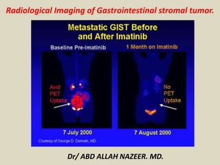
  • 6. US: Typically hypoechoic, Variable vascularity on color Doppler and usually difficult to identify organ of origin. MRI Presence of necrosis, hemorrhagic and cystic change make appearances variable: T1: low signal intensity solid component enhancement is usually present, and predominantly peripheral in larger lesions T2: high signal intensity solid component. T1 post-contrast: Enhancement of solid areas and non-enhancing regions consistent with necrosis or hemorrhage. Angiography: SMA angiogram show hypervascular GIST and the tumour blush on arteriogram. 18 FDG-PET, PET/CT: Metabolic responses to imatinib treatment can be followed using positron emission tomography(PET) using fluorine-18-flourodeoxyglucose(18 FDG). Significant reduction in tumour 18FDG uptake in imatinib –responsive tumors as early as 24 hours later and by 1 month after initiating imatinib therapy. Lack of metabolic response on 18FD-PET indicates primary or secondary resistance to chromo therapeutic agent.
  • 7. Ilial perivesical GIST, appearing on ultrasound as an hypoechoic mass, markedly heterogeneous and slightly lobulated.
  • 8. Endoscopic Ultrasonography (EU) of a duodenal bulb GIST, with calcifications on its deeper segment.
  • 9. EU of a large gastric body GIST, with areas of necrosis and hemorrhage.
  • 10. Cystic GIST. Longitudinal USG scan of the abdomen (A) in a 41-year-old male shows a predominantly cystic mass with septae (arrows) in the left hypochondrium. Axial contrast-enhanced CT scan (B) shows a peripherally enhancing cystic mass (arrow) in the lesser sac region along the tail of the pancreas.
  • 11. Gastric gastrointestinal stromal tumor (GIST) en face. Upper GI image obtained during the single contrast enhancement portion shows an incidentally found mass. The smooth borders suggest a submucosal process. At surgery, a GIST was found.
  • 12. Multifocal gastrointestinal stromal tumors. Barium examination and CT Scan reveals a smooth mass that causes narrowing in the second portion of the duodenum (arrows).
  • 13. Malignant GIST. Barium meal study (A) shows an irregular filling defect along the fundus and greater curvature of the stomach, suggestive of ulceration (arrow). Axial contrast-enhanced CT scan (B) shows an exophytic heterogeneously enhancing mass (arrowhead) arising from the stomach (small arrow) with areas of ulceration (long arrow)
  • 14. Small-bowel gastrointestinal stromal tumor with a diffusely thickened bowel wall.
  • 15. Gastric gastrointestinal stromal tumor (GIST) en face. Upper GI image obtained during the single contrast enhancement portion shows an incidentally found mass. The smooth borders suggest a submucosal process. At surgery, a GIST was found.
  • 16. Gastric gastrointestinal stromal tumor with huge exophytic component, which has become ulcerated. Barium collects in the exophytic ulcer crater (arrows). CT demonstrates the GIST with large exophytic ulceration (arrows).
  • 17. Proximal jejunal gastrointestinal stromal tumor that is completely exophytic and not visible at small- bowel barium examination. Contrast-enhanced CT obtained in the same patient as in the previous image demonstrates a peripherally enhancing mass immediately adjacent to the pancreas that was thought to be a pancreatic neoplasm. However, at surgery, this proved to be a proximal exophytic jejunal GIST.
  • 18. Gastrointestinal stromal tumor. Enteroclysis results were surprisingly normal. No abnormalities are seen, and no significant mass effect suggests the presence of the mass. Gastrointestinal stromal tumor (GIST). CT demonstrates a complex mass originating from the small bowel with characteristics of a GIST.
  • 19.
  • 20.
  • 21.
  • 22.
  • 23.
  • 24.
  • 25.
  • 26.
  • 27.
  • 28.
  • 29.
  • 30.
  • 31.
  • 32. Massive ileal GIST, with invasion of adjacent bowel loops. Note the small calcification at the bottom portion of the mass.
  • 33. Large gastric GIST on a T2-weighted MRI, with areas of hemorrhage and necrosis.
  • 34. T2-weighted MRI: large rectal GIST with mild hyperintensity relative to skeletal muscle.
  • 35. Gastrointestinal stromal tumor (GIST). Patient in the previous image then underwent angiography, which demonstrates a mass (white arrows) in the same location as the small-bowel bleed. The blood supply is from the superior mesenteric artery (red arrow). Minimal mass effect on the bladder is noted (yellow arrows). Findings are consistent with a small bowel mass, which stained positive for CD117; this result is consistent with a GIST.
  • 36. GIST of the jejunum in a 53-year-old woman with acute GI bleeding. CT show prominent feeding and draining veins (c) Digital subtraction angiography shows a hypervascular mass (arrows) fed by branches of the superior mesenteric artery (arrowhead).
  • 37. Larger and multiple small bowel gastrointestinal stromal tumors. A 52-year-old woman with 2 gastrointestinal stromal tumors in the middle jejunum. The tumors (white arrows) were 5.5 cm and 1.5 cm in diameter, respectively, and were also hypervascular and homogeneous in the early arterial phrase. The feeding artery of the large tumor (A: white arrowhead) was slightly thickened. The draining veins of the tumors (B: black arrowheads) were also clear. The pathological risk classification was high (large) and very low (small).
  • 38. Small bowel gastrointestinal stromal tumor. A 70-year-old man with a 1.2 cm gastrointestinal stromal tumor of very low risk classification in the upper jejunum. The tumor (white arrows) was round, well-defined and homogeneous in the early arterial phase (A, B). The feeding arteries were not significantly enlarged, and the draining veins (B: black arrowheads) developed clearly and early, merging into the portal vein system (B, asterisk).
  • 39. Small bowel gastrointestinal stromal tumor case with active bleeding. A 35-year-old woman with a 3.2 cm gastrointestinal stromal tumor in the middle jejunum. The hemoglobin was 34 g/L. Digital subtraction angiography showed many draining veins (arrows) on the tumor merging into the portal vein system (arrowheads) without signs of active bleeding. The tumor was slightly lobular and was low risk based on the pathological classification.
  • 40. Gastrointestinal stromal tumor. A 62-year-old women presented with 2 episodes of massive GI bleeding. Nuclear medicine GI bleed scanning demonstrates increased tracer activity in the mid abdomen (arrows) consistent with a small bowel active bleed.
  • 41.
  • 42. This correlates with computed tomography (CT) scans, on this side shrinking. On this side, you see the PET scan is very hot, there is lots of uptake, then it goes cold. The speed of this is what has been amazing to all of us.
  • 43.
  • 44.
  • 45.
  • 46. A 52-year-old man with suspected recurrence of gastrointestinal stromal tumors (GISTs). (A) An upper abdominal image reveals a 10- x 7-cm mass in the right liver lobe (arrow). (B) Mid abdominal image shows a 12- x 6-cm subhepatic mass (arrow). (C) A necrotic pelvic lymph node (arrow) measuring 3 x 3 cm was also noted. (D) A positron emission tomography scan with 18F-fluorodeoxyglucose (FDG) shows markedly increased uptake in the liver (short arrow) and the subhepatic masses (long arrow). Lack of FDG uptake in the pelvis excludes this mass as a GIST. Positron emission tomography helped in accurate restaging by revealing the malignant nature of the hepatic and subhepatic masses and excluding spread to the pelvis.
  • 47. A 42-year-old woman with a history of surgical removal of gastrointestinal stromal tumor had a PET scan with 18F-fluorodeoxyglucose for restaging. (A) The positron emission tomography (PET) scan showed a small focus of markedly increased FDG uptake in the right lower neck (black arrow). (B) An ultrasound image revealed a hypoechoic lesion (white arrow) in the right lobe of the thyroid gland correlating with the abnormal uptake noted on the PET scan. Biopsy revealed an unsuspected incidental papillary carcinoma.
  • 48. 41-year-old woman following partial resection of gastrointestinal stromal tumors. (A) Before therapy with Gleevec (Novartis Pharmaceuticals Corp., East Hanover, NJ), positron emission tomography (PET) shows markedly increased uptake in the left abdomen and 3 liver lesions (arrows). (B) A PET scan performed 5 weeks after the initiation of therapy was normal, with no abnormal uptake in the abdomen or liver, indicating an excellent tumor response to therapy. The uptake of 18F-fluorodeoxyglucose in the heart, renal collecting systems, and bladder is physiologic.