SlideShare a Scribd company logo
Radiological Imaging of Gastrointestinal tract Schwannoma.
Dr/ ABD ALLAH NAZEER.MD.
Schwannoma represents 1% of tumors of the
gastrointestinal tract and parts 2-8%
mesenchymal tumors of the gastrointestinal. In
patients with neurofibromatosis type 1 there
gastrointestinal involvement in 10-25% of cases
including: neurofibroma solitary leiomyoma and
rarely plexiform neurofibroma.
More and more frequently diagnosed by
immunohistochemical stains.
The average lesion size is 6-7 cm (range, 0.5 to 14
cm). They occur most often in the stomach (60-70%
of cases), followed by colorectal, being exceptional
in the small intestine and esophagus
Gastric
Represent 0.2% of gastric tumors, 4% of all benign gastric tumors
with peak incidence between the fourth and fifth decade of life. It is
usually asymptomatic, with incidental finding, although they are
sometimes shaped symptomatic gastrointestinal ulceration and
bleeding.
Gastrointestinal schwannomas are distinctly different neoplasms
from conventional soft-tissue and central nervous system
schwannomas, some of which may be associated with
neurofibromatosis 2.
Most injuries TC are hypodense, well defined and standardized with
homogeneous enhancement contrast, endoscopy can be checked
exophytic nature of the injury. For RM, most of the lesions are
iso/ hypointense on T1-weighted sequences and hyperintense on
T2-weighted sequences. The discrepancy in signal intensity between
our T2-weight-ed images and those described in literature might be
caused by the presence of melanin in the schwannoma.
A 41-year-old female with a Schwannoma of the stomach. A. Unenhanced CT
shows a well-defined extraluminal soft tissue mass with homogeneous density;
B. Enhanced CT shows the neoplasm is of homogeneous enhancement.
Schwannoma, Large exophytic soft
tissue mass arising from the
greater curvature of the stomach.
Abdominopelvic computed tomography scans show a 1.5×1.0 cm sized
homogenously-enhancing mass in the anterior wall of the duodenal
bulb (arrow). (A) Axial section view. (B) Coronal section view.
Schwannoma of the stomach.
Pancreatic schwannoma is extremely rare with similar incidence in both
genders. Pancreatic schwannoma is a slowly growing, encapsulated, benign
neoplasm that typically arises in the peripheral epineurium of either the
sympathetic or parasympathetic autonomic fibers or branches of vagus
nerve that extend to the pancreas. Pancreatic schwannomas most
frequently involve the pancreas head (40%), followed by body (21%), neck
(6%), tail (15%), and uncinate process (13%), respectively. Patients with
pancreatic schwannoma are usually asymptomatic, or have abdominal
pain, nausea, and vomiting. Weight loss and jaundice may sometimes be
seen. CT or MRI can help distinguish if a lesion is solid or cystic, can define
the anatomic location of the lesion, delineate its relation to mesenteric
vessels, and determine the lesion causes pancreatic duct dilation. The
features of pancreatic schwannomas on CT scan include low-density
and/or cystic degenerative areas. MR imaging usually shows hypointensity
on T1-weighted images and hyperintensity on T2-weighted images but
like the CT features, these findings are nonspecific. Two-thirds of
pancreatic schwannomas undergo degenerative changes such as cyst
formation, necrosis, calcification, and hemorrhage, and these changes
can mimic pancreatic cystic tumors.
60-year-old male with pancreatic schwannoma. A, B There is heterogeneously enhancing
mass (arrows) in the pancreas uncinated process on arterial axial (A) and venous phase
coronal (D) images. C, D On pathology, the mass showed cystic degeneration with
hemorrhage (C H&E stain ×4) and was positive for S100 on immunohistostain (×10).
40-year-old female with pancreatic schwannoma. A On venous phase axial
CT, there is a round low-density cystic mass (arrow) in the pancreas neck. B
On MRI axial T2WI, the cystic mass (arrow) shows small septations. C On
post-contrast axial image of MRI, there is mild septal enhancement (arrow).
Pancreatic schwannoma.
Well-defined cystic lesion located in the retroperitoneal space under the cauda pancreatis
slightly pushing it back and the adjacent jejunal loops. Distinct liquid T2 hypersignal of the
tumour. a: injected CT-scan, coronal format; b: injected CT-scan, axial section; c: MRI in T2
weighting, axial plane.
Pancreatic schwannoma
showing increased FDG
uptake on PET/CT.
60-year-old female with peripancreatic schwannoma. A–C There is a well-circumscribed
low-density solid mass (arrows) in the region of pancreas head/neck with central cystic
degeneration/necrosis on arterial phase (A) and venous phase (B, C). Note vascularity in
the solid portion of the mass seen inferiorly and mild upstream pancreatic duct dilation (C).
Schwannomas that arise in the adrenal medulla are very rare. Often,
schwannomas of the retroperitoneum, especially in the juxta-adrenal
space, can be misdiagnosed as they can mimic more common primary
adrenal lesions. Studies have shown that approximately 0.5% to 5% of
schwannomas are retroperitoneal, constituting <0.2% of adrenal
incidental tumors. Most of the patients do not have any symptoms and
delayed diagnosis results in a significant size at the time of diagnosis.
Both CT and MRI are nonspecific in diagnosis of adrenal schwannomas.
Adrenal schwannomas often show septa and cystic change. Cystic changes
are rare in other retroperitoneal tumors. Characteristic enhancement
patterns of primary adrenal schwannomas are mild heterogeneous
enhancement on arterial phase and progressive enhancement during the
portal venous and equilibrium phases, which are likely due to variable
degree of tumor cellularity or degenerative changes such as cystic
degeneration, necrosis, and hemorrhage. Minimal contrast enhancement
may be seen in neoplasms with low cellularity and edema. On MRI,
schwannomas exhibit low-signal intensity on T1-weighted images and
heterogeneous high-signal intensity on T2-weighted images
50-year-old female with
retroperitoneal/adrenal
schwannoma. The mass
(arrows) shows low-
signal intensity on T1WI
(A), high-signal intensity
on T2WI (B, C), with
cystic component,
multiple internal septa,
and surrounding
capsule. After contrast
enhancement (D, E), the
mass (arrows) shows
peripheral enhancing
solid component.
A giant spinal schwannoma mimicking a renal mass.
70-year-old male with small bowel schwannoma. A, B There is an incidentally found
homogeneously enhancing mass (arrows) in the ileal loop on venous phase CT (A) and
contrast-enhanced MR (B). C FDG uptake is seen on fusion image of PET/CT. D, E On
pathology, the neoplasm (arrow) abuts the jejunal wall on H&E stain (D ×4). The neoplasm
involved the submucosa and muscularis propria. The mass was composed of a bland
spindle cell proliferation with a prominent lymphoid cuff. Neoplastic cells are diffusely
positive for S-100 protein (E ×20). The mass was negative for c-Kit, DOG1, and SMA
Abdominal CT and MRI findings
of the patient. a Plain CT scan
revealed a 4.0 × 3.2 × 3.0-cm
hypodense mass in the porta
hepatis (black arrow). b Enhanced
CT scan revealed a mildly
enhanced mass in arterial phase
(black arrow). c Moderately
enhanced mass in portal phase
(black arrow). d MRI revealed a
4.0 × 3.0-cm mass in the sagittal
section (black arrow). e Low signal
intensity in T1-weighted imaging
(black arrow). f High signal
intensity in T2-weighted imaging
(black arrow).
Multiple
schwannomas
synchronously
occurring in
the porta
hepatis, liver,
and
gallbladder:
Incidental finding in patients with a history of splenectomy. in hilum loculated liver, hypodense, cystic
appearance without mass effect or biliary dilatation associated injury. Schwannoma hepatic hilum.
Retroperitoneal schwannoma.
Schwannoma, well-defined, retroperitoneal soft tissue mass in close proximity to
the superior mesenteric vessels with a feeding vessel from the superior mesenteric
artery (SMA) and venous drainage to the superior mesenteric vein (SMV).
CT injected at portal time revealing this mass along the retroperitoneal vessels and the
independent and slightly retaining aspect of the intestinal loops. Presence of a
central necrosis and several microcalcifications. a: oblique coronal format; b: axial section.
Retroperitoneal schwannoma.
Giant retroperitoneal
ancient schwannoma
Giant ossified
benign
mesenteric
schwannoma
Abdominal wall Schwannoma.
(a) Color flow showing
hypovascularity of the mass.
(b) B-mode ultrasound shows
the well-encapsulated mass in
anterior abdominal wall (arrow).
Abdominal Wall
Schwannoma
MRI and CT analysis confirmed the
retroperitoneal Schwannoma.
PET (first line), CT (second line) images and PET-scan fusion (third line)
objectifying the fixation of 18-FDG within the tissue zones of the mass.
Pelvic MRI revealing the tumour in distinct T2 hypersignal (a) enhanced
after injection (b), the lesion appearing well defined developed at the
expense of the right L5 root. a: coronal section in T2 weighting; b: axial
section in T1 fat-sat weighting after injection of gadolinium.
Retrorectal tumor: a case report of a patient with "schwannoma.
Ancient schwannoma. (a) Axial T2-weighted MR image shows a well-defined, complex
cystic lesion with internal septa and solid components (arrow) in the left hemipelvis. (b)
Axial postcontrast T1-weighted MR image reveals heterogeneous enhancement
of the internal septa and solid components (arrow). Surgical excision and
histopathologic analysis helped confirm the diagnosis of ancient schwannoma.
Pelvic schwannoma, Fat-suppressed T2-weighted coronal (A), sagittal (B),
axial (C) postoperative tumor image. There was remained tumor and
capsule on magnetic resonance image in the post-operative 7 months.
Pelvic schwannoma. (a) Axial T2W fat-suppressed and (b) post-contrast
fat-suppressed T1W images show a heterogeneously hyperintense area in
T2W image due to microcytic spaces (arrowheads). Peripheral contrast
enhancement is observed in post-contrast T1W image (curved arrows).
Rectal schwannoma in 67-year-old woman. Sagittal T2-weighted
image (A) and axial T1-weighted image (B) show well-circumscribed
homogeneous submucosal mass (arrows) in lower rectum.
Malignant
Schwannoma
of Anterior
Abdominal Wall
Thank You.

More Related Content

What's hot

imaging of benign hepatic masses
imaging of benign hepatic massesimaging of benign hepatic masses
imaging of benign hepatic masses
Navni Garg
 
Retroperitoneal masses radiology
Retroperitoneal masses radiologyRetroperitoneal masses radiology
Retroperitoneal masses radiology
Dr. Mohit Goel
 
CT Imaging of CA Esophagus
CT Imaging of CA EsophagusCT Imaging of CA Esophagus
CT Imaging of CA Esophagus
Dr. Yash Kumar Achantani
 
Imaging of portal hypertension
Imaging of portal hypertensionImaging of portal hypertension
Imaging of portal hypertension
Dev Lakhera
 
larynx Imaging 1st part laryngeal anatomy CT MRI Dr Ahmed Esawy
larynx Imaging 1st part laryngeal anatomy CT MRI Dr Ahmed Esawylarynx Imaging 1st part laryngeal anatomy CT MRI Dr Ahmed Esawy
larynx Imaging 1st part laryngeal anatomy CT MRI Dr Ahmed Esawy
AHMED ESAWY
 
Diagnostic Imaging of Renal Tumors
Diagnostic Imaging of Renal TumorsDiagnostic Imaging of Renal Tumors
Diagnostic Imaging of Renal Tumors
Mohamed M.A. Zaitoun
 
Presentation1, radiological imaging of gastro intestinal stromal tumour(gist).
Presentation1, radiological imaging of gastro intestinal stromal tumour(gist).Presentation1, radiological imaging of gastro intestinal stromal tumour(gist).
Presentation1, radiological imaging of gastro intestinal stromal tumour(gist).
Abdellah Nazeer
 
Presentation1.pptx, radiological imaging of large bowel diseases
Presentation1.pptx, radiological imaging of large bowel diseasesPresentation1.pptx, radiological imaging of large bowel diseases
Presentation1.pptx, radiological imaging of large bowel diseasesAbdellah Nazeer
 
Retroperitoneal masses
Retroperitoneal massesRetroperitoneal masses
Retroperitoneal masses
Anish Choudhary
 
Larynx anatomy ct and mri
Larynx anatomy ct and mriLarynx anatomy ct and mri
Larynx anatomy ct and mri
Anish Choudhary
 
Larynx anatomy CT and MRI
Larynx anatomy CT and MRILarynx anatomy CT and MRI
Larynx anatomy CT and MRI
Dr. Mohit Goel
 
Presentation1.pptx, radiological imaging of uterine cervix diseases.
Presentation1.pptx, radiological imaging of uterine cervix diseases.Presentation1.pptx, radiological imaging of uterine cervix diseases.
Presentation1.pptx, radiological imaging of uterine cervix diseases.Abdellah Nazeer
 
Presentation2.pptx, radiological imaging of the rectal diseases.
Presentation2.pptx, radiological imaging of the rectal diseases.Presentation2.pptx, radiological imaging of the rectal diseases.
Presentation2.pptx, radiological imaging of the rectal diseases.Abdellah Nazeer
 
Diagnostic Imaging of Mediastinal Masses
Diagnostic Imaging of Mediastinal MassesDiagnostic Imaging of Mediastinal Masses
Diagnostic Imaging of Mediastinal Masses
Mohamed M.A. Zaitoun
 
HEPATOCELLULAR CARCINOMA RADIOLOGY
HEPATOCELLULAR CARCINOMA RADIOLOGYHEPATOCELLULAR CARCINOMA RADIOLOGY
HEPATOCELLULAR CARCINOMA RADIOLOGY
RMLIMS
 
Thyroid ultrasound
Thyroid ultrasoundThyroid ultrasound
Thyroid ultrasound
Doaa Gadalla
 
Presentation1, mri imaging of the prostate
Presentation1, mri imaging of the prostatePresentation1, mri imaging of the prostate
Presentation1, mri imaging of the prostate
Abdellah Nazeer
 
Radiological Approach To Diffuse Lung Diseases
Radiological Approach To Diffuse Lung DiseasesRadiological Approach To Diffuse Lung Diseases
Radiological Approach To Diffuse Lung Diseases
Dr. Soe Moe Htoo
 
Diagnostic Imaging of Intracranial Vascular malformations
Diagnostic Imaging of Intracranial Vascular malformationsDiagnostic Imaging of Intracranial Vascular malformations
Diagnostic Imaging of Intracranial Vascular malformations
Mohamed M.A. Zaitoun
 
Adrenal gland tumors (Radiology)
Adrenal gland tumors (Radiology)Adrenal gland tumors (Radiology)
Adrenal gland tumors (Radiology)
Dr Abdalla M. Gamal
 

What's hot (20)

imaging of benign hepatic masses
imaging of benign hepatic massesimaging of benign hepatic masses
imaging of benign hepatic masses
 
Retroperitoneal masses radiology
Retroperitoneal masses radiologyRetroperitoneal masses radiology
Retroperitoneal masses radiology
 
CT Imaging of CA Esophagus
CT Imaging of CA EsophagusCT Imaging of CA Esophagus
CT Imaging of CA Esophagus
 
Imaging of portal hypertension
Imaging of portal hypertensionImaging of portal hypertension
Imaging of portal hypertension
 
larynx Imaging 1st part laryngeal anatomy CT MRI Dr Ahmed Esawy
larynx Imaging 1st part laryngeal anatomy CT MRI Dr Ahmed Esawylarynx Imaging 1st part laryngeal anatomy CT MRI Dr Ahmed Esawy
larynx Imaging 1st part laryngeal anatomy CT MRI Dr Ahmed Esawy
 
Diagnostic Imaging of Renal Tumors
Diagnostic Imaging of Renal TumorsDiagnostic Imaging of Renal Tumors
Diagnostic Imaging of Renal Tumors
 
Presentation1, radiological imaging of gastro intestinal stromal tumour(gist).
Presentation1, radiological imaging of gastro intestinal stromal tumour(gist).Presentation1, radiological imaging of gastro intestinal stromal tumour(gist).
Presentation1, radiological imaging of gastro intestinal stromal tumour(gist).
 
Presentation1.pptx, radiological imaging of large bowel diseases
Presentation1.pptx, radiological imaging of large bowel diseasesPresentation1.pptx, radiological imaging of large bowel diseases
Presentation1.pptx, radiological imaging of large bowel diseases
 
Retroperitoneal masses
Retroperitoneal massesRetroperitoneal masses
Retroperitoneal masses
 
Larynx anatomy ct and mri
Larynx anatomy ct and mriLarynx anatomy ct and mri
Larynx anatomy ct and mri
 
Larynx anatomy CT and MRI
Larynx anatomy CT and MRILarynx anatomy CT and MRI
Larynx anatomy CT and MRI
 
Presentation1.pptx, radiological imaging of uterine cervix diseases.
Presentation1.pptx, radiological imaging of uterine cervix diseases.Presentation1.pptx, radiological imaging of uterine cervix diseases.
Presentation1.pptx, radiological imaging of uterine cervix diseases.
 
Presentation2.pptx, radiological imaging of the rectal diseases.
Presentation2.pptx, radiological imaging of the rectal diseases.Presentation2.pptx, radiological imaging of the rectal diseases.
Presentation2.pptx, radiological imaging of the rectal diseases.
 
Diagnostic Imaging of Mediastinal Masses
Diagnostic Imaging of Mediastinal MassesDiagnostic Imaging of Mediastinal Masses
Diagnostic Imaging of Mediastinal Masses
 
HEPATOCELLULAR CARCINOMA RADIOLOGY
HEPATOCELLULAR CARCINOMA RADIOLOGYHEPATOCELLULAR CARCINOMA RADIOLOGY
HEPATOCELLULAR CARCINOMA RADIOLOGY
 
Thyroid ultrasound
Thyroid ultrasoundThyroid ultrasound
Thyroid ultrasound
 
Presentation1, mri imaging of the prostate
Presentation1, mri imaging of the prostatePresentation1, mri imaging of the prostate
Presentation1, mri imaging of the prostate
 
Radiological Approach To Diffuse Lung Diseases
Radiological Approach To Diffuse Lung DiseasesRadiological Approach To Diffuse Lung Diseases
Radiological Approach To Diffuse Lung Diseases
 
Diagnostic Imaging of Intracranial Vascular malformations
Diagnostic Imaging of Intracranial Vascular malformationsDiagnostic Imaging of Intracranial Vascular malformations
Diagnostic Imaging of Intracranial Vascular malformations
 
Adrenal gland tumors (Radiology)
Adrenal gland tumors (Radiology)Adrenal gland tumors (Radiology)
Adrenal gland tumors (Radiology)
 

Similar to Presentation2, radiological imaging of gastrointestinal schwannoma.

Mediastinum masses
Mediastinum massesMediastinum masses
Mediastinum massesNavdeep Shah
 
Case Report_2.pptx
Case Report_2.pptxCase Report_2.pptx
Case Report_2.pptx
Zahra1373
 
Imaging of non endocrine tumour of pancreas
Imaging of non endocrine tumour of pancreasImaging of non endocrine tumour of pancreas
Imaging of non endocrine tumour of pancreas
Dev Lakhera
 
Carcinoma oesophagus
Carcinoma oesophagusCarcinoma oesophagus
Carcinoma oesophagus
Dr Vandana Singh Kushwaha
 
Case of pancreatic adenocarcinoma and other pancreatic tumors
Case of pancreatic adenocarcinoma and other pancreatic tumorsCase of pancreatic adenocarcinoma and other pancreatic tumors
Case of pancreatic adenocarcinoma and other pancreatic tumors
Alex Allu
 
Imaging of urinary bladder malignancy
Imaging of urinary bladder malignancyImaging of urinary bladder malignancy
Imaging of urinary bladder malignancy
PrasunDas31
 
Presentation1, radiological imaging of anal carcinoma.
Presentation1, radiological imaging of anal carcinoma.Presentation1, radiological imaging of anal carcinoma.
Presentation1, radiological imaging of anal carcinoma.
Abdellah Nazeer
 
CNS TUMOR (Radiology) By Oconer
CNS TUMOR (Radiology) By OconerCNS TUMOR (Radiology) By Oconer
CNS TUMOR (Radiology) By Oconer
Eunice Jade
 
Pancreatic neoplasms
Pancreatic neoplasmsPancreatic neoplasms
Pancreatic neoplasms
drksreenath
 
Retroperitoneal mass
Retroperitoneal  massRetroperitoneal  mass
Giant Thoracoabdominal Ganglioneuromma : an unusual mimicker of ruptured Aort...
Giant Thoracoabdominal Ganglioneuromma : an unusual mimicker of ruptured Aort...Giant Thoracoabdominal Ganglioneuromma : an unusual mimicker of ruptured Aort...
Giant Thoracoabdominal Ganglioneuromma : an unusual mimicker of ruptured Aort...
WCER 2021
 
Carcinoma stomach seminar
Carcinoma stomach seminarCarcinoma stomach seminar
Carcinoma stomach seminar
Rushabh Shah
 
3069576
30695763069576
3069576
adinatasatria
 
Carcinoma of Esophagus
Carcinoma of  EsophagusCarcinoma of  Esophagus
Carcinoma of Esophagus
Dr. Naveed Quetta
 
Coloretcal carcinoma
Coloretcal carcinomaColoretcal carcinoma
Coloretcal carcinoma
Dr. Naveed Quetta
 
Imaging in Benign hepatic lesions
Imaging in Benign hepatic lesionsImaging in Benign hepatic lesions
Imaging in Benign hepatic lesions
Girendra Shankar
 
Presentation1.pptx, radiological imaging of the nasopharyngeal diseases.
Presentation1.pptx, radiological imaging of the nasopharyngeal diseases.Presentation1.pptx, radiological imaging of the nasopharyngeal diseases.
Presentation1.pptx, radiological imaging of the nasopharyngeal diseases.Abdellah Nazeer
 
Veeru ca pancreas
Veeru ca pancreasVeeru ca pancreas
Veeru ca pancreas
Veeru Reddy
 
Presentation1.pptx, radiological imaging of extra nodal lymphoma.
Presentation1.pptx, radiological imaging of extra nodal lymphoma.Presentation1.pptx, radiological imaging of extra nodal lymphoma.
Presentation1.pptx, radiological imaging of extra nodal lymphoma.Abdellah Nazeer
 
Biliary Tract Tumours(Cholangiocarcinoma,Cystadenoma/cystadenocarcinoma,Gall ...
Biliary Tract Tumours(Cholangiocarcinoma,Cystadenoma/cystadenocarcinoma,Gall ...Biliary Tract Tumours(Cholangiocarcinoma,Cystadenoma/cystadenocarcinoma,Gall ...
Biliary Tract Tumours(Cholangiocarcinoma,Cystadenoma/cystadenocarcinoma,Gall ...
Abdul Abasi
 

Similar to Presentation2, radiological imaging of gastrointestinal schwannoma. (20)

Mediastinum masses
Mediastinum massesMediastinum masses
Mediastinum masses
 
Case Report_2.pptx
Case Report_2.pptxCase Report_2.pptx
Case Report_2.pptx
 
Imaging of non endocrine tumour of pancreas
Imaging of non endocrine tumour of pancreasImaging of non endocrine tumour of pancreas
Imaging of non endocrine tumour of pancreas
 
Carcinoma oesophagus
Carcinoma oesophagusCarcinoma oesophagus
Carcinoma oesophagus
 
Case of pancreatic adenocarcinoma and other pancreatic tumors
Case of pancreatic adenocarcinoma and other pancreatic tumorsCase of pancreatic adenocarcinoma and other pancreatic tumors
Case of pancreatic adenocarcinoma and other pancreatic tumors
 
Imaging of urinary bladder malignancy
Imaging of urinary bladder malignancyImaging of urinary bladder malignancy
Imaging of urinary bladder malignancy
 
Presentation1, radiological imaging of anal carcinoma.
Presentation1, radiological imaging of anal carcinoma.Presentation1, radiological imaging of anal carcinoma.
Presentation1, radiological imaging of anal carcinoma.
 
CNS TUMOR (Radiology) By Oconer
CNS TUMOR (Radiology) By OconerCNS TUMOR (Radiology) By Oconer
CNS TUMOR (Radiology) By Oconer
 
Pancreatic neoplasms
Pancreatic neoplasmsPancreatic neoplasms
Pancreatic neoplasms
 
Retroperitoneal mass
Retroperitoneal  massRetroperitoneal  mass
Retroperitoneal mass
 
Giant Thoracoabdominal Ganglioneuromma : an unusual mimicker of ruptured Aort...
Giant Thoracoabdominal Ganglioneuromma : an unusual mimicker of ruptured Aort...Giant Thoracoabdominal Ganglioneuromma : an unusual mimicker of ruptured Aort...
Giant Thoracoabdominal Ganglioneuromma : an unusual mimicker of ruptured Aort...
 
Carcinoma stomach seminar
Carcinoma stomach seminarCarcinoma stomach seminar
Carcinoma stomach seminar
 
3069576
30695763069576
3069576
 
Carcinoma of Esophagus
Carcinoma of  EsophagusCarcinoma of  Esophagus
Carcinoma of Esophagus
 
Coloretcal carcinoma
Coloretcal carcinomaColoretcal carcinoma
Coloretcal carcinoma
 
Imaging in Benign hepatic lesions
Imaging in Benign hepatic lesionsImaging in Benign hepatic lesions
Imaging in Benign hepatic lesions
 
Presentation1.pptx, radiological imaging of the nasopharyngeal diseases.
Presentation1.pptx, radiological imaging of the nasopharyngeal diseases.Presentation1.pptx, radiological imaging of the nasopharyngeal diseases.
Presentation1.pptx, radiological imaging of the nasopharyngeal diseases.
 
Veeru ca pancreas
Veeru ca pancreasVeeru ca pancreas
Veeru ca pancreas
 
Presentation1.pptx, radiological imaging of extra nodal lymphoma.
Presentation1.pptx, radiological imaging of extra nodal lymphoma.Presentation1.pptx, radiological imaging of extra nodal lymphoma.
Presentation1.pptx, radiological imaging of extra nodal lymphoma.
 
Biliary Tract Tumours(Cholangiocarcinoma,Cystadenoma/cystadenocarcinoma,Gall ...
Biliary Tract Tumours(Cholangiocarcinoma,Cystadenoma/cystadenocarcinoma,Gall ...Biliary Tract Tumours(Cholangiocarcinoma,Cystadenoma/cystadenocarcinoma,Gall ...
Biliary Tract Tumours(Cholangiocarcinoma,Cystadenoma/cystadenocarcinoma,Gall ...
 

More from Abdellah Nazeer

Muculoskeletal Pediatic Imaging..pptx
Muculoskeletal Pediatic Imaging..pptxMuculoskeletal Pediatic Imaging..pptx
Muculoskeletal Pediatic Imaging..pptx
Abdellah Nazeer
 
Presentation1, Ultrasound of the bowel loops and the lymph nodes..pptx
Presentation1, Ultrasound of the bowel loops and the lymph nodes..pptxPresentation1, Ultrasound of the bowel loops and the lymph nodes..pptx
Presentation1, Ultrasound of the bowel loops and the lymph nodes..pptx
Abdellah Nazeer
 
Presentation1 Short cases MD..pptx
Presentation1 Short cases MD..pptxPresentation1 Short cases MD..pptx
Presentation1 Short cases MD..pptx
Abdellah Nazeer
 
Presentation1, MD MCQ Cases..pptx
Presentation1, MD MCQ Cases..pptxPresentation1, MD MCQ Cases..pptx
Presentation1, MD MCQ Cases..pptx
Abdellah Nazeer
 
Presentation1, Short Cases Quiz..pptx
Presentation1, Short Cases Quiz..pptxPresentation1, Short Cases Quiz..pptx
Presentation1, Short Cases Quiz..pptx
Abdellah Nazeer
 
Presentation1, radiological imaging of lateral hindfoot impingement.
Presentation1, radiological imaging of lateral hindfoot impingement.Presentation1, radiological imaging of lateral hindfoot impingement.
Presentation1, radiological imaging of lateral hindfoot impingement.
Abdellah Nazeer
 
Presentation2, radiological anatomy of the liver and spleen.
Presentation2, radiological anatomy of the liver and spleen.Presentation2, radiological anatomy of the liver and spleen.
Presentation2, radiological anatomy of the liver and spleen.
Abdellah Nazeer
 
Presentation1, artifacts and pitfalls of the wrist and elbow joints.
Presentation1, artifacts and pitfalls of the wrist and elbow joints.Presentation1, artifacts and pitfalls of the wrist and elbow joints.
Presentation1, artifacts and pitfalls of the wrist and elbow joints.
Abdellah Nazeer
 
Presentation1, artifact and pitfalls of the knee, hip and ankle joints.
Presentation1, artifact and pitfalls of the knee, hip and ankle joints.Presentation1, artifact and pitfalls of the knee, hip and ankle joints.
Presentation1, artifact and pitfalls of the knee, hip and ankle joints.
Abdellah Nazeer
 
Presentation1, radiological imaging of artifact and pitfalls in shoulder join...
Presentation1, radiological imaging of artifact and pitfalls in shoulder join...Presentation1, radiological imaging of artifact and pitfalls in shoulder join...
Presentation1, radiological imaging of artifact and pitfalls in shoulder join...
Abdellah Nazeer
 
Presentation1, radiological imaging of internal abdominal hernia.
Presentation1, radiological imaging of internal abdominal hernia.Presentation1, radiological imaging of internal abdominal hernia.
Presentation1, radiological imaging of internal abdominal hernia.
Abdellah Nazeer
 
Presentation11, radiological imaging of ovarian torsion.
Presentation11, radiological imaging of ovarian torsion.Presentation11, radiological imaging of ovarian torsion.
Presentation11, radiological imaging of ovarian torsion.
Abdellah Nazeer
 
Presentation1, musculoskeletal anatomy.
Presentation1, musculoskeletal anatomy.Presentation1, musculoskeletal anatomy.
Presentation1, musculoskeletal anatomy.
Abdellah Nazeer
 
Presentation1, new mri techniques in the diagnosis and monitoring of multiple...
Presentation1, new mri techniques in the diagnosis and monitoring of multiple...Presentation1, new mri techniques in the diagnosis and monitoring of multiple...
Presentation1, new mri techniques in the diagnosis and monitoring of multiple...
Abdellah Nazeer
 
Presentation1, radiological application of diffusion weighted mri in neck mas...
Presentation1, radiological application of diffusion weighted mri in neck mas...Presentation1, radiological application of diffusion weighted mri in neck mas...
Presentation1, radiological application of diffusion weighted mri in neck mas...
Abdellah Nazeer
 
Presentation1, radiological application of diffusion weighted images in breas...
Presentation1, radiological application of diffusion weighted images in breas...Presentation1, radiological application of diffusion weighted images in breas...
Presentation1, radiological application of diffusion weighted images in breas...
Abdellah Nazeer
 
Presentation1, radiological application of diffusion weighted images in abdom...
Presentation1, radiological application of diffusion weighted images in abdom...Presentation1, radiological application of diffusion weighted images in abdom...
Presentation1, radiological application of diffusion weighted images in abdom...
Abdellah Nazeer
 
Presentation1, radiological application of diffusion weighted imges in neuror...
Presentation1, radiological application of diffusion weighted imges in neuror...Presentation1, radiological application of diffusion weighted imges in neuror...
Presentation1, radiological application of diffusion weighted imges in neuror...
Abdellah Nazeer
 
Presentation1, mr physics.
Presentation1, mr physics.Presentation1, mr physics.
Presentation1, mr physics.
Abdellah Nazeer
 
Presentation1. ct physics.
Presentation1. ct physics.Presentation1. ct physics.
Presentation1. ct physics.
Abdellah Nazeer
 

More from Abdellah Nazeer (20)

Muculoskeletal Pediatic Imaging..pptx
Muculoskeletal Pediatic Imaging..pptxMuculoskeletal Pediatic Imaging..pptx
Muculoskeletal Pediatic Imaging..pptx
 
Presentation1, Ultrasound of the bowel loops and the lymph nodes..pptx
Presentation1, Ultrasound of the bowel loops and the lymph nodes..pptxPresentation1, Ultrasound of the bowel loops and the lymph nodes..pptx
Presentation1, Ultrasound of the bowel loops and the lymph nodes..pptx
 
Presentation1 Short cases MD..pptx
Presentation1 Short cases MD..pptxPresentation1 Short cases MD..pptx
Presentation1 Short cases MD..pptx
 
Presentation1, MD MCQ Cases..pptx
Presentation1, MD MCQ Cases..pptxPresentation1, MD MCQ Cases..pptx
Presentation1, MD MCQ Cases..pptx
 
Presentation1, Short Cases Quiz..pptx
Presentation1, Short Cases Quiz..pptxPresentation1, Short Cases Quiz..pptx
Presentation1, Short Cases Quiz..pptx
 
Presentation1, radiological imaging of lateral hindfoot impingement.
Presentation1, radiological imaging of lateral hindfoot impingement.Presentation1, radiological imaging of lateral hindfoot impingement.
Presentation1, radiological imaging of lateral hindfoot impingement.
 
Presentation2, radiological anatomy of the liver and spleen.
Presentation2, radiological anatomy of the liver and spleen.Presentation2, radiological anatomy of the liver and spleen.
Presentation2, radiological anatomy of the liver and spleen.
 
Presentation1, artifacts and pitfalls of the wrist and elbow joints.
Presentation1, artifacts and pitfalls of the wrist and elbow joints.Presentation1, artifacts and pitfalls of the wrist and elbow joints.
Presentation1, artifacts and pitfalls of the wrist and elbow joints.
 
Presentation1, artifact and pitfalls of the knee, hip and ankle joints.
Presentation1, artifact and pitfalls of the knee, hip and ankle joints.Presentation1, artifact and pitfalls of the knee, hip and ankle joints.
Presentation1, artifact and pitfalls of the knee, hip and ankle joints.
 
Presentation1, radiological imaging of artifact and pitfalls in shoulder join...
Presentation1, radiological imaging of artifact and pitfalls in shoulder join...Presentation1, radiological imaging of artifact and pitfalls in shoulder join...
Presentation1, radiological imaging of artifact and pitfalls in shoulder join...
 
Presentation1, radiological imaging of internal abdominal hernia.
Presentation1, radiological imaging of internal abdominal hernia.Presentation1, radiological imaging of internal abdominal hernia.
Presentation1, radiological imaging of internal abdominal hernia.
 
Presentation11, radiological imaging of ovarian torsion.
Presentation11, radiological imaging of ovarian torsion.Presentation11, radiological imaging of ovarian torsion.
Presentation11, radiological imaging of ovarian torsion.
 
Presentation1, musculoskeletal anatomy.
Presentation1, musculoskeletal anatomy.Presentation1, musculoskeletal anatomy.
Presentation1, musculoskeletal anatomy.
 
Presentation1, new mri techniques in the diagnosis and monitoring of multiple...
Presentation1, new mri techniques in the diagnosis and monitoring of multiple...Presentation1, new mri techniques in the diagnosis and monitoring of multiple...
Presentation1, new mri techniques in the diagnosis and monitoring of multiple...
 
Presentation1, radiological application of diffusion weighted mri in neck mas...
Presentation1, radiological application of diffusion weighted mri in neck mas...Presentation1, radiological application of diffusion weighted mri in neck mas...
Presentation1, radiological application of diffusion weighted mri in neck mas...
 
Presentation1, radiological application of diffusion weighted images in breas...
Presentation1, radiological application of diffusion weighted images in breas...Presentation1, radiological application of diffusion weighted images in breas...
Presentation1, radiological application of diffusion weighted images in breas...
 
Presentation1, radiological application of diffusion weighted images in abdom...
Presentation1, radiological application of diffusion weighted images in abdom...Presentation1, radiological application of diffusion weighted images in abdom...
Presentation1, radiological application of diffusion weighted images in abdom...
 
Presentation1, radiological application of diffusion weighted imges in neuror...
Presentation1, radiological application of diffusion weighted imges in neuror...Presentation1, radiological application of diffusion weighted imges in neuror...
Presentation1, radiological application of diffusion weighted imges in neuror...
 
Presentation1, mr physics.
Presentation1, mr physics.Presentation1, mr physics.
Presentation1, mr physics.
 
Presentation1. ct physics.
Presentation1. ct physics.Presentation1. ct physics.
Presentation1. ct physics.
 

Recently uploaded

Tom Selleck Health: A Comprehensive Look at the Iconic Actor’s Wellness Journey
Tom Selleck Health: A Comprehensive Look at the Iconic Actor’s Wellness JourneyTom Selleck Health: A Comprehensive Look at the Iconic Actor’s Wellness Journey
Tom Selleck Health: A Comprehensive Look at the Iconic Actor’s Wellness Journey
greendigital
 
Phone Us ❤85270-49040❤ #ℂall #gIRLS In Surat By Surat @ℂall @Girls Hotel With...
Phone Us ❤85270-49040❤ #ℂall #gIRLS In Surat By Surat @ℂall @Girls Hotel With...Phone Us ❤85270-49040❤ #ℂall #gIRLS In Surat By Surat @ℂall @Girls Hotel With...
Phone Us ❤85270-49040❤ #ℂall #gIRLS In Surat By Surat @ℂall @Girls Hotel With...
Savita Shen $i11
 
Are There Any Natural Remedies To Treat Syphilis.pdf
Are There Any Natural Remedies To Treat Syphilis.pdfAre There Any Natural Remedies To Treat Syphilis.pdf
Are There Any Natural Remedies To Treat Syphilis.pdf
Little Cross Family Clinic
 
How to Give Better Lectures: Some Tips for Doctors
How to Give Better Lectures: Some Tips for DoctorsHow to Give Better Lectures: Some Tips for Doctors
How to Give Better Lectures: Some Tips for Doctors
LanceCatedral
 
ARTIFICIAL INTELLIGENCE IN HEALTHCARE.pdf
ARTIFICIAL INTELLIGENCE IN  HEALTHCARE.pdfARTIFICIAL INTELLIGENCE IN  HEALTHCARE.pdf
ARTIFICIAL INTELLIGENCE IN HEALTHCARE.pdf
Anujkumaranit
 
Non-respiratory Functions of the Lungs.pdf
Non-respiratory Functions of the Lungs.pdfNon-respiratory Functions of the Lungs.pdf
Non-respiratory Functions of the Lungs.pdf
MedicoseAcademics
 
Triangles of Neck and Clinical Correlation by Dr. RIG.pptx
Triangles of Neck and Clinical Correlation by Dr. RIG.pptxTriangles of Neck and Clinical Correlation by Dr. RIG.pptx
Triangles of Neck and Clinical Correlation by Dr. RIG.pptx
Dr. Rabia Inam Gandapore
 
basicmodesofventilation2022-220313203758.pdf
basicmodesofventilation2022-220313203758.pdfbasicmodesofventilation2022-220313203758.pdf
basicmodesofventilation2022-220313203758.pdf
aljamhori teaching hospital
 
Novas diretrizes da OMS para os cuidados perinatais de mais qualidade
Novas diretrizes da OMS para os cuidados perinatais de mais qualidadeNovas diretrizes da OMS para os cuidados perinatais de mais qualidade
Novas diretrizes da OMS para os cuidados perinatais de mais qualidade
Prof. Marcus Renato de Carvalho
 
Flu Vaccine Alert in Bangalore Karnataka
Flu Vaccine Alert in Bangalore KarnatakaFlu Vaccine Alert in Bangalore Karnataka
Flu Vaccine Alert in Bangalore Karnataka
addon Scans
 
Couples presenting to the infertility clinic- Do they really have infertility...
Couples presenting to the infertility clinic- Do they really have infertility...Couples presenting to the infertility clinic- Do they really have infertility...
Couples presenting to the infertility clinic- Do they really have infertility...
Sujoy Dasgupta
 
HOT NEW PRODUCT! BIG SALES FAST SHIPPING NOW FROM CHINA!! EU KU DB BK substit...
HOT NEW PRODUCT! BIG SALES FAST SHIPPING NOW FROM CHINA!! EU KU DB BK substit...HOT NEW PRODUCT! BIG SALES FAST SHIPPING NOW FROM CHINA!! EU KU DB BK substit...
HOT NEW PRODUCT! BIG SALES FAST SHIPPING NOW FROM CHINA!! EU KU DB BK substit...
GL Anaacs
 
ACUTE SCROTUM.....pdf. ACUTE SCROTAL CONDITIOND
ACUTE SCROTUM.....pdf. ACUTE SCROTAL CONDITIONDACUTE SCROTUM.....pdf. ACUTE SCROTAL CONDITIOND
ACUTE SCROTUM.....pdf. ACUTE SCROTAL CONDITIOND
DR SETH JOTHAM
 
Maxilla, Mandible & Hyoid Bone & Clinical Correlations by Dr. RIG.pptx
Maxilla, Mandible & Hyoid Bone & Clinical Correlations by Dr. RIG.pptxMaxilla, Mandible & Hyoid Bone & Clinical Correlations by Dr. RIG.pptx
Maxilla, Mandible & Hyoid Bone & Clinical Correlations by Dr. RIG.pptx
Dr. Rabia Inam Gandapore
 
Surat @ℂall @Girls ꧁❤8527049040❤꧂@ℂall @Girls Service Vip Top Model Safe
Surat @ℂall @Girls ꧁❤8527049040❤꧂@ℂall @Girls Service Vip Top Model SafeSurat @ℂall @Girls ꧁❤8527049040❤꧂@ℂall @Girls Service Vip Top Model Safe
Surat @ℂall @Girls ꧁❤8527049040❤꧂@ℂall @Girls Service Vip Top Model Safe
Savita Shen $i11
 
Physiology of Special Chemical Sensation of Taste
Physiology of Special Chemical Sensation of TastePhysiology of Special Chemical Sensation of Taste
Physiology of Special Chemical Sensation of Taste
MedicoseAcademics
 
Ozempic: Preoperative Management of Patients on GLP-1 Receptor Agonists
Ozempic: Preoperative Management of Patients on GLP-1 Receptor Agonists  Ozempic: Preoperative Management of Patients on GLP-1 Receptor Agonists
Ozempic: Preoperative Management of Patients on GLP-1 Receptor Agonists
Saeid Safari
 
TEST BANK for Operations Management, 14th Edition by William J. Stevenson, Ve...
TEST BANK for Operations Management, 14th Edition by William J. Stevenson, Ve...TEST BANK for Operations Management, 14th Edition by William J. Stevenson, Ve...
TEST BANK for Operations Management, 14th Edition by William J. Stevenson, Ve...
kevinkariuki227
 
For Better Surat #ℂall #Girl Service ❤85270-49040❤ Surat #ℂall #Girls
For Better Surat #ℂall #Girl Service ❤85270-49040❤ Surat #ℂall #GirlsFor Better Surat #ℂall #Girl Service ❤85270-49040❤ Surat #ℂall #Girls
For Better Surat #ℂall #Girl Service ❤85270-49040❤ Surat #ℂall #Girls
Savita Shen $i11
 
POST OPERATIVE OLIGURIA and its management
POST OPERATIVE OLIGURIA and its managementPOST OPERATIVE OLIGURIA and its management
POST OPERATIVE OLIGURIA and its management
touseefaziz1
 

Recently uploaded (20)

Tom Selleck Health: A Comprehensive Look at the Iconic Actor’s Wellness Journey
Tom Selleck Health: A Comprehensive Look at the Iconic Actor’s Wellness JourneyTom Selleck Health: A Comprehensive Look at the Iconic Actor’s Wellness Journey
Tom Selleck Health: A Comprehensive Look at the Iconic Actor’s Wellness Journey
 
Phone Us ❤85270-49040❤ #ℂall #gIRLS In Surat By Surat @ℂall @Girls Hotel With...
Phone Us ❤85270-49040❤ #ℂall #gIRLS In Surat By Surat @ℂall @Girls Hotel With...Phone Us ❤85270-49040❤ #ℂall #gIRLS In Surat By Surat @ℂall @Girls Hotel With...
Phone Us ❤85270-49040❤ #ℂall #gIRLS In Surat By Surat @ℂall @Girls Hotel With...
 
Are There Any Natural Remedies To Treat Syphilis.pdf
Are There Any Natural Remedies To Treat Syphilis.pdfAre There Any Natural Remedies To Treat Syphilis.pdf
Are There Any Natural Remedies To Treat Syphilis.pdf
 
How to Give Better Lectures: Some Tips for Doctors
How to Give Better Lectures: Some Tips for DoctorsHow to Give Better Lectures: Some Tips for Doctors
How to Give Better Lectures: Some Tips for Doctors
 
ARTIFICIAL INTELLIGENCE IN HEALTHCARE.pdf
ARTIFICIAL INTELLIGENCE IN  HEALTHCARE.pdfARTIFICIAL INTELLIGENCE IN  HEALTHCARE.pdf
ARTIFICIAL INTELLIGENCE IN HEALTHCARE.pdf
 
Non-respiratory Functions of the Lungs.pdf
Non-respiratory Functions of the Lungs.pdfNon-respiratory Functions of the Lungs.pdf
Non-respiratory Functions of the Lungs.pdf
 
Triangles of Neck and Clinical Correlation by Dr. RIG.pptx
Triangles of Neck and Clinical Correlation by Dr. RIG.pptxTriangles of Neck and Clinical Correlation by Dr. RIG.pptx
Triangles of Neck and Clinical Correlation by Dr. RIG.pptx
 
basicmodesofventilation2022-220313203758.pdf
basicmodesofventilation2022-220313203758.pdfbasicmodesofventilation2022-220313203758.pdf
basicmodesofventilation2022-220313203758.pdf
 
Novas diretrizes da OMS para os cuidados perinatais de mais qualidade
Novas diretrizes da OMS para os cuidados perinatais de mais qualidadeNovas diretrizes da OMS para os cuidados perinatais de mais qualidade
Novas diretrizes da OMS para os cuidados perinatais de mais qualidade
 
Flu Vaccine Alert in Bangalore Karnataka
Flu Vaccine Alert in Bangalore KarnatakaFlu Vaccine Alert in Bangalore Karnataka
Flu Vaccine Alert in Bangalore Karnataka
 
Couples presenting to the infertility clinic- Do they really have infertility...
Couples presenting to the infertility clinic- Do they really have infertility...Couples presenting to the infertility clinic- Do they really have infertility...
Couples presenting to the infertility clinic- Do they really have infertility...
 
HOT NEW PRODUCT! BIG SALES FAST SHIPPING NOW FROM CHINA!! EU KU DB BK substit...
HOT NEW PRODUCT! BIG SALES FAST SHIPPING NOW FROM CHINA!! EU KU DB BK substit...HOT NEW PRODUCT! BIG SALES FAST SHIPPING NOW FROM CHINA!! EU KU DB BK substit...
HOT NEW PRODUCT! BIG SALES FAST SHIPPING NOW FROM CHINA!! EU KU DB BK substit...
 
ACUTE SCROTUM.....pdf. ACUTE SCROTAL CONDITIOND
ACUTE SCROTUM.....pdf. ACUTE SCROTAL CONDITIONDACUTE SCROTUM.....pdf. ACUTE SCROTAL CONDITIOND
ACUTE SCROTUM.....pdf. ACUTE SCROTAL CONDITIOND
 
Maxilla, Mandible & Hyoid Bone & Clinical Correlations by Dr. RIG.pptx
Maxilla, Mandible & Hyoid Bone & Clinical Correlations by Dr. RIG.pptxMaxilla, Mandible & Hyoid Bone & Clinical Correlations by Dr. RIG.pptx
Maxilla, Mandible & Hyoid Bone & Clinical Correlations by Dr. RIG.pptx
 
Surat @ℂall @Girls ꧁❤8527049040❤꧂@ℂall @Girls Service Vip Top Model Safe
Surat @ℂall @Girls ꧁❤8527049040❤꧂@ℂall @Girls Service Vip Top Model SafeSurat @ℂall @Girls ꧁❤8527049040❤꧂@ℂall @Girls Service Vip Top Model Safe
Surat @ℂall @Girls ꧁❤8527049040❤꧂@ℂall @Girls Service Vip Top Model Safe
 
Physiology of Special Chemical Sensation of Taste
Physiology of Special Chemical Sensation of TastePhysiology of Special Chemical Sensation of Taste
Physiology of Special Chemical Sensation of Taste
 
Ozempic: Preoperative Management of Patients on GLP-1 Receptor Agonists
Ozempic: Preoperative Management of Patients on GLP-1 Receptor Agonists  Ozempic: Preoperative Management of Patients on GLP-1 Receptor Agonists
Ozempic: Preoperative Management of Patients on GLP-1 Receptor Agonists
 
TEST BANK for Operations Management, 14th Edition by William J. Stevenson, Ve...
TEST BANK for Operations Management, 14th Edition by William J. Stevenson, Ve...TEST BANK for Operations Management, 14th Edition by William J. Stevenson, Ve...
TEST BANK for Operations Management, 14th Edition by William J. Stevenson, Ve...
 
For Better Surat #ℂall #Girl Service ❤85270-49040❤ Surat #ℂall #Girls
For Better Surat #ℂall #Girl Service ❤85270-49040❤ Surat #ℂall #GirlsFor Better Surat #ℂall #Girl Service ❤85270-49040❤ Surat #ℂall #Girls
For Better Surat #ℂall #Girl Service ❤85270-49040❤ Surat #ℂall #Girls
 
POST OPERATIVE OLIGURIA and its management
POST OPERATIVE OLIGURIA and its managementPOST OPERATIVE OLIGURIA and its management
POST OPERATIVE OLIGURIA and its management
 

Presentation2, radiological imaging of gastrointestinal schwannoma.

  • 1. Radiological Imaging of Gastrointestinal tract Schwannoma. Dr/ ABD ALLAH NAZEER.MD.
  • 2. Schwannoma represents 1% of tumors of the gastrointestinal tract and parts 2-8% mesenchymal tumors of the gastrointestinal. In patients with neurofibromatosis type 1 there gastrointestinal involvement in 10-25% of cases including: neurofibroma solitary leiomyoma and rarely plexiform neurofibroma. More and more frequently diagnosed by immunohistochemical stains. The average lesion size is 6-7 cm (range, 0.5 to 14 cm). They occur most often in the stomach (60-70% of cases), followed by colorectal, being exceptional in the small intestine and esophagus
  • 3. Gastric Represent 0.2% of gastric tumors, 4% of all benign gastric tumors with peak incidence between the fourth and fifth decade of life. It is usually asymptomatic, with incidental finding, although they are sometimes shaped symptomatic gastrointestinal ulceration and bleeding. Gastrointestinal schwannomas are distinctly different neoplasms from conventional soft-tissue and central nervous system schwannomas, some of which may be associated with neurofibromatosis 2. Most injuries TC are hypodense, well defined and standardized with homogeneous enhancement contrast, endoscopy can be checked exophytic nature of the injury. For RM, most of the lesions are iso/ hypointense on T1-weighted sequences and hyperintense on T2-weighted sequences. The discrepancy in signal intensity between our T2-weight-ed images and those described in literature might be caused by the presence of melanin in the schwannoma.
  • 4. A 41-year-old female with a Schwannoma of the stomach. A. Unenhanced CT shows a well-defined extraluminal soft tissue mass with homogeneous density; B. Enhanced CT shows the neoplasm is of homogeneous enhancement.
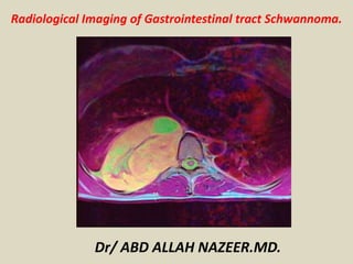
  • 5. Schwannoma, Large exophytic soft tissue mass arising from the greater curvature of the stomach.
  • 6. Abdominopelvic computed tomography scans show a 1.5×1.0 cm sized homogenously-enhancing mass in the anterior wall of the duodenal bulb (arrow). (A) Axial section view. (B) Coronal section view.
  • 7. Schwannoma of the stomach.
  • 8. Pancreatic schwannoma is extremely rare with similar incidence in both genders. Pancreatic schwannoma is a slowly growing, encapsulated, benign neoplasm that typically arises in the peripheral epineurium of either the sympathetic or parasympathetic autonomic fibers or branches of vagus nerve that extend to the pancreas. Pancreatic schwannomas most frequently involve the pancreas head (40%), followed by body (21%), neck (6%), tail (15%), and uncinate process (13%), respectively. Patients with pancreatic schwannoma are usually asymptomatic, or have abdominal pain, nausea, and vomiting. Weight loss and jaundice may sometimes be seen. CT or MRI can help distinguish if a lesion is solid or cystic, can define the anatomic location of the lesion, delineate its relation to mesenteric vessels, and determine the lesion causes pancreatic duct dilation. The features of pancreatic schwannomas on CT scan include low-density and/or cystic degenerative areas. MR imaging usually shows hypointensity on T1-weighted images and hyperintensity on T2-weighted images but like the CT features, these findings are nonspecific. Two-thirds of pancreatic schwannomas undergo degenerative changes such as cyst formation, necrosis, calcification, and hemorrhage, and these changes can mimic pancreatic cystic tumors.
  • 9. 60-year-old male with pancreatic schwannoma. A, B There is heterogeneously enhancing mass (arrows) in the pancreas uncinated process on arterial axial (A) and venous phase coronal (D) images. C, D On pathology, the mass showed cystic degeneration with hemorrhage (C H&E stain ×4) and was positive for S100 on immunohistostain (×10).
  • 10. 40-year-old female with pancreatic schwannoma. A On venous phase axial CT, there is a round low-density cystic mass (arrow) in the pancreas neck. B On MRI axial T2WI, the cystic mass (arrow) shows small septations. C On post-contrast axial image of MRI, there is mild septal enhancement (arrow).
  • 12. Well-defined cystic lesion located in the retroperitoneal space under the cauda pancreatis slightly pushing it back and the adjacent jejunal loops. Distinct liquid T2 hypersignal of the tumour. a: injected CT-scan, coronal format; b: injected CT-scan, axial section; c: MRI in T2 weighting, axial plane.
  • 14. 60-year-old female with peripancreatic schwannoma. A–C There is a well-circumscribed low-density solid mass (arrows) in the region of pancreas head/neck with central cystic degeneration/necrosis on arterial phase (A) and venous phase (B, C). Note vascularity in the solid portion of the mass seen inferiorly and mild upstream pancreatic duct dilation (C).
  • 15. Schwannomas that arise in the adrenal medulla are very rare. Often, schwannomas of the retroperitoneum, especially in the juxta-adrenal space, can be misdiagnosed as they can mimic more common primary adrenal lesions. Studies have shown that approximately 0.5% to 5% of schwannomas are retroperitoneal, constituting <0.2% of adrenal incidental tumors. Most of the patients do not have any symptoms and delayed diagnosis results in a significant size at the time of diagnosis. Both CT and MRI are nonspecific in diagnosis of adrenal schwannomas. Adrenal schwannomas often show septa and cystic change. Cystic changes are rare in other retroperitoneal tumors. Characteristic enhancement patterns of primary adrenal schwannomas are mild heterogeneous enhancement on arterial phase and progressive enhancement during the portal venous and equilibrium phases, which are likely due to variable degree of tumor cellularity or degenerative changes such as cystic degeneration, necrosis, and hemorrhage. Minimal contrast enhancement may be seen in neoplasms with low cellularity and edema. On MRI, schwannomas exhibit low-signal intensity on T1-weighted images and heterogeneous high-signal intensity on T2-weighted images
  • 16. 50-year-old female with retroperitoneal/adrenal schwannoma. The mass (arrows) shows low- signal intensity on T1WI (A), high-signal intensity on T2WI (B, C), with cystic component, multiple internal septa, and surrounding capsule. After contrast enhancement (D, E), the mass (arrows) shows peripheral enhancing solid component.
  • 17. A giant spinal schwannoma mimicking a renal mass.
  • 18. 70-year-old male with small bowel schwannoma. A, B There is an incidentally found homogeneously enhancing mass (arrows) in the ileal loop on venous phase CT (A) and contrast-enhanced MR (B). C FDG uptake is seen on fusion image of PET/CT. D, E On pathology, the neoplasm (arrow) abuts the jejunal wall on H&E stain (D ×4). The neoplasm involved the submucosa and muscularis propria. The mass was composed of a bland spindle cell proliferation with a prominent lymphoid cuff. Neoplastic cells are diffusely positive for S-100 protein (E ×20). The mass was negative for c-Kit, DOG1, and SMA
  • 19. Abdominal CT and MRI findings of the patient. a Plain CT scan revealed a 4.0 × 3.2 × 3.0-cm hypodense mass in the porta hepatis (black arrow). b Enhanced CT scan revealed a mildly enhanced mass in arterial phase (black arrow). c Moderately enhanced mass in portal phase (black arrow). d MRI revealed a 4.0 × 3.0-cm mass in the sagittal section (black arrow). e Low signal intensity in T1-weighted imaging (black arrow). f High signal intensity in T2-weighted imaging (black arrow).
  • 21. Incidental finding in patients with a history of splenectomy. in hilum loculated liver, hypodense, cystic appearance without mass effect or biliary dilatation associated injury. Schwannoma hepatic hilum.
  • 23. Schwannoma, well-defined, retroperitoneal soft tissue mass in close proximity to the superior mesenteric vessels with a feeding vessel from the superior mesenteric artery (SMA) and venous drainage to the superior mesenteric vein (SMV).
  • 24. CT injected at portal time revealing this mass along the retroperitoneal vessels and the independent and slightly retaining aspect of the intestinal loops. Presence of a central necrosis and several microcalcifications. a: oblique coronal format; b: axial section.
  • 28. Abdominal wall Schwannoma. (a) Color flow showing hypovascularity of the mass. (b) B-mode ultrasound shows the well-encapsulated mass in anterior abdominal wall (arrow).
  • 30. MRI and CT analysis confirmed the retroperitoneal Schwannoma.
  • 31. PET (first line), CT (second line) images and PET-scan fusion (third line) objectifying the fixation of 18-FDG within the tissue zones of the mass.
  • 32. Pelvic MRI revealing the tumour in distinct T2 hypersignal (a) enhanced after injection (b), the lesion appearing well defined developed at the expense of the right L5 root. a: coronal section in T2 weighting; b: axial section in T1 fat-sat weighting after injection of gadolinium.
  • 33. Retrorectal tumor: a case report of a patient with "schwannoma.
  • 34. Ancient schwannoma. (a) Axial T2-weighted MR image shows a well-defined, complex cystic lesion with internal septa and solid components (arrow) in the left hemipelvis. (b) Axial postcontrast T1-weighted MR image reveals heterogeneous enhancement of the internal septa and solid components (arrow). Surgical excision and histopathologic analysis helped confirm the diagnosis of ancient schwannoma.
  • 35.
  • 36. Pelvic schwannoma, Fat-suppressed T2-weighted coronal (A), sagittal (B), axial (C) postoperative tumor image. There was remained tumor and capsule on magnetic resonance image in the post-operative 7 months.
  • 37. Pelvic schwannoma. (a) Axial T2W fat-suppressed and (b) post-contrast fat-suppressed T1W images show a heterogeneously hyperintense area in T2W image due to microcytic spaces (arrowheads). Peripheral contrast enhancement is observed in post-contrast T1W image (curved arrows).
  • 38. Rectal schwannoma in 67-year-old woman. Sagittal T2-weighted image (A) and axial T1-weighted image (B) show well-circumscribed homogeneous submucosal mass (arrows) in lower rectum.