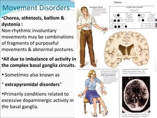
Lecture Outline  Huntington’s Chorea Dystonia Athetosis Ballismus Myoclonus Wilson’s disease Tardive dyskinesia Essential Tremor Tourette’s Syndrome (tics) Exam prep-what to know!
Movement Disorders Chorea, athetosis, ballism & dystonia : Non-rhythmic involuntary movements may be combinations of fragments of purposeful movements & abnormal postures.  All due to imbalance of activity in the complex basal ganglia circuits.  Sometimes also known as  “  extrapyramidal disorders ” Primarily conditions related to excessive dopaminergic activity in the basal ganglia.
These should NOT be thought of as separate entities amenable to specific definition but rather as a  SPECTRUM  of movements that blend into one-another WHY?
They often co-exist  Even neurologists may often not be able to agree as to how a particular movement should be classified!
Ballismus Dystonia Chorea Athetosis Movements become  - Less violent / explosive / jerky - Smoother and more flowing - More sustained They differ from tics in that they cannot be suppressed by voluntary control Myoclonus
Chorea(latin  choreus , dance)  Jerky semi-purposive uncontrollable movements of limbs , face & trunk , increase with anxiety & disappear during sleep. In the limbs they resemble fidgety movements, & in the face, grimaces. Patients often attempt to conceal involuntary movements by superimposing voluntary movements onto them e.g. an involuntary movement of arm towards face may be adapted to look-like an attempt to look at watch Pathophysiology-Structural lesions putamen, globus pallidus and subthalamic nuclei-Balance is critical  between the direct and indirect motor pathways to produce normal movement patterns. HD Video: http://www.youtube.com/watch?v=OveGZdZ_sVs&feature=rela http://www.youtube.com/watch?v=IjFWOJKRLQ0&feature=related Chorea Video: http://www.youtube.com/watch?v=iKwRBVkEwkY&feature=related http://www.youtube.com/watch?v=xW6LciTLoEA&feature=related
Huntington’s Chorea Clinical manifestations: Involuntary choreiform Diminished during sleep Facial tics/grimacing Dementia Paranoia & hallucinations Appetite Ravenous Emotions Labile George Huntington He wrote this paper when he was 22, a year after receiving his medical degree from Columbia University in New York. He first read the paper before the Meigs and Mason Academy of Medicine in Middleport, Ohio on February 15, 1872 and then published it in the Medical and Surgical Reporter of Philadelphia on April 13, 1872.
Huntington’s Chorea Prevalence 1 in 10000, average onset 40 and progresses over around 10-15 years. Etiology  Autosomal Dominant inherited. Gene chromosome 4p. excess number of  CAG tri-nucleotide repeats (>35) .  Diagnosis-DNA testing. Mutant protein product called Huntington with 40-150 glutamine residues.  Usually manifests at middle age – severity related to the number of tri-nucleotide repeats.
Huntington’s Chorea pathology Grossly characterized as  degeneration of the cerebral cortex and the basal ganglia Loss of GABAergic neurons in the striatum.  Severe striatal atrophy with resulting enlargement of ventricles.
Huntington’s Chorea Treatment Loss of GABA mediated inhibition on Substantia Nigra causes: hyperactivity of dopaminergic synapses Medical management No cure Dopamine receptor blocking drugs (antipsychotics), dopamine depleting drugs. Chlorpromazine (Thorazine) Haloperidol (Haldol) Nursing Management Family support Diet Ambulatory Safety
The other most recognised chorea. Mainly children / adolescents patients  5-15 yo  Etiology: Autoimmune response following infection with  group A  β -hemolytic Streptococcal infection Rare in developed countries-antibiotics! Initially characterised by  pharyngitis  (sore throat) followed within approx 1 to 5 weeks by the sudden onset of  acute rheumatic ferver . Chorea usually only occurs in and usually 1-6 months after the onset of sore throat.  Usually generalized chorea that disappears with sleep Usually remits spontaneously within 9 months (on  average) to 2 years Treatment:  Dopamine antagonists, valproic acid. Severe Cases-immunosupression, plasmapheresis or  immunoglobulin  IV Sydenham’s chorea Video: http://www.youtube.com/watch?v=RnxqqW_nH0k&feature=related
Dystonia: Clinical features Dystonias are sustained abnormal postures of limbs , neck , trunk, tongue protrusion or fixed upward deviation of the eyes (occulgyric crisis).  Due to co-contraction of agonist and antagonist muscles in part of body Classification: Most common dystonias are  focal  (single body part);  affect one part of the body such as eyes, neck, arm or vocal cords. Usually idopahic Multifocal dystonia:  affects many different parts of the body.  Segmental dystonias  affect two adjoining parts of the body usually symptomatic: Hemidystonia  affects an arm and a leg on one side of the body.  Generalized dystonia  affects most of the body, frequently involving the legs and back.
Blepharospasm means the involuntary contraction of the eyelids, leading to uncontrollable blinking and closure of the eyelids. Affects  women> men  Torticollis, commonly called wry neck, is the condition of spasm affecting the muscles of the neck, causing the head to assume unnatural postures or turn uncontrollably. Spasmodic torticollis, also known as cervical dystonia, is the most common of the focal dystonias.  Writer’s cramp: -  Dystonic posturing of arm when hand used to perform  specific tasks e.g. writing, playing piano  Dystonia Videos: http://www.youtube.com/watch?v=eKhOVaY-YNo&feature=related http://www.youtube.com/watch?v=EA2HU6rb9UM&feature=related http://www.youtube.com/watch?v=qpd1BxUxn8E&feature=related
Anticholinergics (benzatropine), antihistamines (diphenhydramine), anti-Parkinsons agents (trihexyphenidyl), and muscle relaxers (diazepam) &  GABA B  receptor agonist (baclofen). The drug resistant dystonias can be treated by botulinum toxin injection to the responsible muscles, to overcome the abnormal distribution of muscle activity for a period of time.  Dystonia Treatment:
Dystonia-drug induced Acute dystonic reaction- occulogyric crisis, sustained upward deviation of the eyes +/- toricollis, jaw opening, tongue protrusion and anxiety. Usually a result of administering dopamine receptor blocking drugs, generally metaclopramide ( antiemetic and gastroprokinetic agent used to treat nausea and vomiting, and to facilitate gastric emptying)  or prochlorperazine (neuroleptic;  antiemetic)   in young. 50% in 48hrs, 90% in 5 days. Treatment anticholinergic drugs, usually benztropine 1-2mg IV
Athetosis is a slow continuous stream of slow, sinuous, writhing movements, typically of the hands and feet.  Most commonly seen together with chorea in dyskinetic motor fluctuations in  PD . Also in  athetoid cerebral palsy  where damage occurs in the basal ganglia. Related to  excessive dopaminergic activity . In PD reducing dopaminergic drugs alleviates.  If Athetosis becomes faster, it sometimes blends with chorea, ie choreoathetosis/ 'choreo-athetoid' movements.  Can be thought of as an athetoid movement that “gets stuck” for a period of time; thus, a patient with choreoathetosis may perform an involuntary movement in which his hand and fingers are twisted behind his head. He may hold this position for a few moments before his hand moves back in front of his body. The part of the movement when the limb was held, unmoving, in an abnormal position would be considered a dystonia (may occur alone). Athetosis Athetosis Video: http://www.youtube.com/watch?v=J_wIDm1_ax4&feature=related
Usually in elderly & ceases within few weeks usually self-limited, lasting 6 to 8 wk . More dramatic ballistic movements of  of the arms & legs on one side of the body ( unilateral)  and therefore use term  “ HEMIBALLISMUS” . Treatment with antipsychotics is often effective. Usually due to a CVA in contralateral subthalamic nucleus Hemiballismus video: http://www.youtube.com/watch?v=hqg2GTUq1k4&feature=related
Structural lesions (subthalamic nucleus)- Hereditary  Huntington's disease /Wilson's/Neuroacanthocytosis  Porphyria  Paroxysmal choreoathetosis  Cerebral birth injury (including kernicterus);  Static Encephalopathy (Cerebral Palsy ) Cerebral trauma Cerebrovascular ( ischaemia, haemorrhage ) Drugs  Levodopa /Dopamine agonists /Phenothiazines /Tricyclics  Oral contraceptive  Pregnancy  Endocrine: Thyrotoxicosis /Hypoparathyroidism /Hypoglycaemia  Infective/inflammatory  Post-streptococcal (Sydenham's chorea)  Henoch-Schönlein purpura  Creutzfeldt-Jakob disease  Antiphospholipid antibody syndrome  SLE  Vascular: Lacunar infarction /Arteriovenous malformation  Extra material to read latter-Causes of Chorea, Dystonia and athetosis
Extra material to read latter-Dystonia: Etiology Causes: Primary dystonia refers to the situation where dystonia is the only sign and there is no identifiable cause or structural abnormality in the central nervous system.  Although the causes of dystonia are not fully known it is currently thought that the condition results from a malfunction in a part of the brain called the basal ganglia. The problem may mainly lie in an area of the basal ganglia called the globus pallidus. If this area of the brain is not functioning correctly then the control of another structure in the brain called the thalamus is affected. The thalamus controls the planning and execution of movement and sends nerves to muscles via the spinal cord. The end result is that muscle co-ordination is not regulated properly. The wrong muscles will contract on movement or all muscles will contract unnecessarily causing abnormal movement and posture.  Secondary dystonia implies there is a clear cause, such as a change in the structure of the brain, an environmental cause, as part of an inherited or acquired neurological disease or due to drugs or toxins.  Environmental causes include head trauma, stroke, a tumour, multiple sclerosis, infections in the brain, injury to the spinal cord, or after chemotherapy, drugs ( neuroleptics-Tardive dystonia ) or toxins that affect the basal ganglia, thalamus or brain stem. They may be associated with other hereditary neurological syndromes. Dystonia may be the first sign in a patient with Huntington's disease, and is secondary to many other neurological diseases including Parkinson's disease, Wilson's disease and Ataxia telangiectasia.
Brief, isolated, random, non-purposeful jerks of muscle groups in the limbs, may occur normally at the onset of sleep (hypnic jerks).  May be caused by active muscle contraction  -  positive myoclonus May be caused by inhibition of ongoing muscle activity - negative myoclonus ( eg. Asterixis )  Generalised - widespread throughout body Focal / segmental – restricted to particular part of body Treatment:Valproic acid is drug of choice  May respond to benzodiazepines  e.g. clonazepam Myoclonus video: http://www.youtube.com/watch?v=HeOn99vJ8s0&NR=1
Physiologic - Nocturnal ( usually on falling asleep ) - Hiccups Essential - Occurs in the absence of other     abnormality - Benign and sometimes inherited Epileptic - Demonstrable cortical source Symptomatic  i.e secondary to disease process - Neurodegenerative eg. Wilson’s disease - Infectious e.g CJD, Viral encephalitis - Toxic e.g. penicillin, antidepressants - Metabolic  - anoxic brain damage - hypoglycemia - hepatic failure ( “ asterixis” ) - renal failure - hyponatremia
Wilson’s disease Autosomal recessive defect of copper excretion in which there is defective copper-binding to ceruloplasmin Clinical presentation:  bradykinesia, dysphagia,dysarth ria, dystonia. Leads to copper deposition in: -  liver  causing cirrhosis - brain ( especially basal ganglia )  leading to movement disorders and  other abnormalities - Cornea leading to the appearance of rusty  brown “ Kayser-Fleischer rings” around  cornea (yellow-brown pigment at the limbus of the cornea ). Diagnosis is via low serum ceruloplasmin, increased urinary Cu, liver biopsy ( excessive Cu) and brain MRI changes Although it is rare it is very important to think-of and diagnose as it is TREATABLE:- Penicillamine is given to chelate the copper and promote excretion. Video WD:http://www.youtube.com/watch?v=JgDvQUwUOo0 Wilson’s disease  Hand Dystonia
Involuntary movements as facial grimacing , chewing movements, tongue movements  (oro-bucco-mandibular dyskinesia) . Appear after weeks, generally years of exposure to  dopamine receptor blocking drugs .  Older or classical ‘typical’ antipsychotics e.g. chlorpromazine, haloperidol With typical antipsychotics prevalence around 20% Thought to be due to receptors super sensitivity to these drugs .  Changes in synapse number and dendrite arborisation also occur. Tardive dyskinesia TD Video: http://www.youtube.com/watch?v=FUr8ltXh1Pc&feature=related
Tardive dyskinesia Newer ‘atypical’ drugs (e.g. clozapine, quetiapine, aripiprazole) Atypical antipsychotics  5-HT 1A  receptor partial/full agonists. Antagonist activity at D 2  receptor mainly in limbic system rather than striatum.  Treatment is difficult but withdrawal of the causative drug.  Withdrawal often worsens symptoms.  Tardive dykinesia disappears with time in 1/3 (generally mild cases). Permanent in 2/3s.
Probably the most common movement disorder.  Hallmark: postural and action tremor affecting the hands, head and/or voice. Presents as a  rhythmic tremor  (4–12 Hz).  Typically bilateral, accentuated with goal-directed movement ( Action tremor ) as opposed to  rest tremor  (PD) Often, it is inherited as an autosomal dominant trait with the tremor becoming apparent by middle age and sometimes as early as childhood.  Diagnosis-Clinical grounds;  No gene or identifiable pathology has been identified for essential tremor.  Abnormalities of  the basal ganglia, the cerebellum, the thalamus, the connections between these structures, or a combination of factors may be causative.  Treatment:  First line- beta-blockers ( propranolol 30-240 mg/d ), or anticonvulsants  primadone (50-1000 mg/d) . Second line-benzodiazepines and methazolamide (Carbonic Anhydrase Inhibitor.  Finally if all else fails- Botox or Surgical therapies such as electronic stimulation of the VIM nucleus of the thalamus or ablation of this structure (thalamotomy), can be effective treatments for those refractory to drug therapy.  Can be confused with: Physiologic tremor  -physical exertion, hyperthyroidism, acute hypoglycemia, and other physical and metabolic stressors. Induced tremor  -Stimulant drugs (including caffeine and the amphetamines), antidepressants, Depakote, and    agonist drugs (used to treat asthma).  Katherine Hepburn
Neuropsychiatric condition characterized by the childhood onset of multiple motor and vocal tics- Repetitive semi-purposeful movements as blinking, winking, grinning or screwing up of the eyes.  Coprolalia  (the inadvertent utterance of obscenities), echolalia (involuntary repetition of other's phrases),  palilalia  (involuntary repetition of one's own utterances) and  echopraxia  (involuntary mimicking of the action of others).  Worsen under stress! Distinguished from other involuntary movements by the ability of the patient to suppress their occurrence, at least for a short time.  Often-times, affected individuals have  co-existing   obsessive-compulsive disorder , learning disabilities, hyperactivity/ attention deficit disorder  and behavioral problems. Although the pathogenic basis is not understood,  Tics are believed to result from dysfunction in the thalamus, basal ganglia, and frontal cortex of the brain, involving abnormal activity of the brain chemical, or neurotransmitter,  dopamine   therefore  Tics can be treated with dopamine receptor blocking or dopamine depleting drugs  such as neuroleptics.  Treatment of tics is symptomatic with  haloperidol  and other neuroleptics.  Some patients benefit from  clonidine , an   -2 agonist, or with  benzodiazepines .    May be transmitted as an autosomal dominant trait
Exam prep-what to know! • Learn the basic terms used to describe abnormal movements (chorea, tremor, athetosis, ballismus, dystonia, tics). • Understand the connections of the basal ganglia and their influence on movement. • Recognize the clinical features of the following movement disorders and be familiar with the appropriate pharmacologic interventions for each condition: • Chorea- Huntington’s; Sydenham’s. • Essential tremor. •  Tourette’s syndrome. • Dystonia. • Myoclonus. • Wilson’s disease. • Tardive Dyskinesia.
Movement disorders lecture

Movement disorders lecture

  • 2.
    Lecture Outline Huntington’s Chorea Dystonia Athetosis Ballismus Myoclonus Wilson’s disease Tardive dyskinesia Essential Tremor Tourette’s Syndrome (tics) Exam prep-what to know!
  • 3.
    Movement Disorders Chorea,athetosis, ballism & dystonia : Non-rhythmic involuntary movements may be combinations of fragments of purposeful movements & abnormal postures. All due to imbalance of activity in the complex basal ganglia circuits. Sometimes also known as “ extrapyramidal disorders ” Primarily conditions related to excessive dopaminergic activity in the basal ganglia.
  • 4.
    These should NOTbe thought of as separate entities amenable to specific definition but rather as a SPECTRUM of movements that blend into one-another WHY?
  • 5.
    They often co-exist Even neurologists may often not be able to agree as to how a particular movement should be classified!
  • 6.
    Ballismus Dystonia ChoreaAthetosis Movements become - Less violent / explosive / jerky - Smoother and more flowing - More sustained They differ from tics in that they cannot be suppressed by voluntary control Myoclonus
  • 7.
    Chorea(latin choreus, dance) Jerky semi-purposive uncontrollable movements of limbs , face & trunk , increase with anxiety & disappear during sleep. In the limbs they resemble fidgety movements, & in the face, grimaces. Patients often attempt to conceal involuntary movements by superimposing voluntary movements onto them e.g. an involuntary movement of arm towards face may be adapted to look-like an attempt to look at watch Pathophysiology-Structural lesions putamen, globus pallidus and subthalamic nuclei-Balance is critical between the direct and indirect motor pathways to produce normal movement patterns. HD Video: http://www.youtube.com/watch?v=OveGZdZ_sVs&feature=rela http://www.youtube.com/watch?v=IjFWOJKRLQ0&feature=related Chorea Video: http://www.youtube.com/watch?v=iKwRBVkEwkY&feature=related http://www.youtube.com/watch?v=xW6LciTLoEA&feature=related
  • 8.
    Huntington’s Chorea Clinicalmanifestations: Involuntary choreiform Diminished during sleep Facial tics/grimacing Dementia Paranoia & hallucinations Appetite Ravenous Emotions Labile George Huntington He wrote this paper when he was 22, a year after receiving his medical degree from Columbia University in New York. He first read the paper before the Meigs and Mason Academy of Medicine in Middleport, Ohio on February 15, 1872 and then published it in the Medical and Surgical Reporter of Philadelphia on April 13, 1872.
  • 9.
    Huntington’s Chorea Prevalence1 in 10000, average onset 40 and progresses over around 10-15 years. Etiology Autosomal Dominant inherited. Gene chromosome 4p. excess number of CAG tri-nucleotide repeats (>35) . Diagnosis-DNA testing. Mutant protein product called Huntington with 40-150 glutamine residues. Usually manifests at middle age – severity related to the number of tri-nucleotide repeats.
  • 10.
    Huntington’s Chorea pathologyGrossly characterized as degeneration of the cerebral cortex and the basal ganglia Loss of GABAergic neurons in the striatum. Severe striatal atrophy with resulting enlargement of ventricles.
  • 11.
    Huntington’s Chorea TreatmentLoss of GABA mediated inhibition on Substantia Nigra causes: hyperactivity of dopaminergic synapses Medical management No cure Dopamine receptor blocking drugs (antipsychotics), dopamine depleting drugs. Chlorpromazine (Thorazine) Haloperidol (Haldol) Nursing Management Family support Diet Ambulatory Safety
  • 12.
    The other mostrecognised chorea. Mainly children / adolescents patients 5-15 yo Etiology: Autoimmune response following infection with group A β -hemolytic Streptococcal infection Rare in developed countries-antibiotics! Initially characterised by pharyngitis (sore throat) followed within approx 1 to 5 weeks by the sudden onset of acute rheumatic ferver . Chorea usually only occurs in and usually 1-6 months after the onset of sore throat. Usually generalized chorea that disappears with sleep Usually remits spontaneously within 9 months (on average) to 2 years Treatment: Dopamine antagonists, valproic acid. Severe Cases-immunosupression, plasmapheresis or immunoglobulin IV Sydenham’s chorea Video: http://www.youtube.com/watch?v=RnxqqW_nH0k&feature=related
  • 13.
    Dystonia: Clinical featuresDystonias are sustained abnormal postures of limbs , neck , trunk, tongue protrusion or fixed upward deviation of the eyes (occulgyric crisis). Due to co-contraction of agonist and antagonist muscles in part of body Classification: Most common dystonias are focal (single body part); affect one part of the body such as eyes, neck, arm or vocal cords. Usually idopahic Multifocal dystonia: affects many different parts of the body. Segmental dystonias affect two adjoining parts of the body usually symptomatic: Hemidystonia affects an arm and a leg on one side of the body. Generalized dystonia affects most of the body, frequently involving the legs and back.
  • 14.
    Blepharospasm means theinvoluntary contraction of the eyelids, leading to uncontrollable blinking and closure of the eyelids. Affects women> men Torticollis, commonly called wry neck, is the condition of spasm affecting the muscles of the neck, causing the head to assume unnatural postures or turn uncontrollably. Spasmodic torticollis, also known as cervical dystonia, is the most common of the focal dystonias. Writer’s cramp: - Dystonic posturing of arm when hand used to perform specific tasks e.g. writing, playing piano Dystonia Videos: http://www.youtube.com/watch?v=eKhOVaY-YNo&feature=related http://www.youtube.com/watch?v=EA2HU6rb9UM&feature=related http://www.youtube.com/watch?v=qpd1BxUxn8E&feature=related
  • 15.
    Anticholinergics (benzatropine), antihistamines(diphenhydramine), anti-Parkinsons agents (trihexyphenidyl), and muscle relaxers (diazepam) & GABA B receptor agonist (baclofen). The drug resistant dystonias can be treated by botulinum toxin injection to the responsible muscles, to overcome the abnormal distribution of muscle activity for a period of time. Dystonia Treatment:
  • 16.
    Dystonia-drug induced Acutedystonic reaction- occulogyric crisis, sustained upward deviation of the eyes +/- toricollis, jaw opening, tongue protrusion and anxiety. Usually a result of administering dopamine receptor blocking drugs, generally metaclopramide ( antiemetic and gastroprokinetic agent used to treat nausea and vomiting, and to facilitate gastric emptying) or prochlorperazine (neuroleptic; antiemetic) in young. 50% in 48hrs, 90% in 5 days. Treatment anticholinergic drugs, usually benztropine 1-2mg IV
  • 17.
    Athetosis is aslow continuous stream of slow, sinuous, writhing movements, typically of the hands and feet. Most commonly seen together with chorea in dyskinetic motor fluctuations in PD . Also in athetoid cerebral palsy where damage occurs in the basal ganglia. Related to excessive dopaminergic activity . In PD reducing dopaminergic drugs alleviates. If Athetosis becomes faster, it sometimes blends with chorea, ie choreoathetosis/ 'choreo-athetoid' movements. Can be thought of as an athetoid movement that “gets stuck” for a period of time; thus, a patient with choreoathetosis may perform an involuntary movement in which his hand and fingers are twisted behind his head. He may hold this position for a few moments before his hand moves back in front of his body. The part of the movement when the limb was held, unmoving, in an abnormal position would be considered a dystonia (may occur alone). Athetosis Athetosis Video: http://www.youtube.com/watch?v=J_wIDm1_ax4&feature=related
  • 18.
    Usually in elderly& ceases within few weeks usually self-limited, lasting 6 to 8 wk . More dramatic ballistic movements of of the arms & legs on one side of the body ( unilateral) and therefore use term “ HEMIBALLISMUS” . Treatment with antipsychotics is often effective. Usually due to a CVA in contralateral subthalamic nucleus Hemiballismus video: http://www.youtube.com/watch?v=hqg2GTUq1k4&feature=related
  • 19.
    Structural lesions (subthalamicnucleus)- Hereditary Huntington's disease /Wilson's/Neuroacanthocytosis Porphyria Paroxysmal choreoathetosis Cerebral birth injury (including kernicterus); Static Encephalopathy (Cerebral Palsy ) Cerebral trauma Cerebrovascular ( ischaemia, haemorrhage ) Drugs Levodopa /Dopamine agonists /Phenothiazines /Tricyclics Oral contraceptive Pregnancy Endocrine: Thyrotoxicosis /Hypoparathyroidism /Hypoglycaemia Infective/inflammatory Post-streptococcal (Sydenham's chorea) Henoch-Schönlein purpura Creutzfeldt-Jakob disease Antiphospholipid antibody syndrome SLE Vascular: Lacunar infarction /Arteriovenous malformation Extra material to read latter-Causes of Chorea, Dystonia and athetosis
  • 20.
    Extra material toread latter-Dystonia: Etiology Causes: Primary dystonia refers to the situation where dystonia is the only sign and there is no identifiable cause or structural abnormality in the central nervous system. Although the causes of dystonia are not fully known it is currently thought that the condition results from a malfunction in a part of the brain called the basal ganglia. The problem may mainly lie in an area of the basal ganglia called the globus pallidus. If this area of the brain is not functioning correctly then the control of another structure in the brain called the thalamus is affected. The thalamus controls the planning and execution of movement and sends nerves to muscles via the spinal cord. The end result is that muscle co-ordination is not regulated properly. The wrong muscles will contract on movement or all muscles will contract unnecessarily causing abnormal movement and posture. Secondary dystonia implies there is a clear cause, such as a change in the structure of the brain, an environmental cause, as part of an inherited or acquired neurological disease or due to drugs or toxins. Environmental causes include head trauma, stroke, a tumour, multiple sclerosis, infections in the brain, injury to the spinal cord, or after chemotherapy, drugs ( neuroleptics-Tardive dystonia ) or toxins that affect the basal ganglia, thalamus or brain stem. They may be associated with other hereditary neurological syndromes. Dystonia may be the first sign in a patient with Huntington's disease, and is secondary to many other neurological diseases including Parkinson's disease, Wilson's disease and Ataxia telangiectasia.
  • 21.
    Brief, isolated, random,non-purposeful jerks of muscle groups in the limbs, may occur normally at the onset of sleep (hypnic jerks). May be caused by active muscle contraction - positive myoclonus May be caused by inhibition of ongoing muscle activity - negative myoclonus ( eg. Asterixis ) Generalised - widespread throughout body Focal / segmental – restricted to particular part of body Treatment:Valproic acid is drug of choice May respond to benzodiazepines e.g. clonazepam Myoclonus video: http://www.youtube.com/watch?v=HeOn99vJ8s0&NR=1
  • 22.
    Physiologic - Nocturnal( usually on falling asleep ) - Hiccups Essential - Occurs in the absence of other abnormality - Benign and sometimes inherited Epileptic - Demonstrable cortical source Symptomatic i.e secondary to disease process - Neurodegenerative eg. Wilson’s disease - Infectious e.g CJD, Viral encephalitis - Toxic e.g. penicillin, antidepressants - Metabolic - anoxic brain damage - hypoglycemia - hepatic failure ( “ asterixis” ) - renal failure - hyponatremia
  • 23.
    Wilson’s disease Autosomalrecessive defect of copper excretion in which there is defective copper-binding to ceruloplasmin Clinical presentation: bradykinesia, dysphagia,dysarth ria, dystonia. Leads to copper deposition in: - liver causing cirrhosis - brain ( especially basal ganglia ) leading to movement disorders and other abnormalities - Cornea leading to the appearance of rusty brown “ Kayser-Fleischer rings” around cornea (yellow-brown pigment at the limbus of the cornea ). Diagnosis is via low serum ceruloplasmin, increased urinary Cu, liver biopsy ( excessive Cu) and brain MRI changes Although it is rare it is very important to think-of and diagnose as it is TREATABLE:- Penicillamine is given to chelate the copper and promote excretion. Video WD:http://www.youtube.com/watch?v=JgDvQUwUOo0 Wilson’s disease Hand Dystonia
  • 24.
    Involuntary movements asfacial grimacing , chewing movements, tongue movements (oro-bucco-mandibular dyskinesia) . Appear after weeks, generally years of exposure to dopamine receptor blocking drugs . Older or classical ‘typical’ antipsychotics e.g. chlorpromazine, haloperidol With typical antipsychotics prevalence around 20% Thought to be due to receptors super sensitivity to these drugs . Changes in synapse number and dendrite arborisation also occur. Tardive dyskinesia TD Video: http://www.youtube.com/watch?v=FUr8ltXh1Pc&feature=related
  • 25.
    Tardive dyskinesia Newer‘atypical’ drugs (e.g. clozapine, quetiapine, aripiprazole) Atypical antipsychotics 5-HT 1A receptor partial/full agonists. Antagonist activity at D 2 receptor mainly in limbic system rather than striatum. Treatment is difficult but withdrawal of the causative drug. Withdrawal often worsens symptoms. Tardive dykinesia disappears with time in 1/3 (generally mild cases). Permanent in 2/3s.
  • 26.
    Probably the mostcommon movement disorder. Hallmark: postural and action tremor affecting the hands, head and/or voice. Presents as a rhythmic tremor (4–12 Hz). Typically bilateral, accentuated with goal-directed movement ( Action tremor ) as opposed to rest tremor (PD) Often, it is inherited as an autosomal dominant trait with the tremor becoming apparent by middle age and sometimes as early as childhood. Diagnosis-Clinical grounds; No gene or identifiable pathology has been identified for essential tremor. Abnormalities of the basal ganglia, the cerebellum, the thalamus, the connections between these structures, or a combination of factors may be causative. Treatment: First line- beta-blockers ( propranolol 30-240 mg/d ), or anticonvulsants primadone (50-1000 mg/d) . Second line-benzodiazepines and methazolamide (Carbonic Anhydrase Inhibitor. Finally if all else fails- Botox or Surgical therapies such as electronic stimulation of the VIM nucleus of the thalamus or ablation of this structure (thalamotomy), can be effective treatments for those refractory to drug therapy. Can be confused with: Physiologic tremor -physical exertion, hyperthyroidism, acute hypoglycemia, and other physical and metabolic stressors. Induced tremor -Stimulant drugs (including caffeine and the amphetamines), antidepressants, Depakote, and  agonist drugs (used to treat asthma). Katherine Hepburn
  • 27.
    Neuropsychiatric condition characterizedby the childhood onset of multiple motor and vocal tics- Repetitive semi-purposeful movements as blinking, winking, grinning or screwing up of the eyes. Coprolalia (the inadvertent utterance of obscenities), echolalia (involuntary repetition of other's phrases), palilalia (involuntary repetition of one's own utterances) and echopraxia (involuntary mimicking of the action of others). Worsen under stress! Distinguished from other involuntary movements by the ability of the patient to suppress their occurrence, at least for a short time. Often-times, affected individuals have co-existing obsessive-compulsive disorder , learning disabilities, hyperactivity/ attention deficit disorder and behavioral problems. Although the pathogenic basis is not understood, Tics are believed to result from dysfunction in the thalamus, basal ganglia, and frontal cortex of the brain, involving abnormal activity of the brain chemical, or neurotransmitter, dopamine therefore Tics can be treated with dopamine receptor blocking or dopamine depleting drugs such as neuroleptics. Treatment of tics is symptomatic with haloperidol and other neuroleptics. Some patients benefit from clonidine , an  -2 agonist, or with benzodiazepines . May be transmitted as an autosomal dominant trait
  • 29.
    Exam prep-what toknow! • Learn the basic terms used to describe abnormal movements (chorea, tremor, athetosis, ballismus, dystonia, tics). • Understand the connections of the basal ganglia and their influence on movement. • Recognize the clinical features of the following movement disorders and be familiar with the appropriate pharmacologic interventions for each condition: • Chorea- Huntington’s; Sydenham’s. • Essential tremor. • Tourette’s syndrome. • Dystonia. • Myoclonus. • Wilson’s disease. • Tardive Dyskinesia.

Editor's Notes

  • #2 VCP3042; 1st August 2008 [email_address]
  • #10 VCP3042; 1st August 2008 [email_address] Early massive loss of striatopallidal and then striatonigral neurones; excitotoxicity