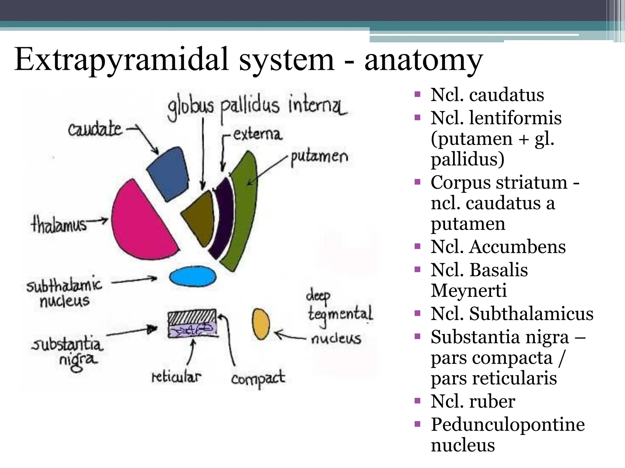
Extrapyramidal system and
cerebellum
Matej Skorvanek MD
Dept. of Neurology
Safarik University in Kosice
Extrapyramidal system - anatomy
 Ncl. caudatus
 Ncl. lentiformis
(putamen + gl.
pallidus)
 Corpus striatum -
ncl. caudatus a
putamen
 Ncl. Accumbens
 Ncl. Basalis
Meynerti
 Ncl. Subthalamicus
 Substantia nigra –
pars compacta /
pars reticularis
 Ncl. ruber
 Pedunculopontine
nucleus
Extrapyramidal syndromes
• Hypokinetic syndrome
• Hyperkinetic syndrome
Hypokinetic syndrome
• Bradykinesia – pathological slowness of movements
• Hypokinesia – lower amplitude of movements
• Akinesia – problem with movement initiation
• Rigidity – increased muscle toneýšenie svalového
tonusu (compared to spasticity resistance is the same
during whole range of passive movement – „lead
pipe phenomenon / cogwheel phenomenon“)
Parkinsonian syndrome
• Bradykinesia (hypokinesia, akinesia) + at least
one of the following:
▫ Rigidity
▫ Resting tremor
Parkinsonian syndrome - aetiology
 Parkinson‘s disease 80%
 Neurodegenerative disorders 10%
 Atypical parkinsonism
 Progressive supranuclear palsy
 Corticobasal degeneration
 Multiple system atrophy
 Dementia with Lewy Bodies
 Secondary 10%
 Drug-induced, vascular, toxic, posttraumatic,
postencephalitic
Hyperkinetic syndrome
• Tremor
• Chorea
• Balismus
• Dystonia
• Myoclonus
• Tics
Tremor
• Rytmical oscillation of a body part around a joint
• Alternating contractions of agonist and
antagonist
• Dif.dg. – rytmical myoclonus, dystonic tremor
Classification
• According to body part affected – most frequently arms
• According to position where tremor is observed
▫ Resting (parkinsonian)
▫ Postural (essential, physiological, drug-induced)
▫ Kinetic/intentional (cerebellar)
• According to frequency
• According to amplitude
History
1. Anatomical localization?
2. Task-specific?
3. Age of onset?
4. Disease course? (progressive – PD, static – ET,
sudden onset and/or sudden remission – functional?)
5. Factors that improve or worsen tremor? – alcohol,
fatigue, anxiety, stress, mental tasks, ...
6. Drug history
7. Bradykinesia/rigidity in history
8. Metabolic diseases? – hyperthyroidism,...
9. Family history
Clinical examination
• Anatomical location – hands, arms, head, chin, legs,
trunk
• Symetrical/asymetrical
• Position – resting, postural, kinetic, intentional
(finger-nose-finger)
• Frequency (low/high), amplitude (big/small)
• Activation maneuvers
• Other extrapyramidal signs – rigidity, dystonia, ...
• Oculomotor abnormalities
• Signs of neuropathy
• Gait abnormalities
Activation maneuvers
• Cognitive task – naming months backwards,
counting (increase of supressed organic tremor/
decrease or character change of functional)
• Archimedes spiral
• Drinking a cup of water
• Writing
Patophysiology of tremor
Helmich 2013
Chorea
• Disorder characterised by involuntary brief jerky or
twisting movements, which appear at random in the
affected body part
• Important is the random nature and unpredictability
of the following movement.
• Movements are brief and brisk, but can be also longer
and more twisting (choreoathetosis), they are most
pronounced at distal body segments
Chorea
• Worsening during speech, movements, emotions
• E.g. „milking maid handshake“, can‘t keep the
tounge protruded, dancing gait (st. Vitus dance)
• Distribution
▫ Focal
▫ Segmental
▫ Hemichorea
▫ Generalized
Chorea - aetiology
• Neurodegenerative – Huntington‘s disease
• Metabolic, toxic and drug-induced (hepatal
encephalopathy, CO poisoning, Mn, tardive
dyskinesias after neuroleptics,...)
• Secondary due to BG lessions (trauma,
stroke, tumors)
• Autoimunne (assoc. with antibodies against
streptococcus – chorea minor assoc. with
rheumatic fever, chorea gravidarum)
Ballism
• Variant of chorea with large amplitude
involuntary movements of proximal parts of the
limbs
• Usually due to lesions of ncl.subthalamicus Luysi
or putamen
• Most frequently hemiballism after contralateral
lesion of the subthalamic nucleus
Dystonia
• Dystonia is defined as a movement disorder
characterized by sustained or intermittent muscle
contractions causing abnormal, often repetitive,
movements, postures, or both.
• Dystonic movements are typically patterned and
twisting, and may be tremulous.
• Dystonia is often initiated or worsened by voluntary
action and associated with overflow muscle
activation.
Albanese et al. 2013
Dystonia
• Fixed dystonia
• Mobile dystonia
• Dystonic myoclonus
• Dystonic tremor
• Dif.dg. Of dystonie
▫ Chorea
▫ Myoclonus
▫ Tics
▫ Stereotypies
Axis I: clinical characteristics
I. Age at onset
I. Infancy (birth to 2 years)
II. Childhood (3–12 years)
III. Adolescence (13–20 years)
IV. Early adulthood (21–40 years)
V. Late adulthood (>40 years)
II. Body distribution
I. Focal
II. Segmental
III. Multifocal
IV. Generalized (with or without leg
involvement)
V. Hemidystonia
III. Temporal pattern
I. Disease course
I. Static
II. Progressive
II. Variability
I. Persistent
II. Action-specific
III. Diurnal
IV. Paroxysmal
IV. Associated features
I. Isolated dystonia or combined with
another movement disorder
II. Occurence of other neurological or
systemic manifestations
Axis II: Etiology
I. Nervous system pathology
I. Evidence of degeneration
II. Evidence of structural (often
static) lesion
III. No evidence of degeneration or
structural lesion
II. Inherited or acquired
I. Inherited
I. AD, AR, X-linked, mitochondrial
II. Acquired
I. Perinatal brain injury
II. Infection
III. Drug-induced
IV. Toxic
V. Vascular
VI. Neoplastic
VII.Posttraumatic
VIII.Psychogenic
III. Idiopathic
I. Sporadic
II. Familial
Albanese et al. 2013
Clinical characteristics
• Body distribution
▫ Focal – only 1 muscle group or body segment
▫ Segmental – 2 neighbouring muscle groups or body
segments (e.g. head + neck)
▫ Multifocal – 2 non-neighbouring muscle groups or body
segments (e.g. right arm + left leg)
▫ Hemidystonia
▫ Generalized – affection of the trunk + at least 2 other
body segments – with or without leg involvement
Age of onset
I. Infancy (<2 years)
II. Childhood (3-12 years)
III.Adolescence (13-20 years)
IV.Early adulthood (21-40 years)
V. Late adulthood (>40 years)
Temporal pattern
I. Disease course
I. Static
II. Progressive
II. Variability
I. Persistent symptoms
II. Action-specific
III.Diurnal fluctuations
IV.Paroxysmal dystonia
Associated features
I. Dystonia isolated or combined with other
extrapyramidal symptom
I. Isolated dystonia
II. Combined dystonia
II. Other neurological or systemic symptoms
Myoclonus
• Very brief involuntary jerky movements caused by
intermitent muscle contractions or relaxations
• Regular / irregular
• Positive / negative (asterixis)
• Focal, segmental, multifocal, generalized
• Stimulus sensitive to sound, touch?
• Cortical / subcortical / spinal
• Dif.dg. Tremor, chorea, epilepsy, fasciculations, psychogenic
Tics
 Fast (clonic) or slow (tonic) irregular repetitive stereotyped
movements or vocalizations
 Preceded by growing inner tension and urge to perform the
tic and followed be relief after performing the tic
 Partially voluntarily supressible
 Simple motor tics
 Complex motor tics
 Simple vocal tics
 Complex motor tics
Cerebellar anatomy
• Archicerebellum
▫ Oldest part
▫ Flocculonodular lobe
▫ Balance, eye movements
• Palleocerebellum
▫ Ant. a post. part of vermis
▫ Modulates sequential movements
• Neocerebellum
▫ Youngest part
▫ Hemispheres and central part of vermis
▫ Fine motor and speech control
Clinical features of cerebellar dysfunction
• Ataxia (appendicular/axial)
▫ Dysmetria (hyper) – disturbance of aiming
▫ Dysdiadochokinesis – disturbance of fast
alternating movements
▫ Dyssynergy – loss of coordination between
involved muscle groups
• Rebound
• Failure/disturbance of inhibition
Clinical features of cerebellar dysfunction
• Tremor (kinetic/intentional)
• Titubations and oscillations around the axis on standing
• Hypotonia
• Dysarthria, Ataxic (scanning) speech
• Dysphagia
• Ocular movement abnormalities (nystagmus, slow saccades, saccadic
dysmetria, occular flutter, opsoclonus, skew deviation)
• Cognition – slow thinking, problems in motor learning
• Palleocerebellar syndrome
▫ Ataxia of stance and gait (archicerebellum)
▫ Trunk assynergz (axial muscles)
▫ Falls (frequent, usually backwards)
• Neocerebellar syndrome
▫ Hypermetria
▫ Adiadochokinesis
▫ Assynergy of fine movements
▫ Intentional tremor
▫ Pasivity (lower muscle tone)

Extrapyramidal system and cerebellum.pdf

  • 1.
    Extrapyramidal system and cerebellum MatejSkorvanek MD Dept. of Neurology Safarik University in Kosice
  • 2.
    Extrapyramidal system -anatomy  Ncl. caudatus  Ncl. lentiformis (putamen + gl. pallidus)  Corpus striatum - ncl. caudatus a putamen  Ncl. Accumbens  Ncl. Basalis Meynerti  Ncl. Subthalamicus  Substantia nigra – pars compacta / pars reticularis  Ncl. ruber  Pedunculopontine nucleus
  • 3.
    Extrapyramidal syndromes • Hypokineticsyndrome • Hyperkinetic syndrome
  • 5.
    Hypokinetic syndrome • Bradykinesia– pathological slowness of movements • Hypokinesia – lower amplitude of movements • Akinesia – problem with movement initiation • Rigidity – increased muscle toneýšenie svalového tonusu (compared to spasticity resistance is the same during whole range of passive movement – „lead pipe phenomenon / cogwheel phenomenon“)
  • 6.
    Parkinsonian syndrome • Bradykinesia(hypokinesia, akinesia) + at least one of the following: ▫ Rigidity ▫ Resting tremor
  • 7.
    Parkinsonian syndrome -aetiology  Parkinson‘s disease 80%  Neurodegenerative disorders 10%  Atypical parkinsonism  Progressive supranuclear palsy  Corticobasal degeneration  Multiple system atrophy  Dementia with Lewy Bodies  Secondary 10%  Drug-induced, vascular, toxic, posttraumatic, postencephalitic
  • 8.
    Hyperkinetic syndrome • Tremor •Chorea • Balismus • Dystonia • Myoclonus • Tics
  • 9.
    Tremor • Rytmical oscillationof a body part around a joint • Alternating contractions of agonist and antagonist • Dif.dg. – rytmical myoclonus, dystonic tremor
  • 10.
    Classification • According tobody part affected – most frequently arms • According to position where tremor is observed ▫ Resting (parkinsonian) ▫ Postural (essential, physiological, drug-induced) ▫ Kinetic/intentional (cerebellar) • According to frequency • According to amplitude
  • 11.
    History 1. Anatomical localization? 2.Task-specific? 3. Age of onset? 4. Disease course? (progressive – PD, static – ET, sudden onset and/or sudden remission – functional?) 5. Factors that improve or worsen tremor? – alcohol, fatigue, anxiety, stress, mental tasks, ... 6. Drug history 7. Bradykinesia/rigidity in history 8. Metabolic diseases? – hyperthyroidism,... 9. Family history
  • 12.
    Clinical examination • Anatomicallocation – hands, arms, head, chin, legs, trunk • Symetrical/asymetrical • Position – resting, postural, kinetic, intentional (finger-nose-finger) • Frequency (low/high), amplitude (big/small) • Activation maneuvers • Other extrapyramidal signs – rigidity, dystonia, ... • Oculomotor abnormalities • Signs of neuropathy • Gait abnormalities
  • 13.
    Activation maneuvers • Cognitivetask – naming months backwards, counting (increase of supressed organic tremor/ decrease or character change of functional) • Archimedes spiral • Drinking a cup of water • Writing
  • 14.
  • 15.
    Chorea • Disorder characterisedby involuntary brief jerky or twisting movements, which appear at random in the affected body part • Important is the random nature and unpredictability of the following movement. • Movements are brief and brisk, but can be also longer and more twisting (choreoathetosis), they are most pronounced at distal body segments
  • 16.
    Chorea • Worsening duringspeech, movements, emotions • E.g. „milking maid handshake“, can‘t keep the tounge protruded, dancing gait (st. Vitus dance) • Distribution ▫ Focal ▫ Segmental ▫ Hemichorea ▫ Generalized
  • 17.
    Chorea - aetiology •Neurodegenerative – Huntington‘s disease • Metabolic, toxic and drug-induced (hepatal encephalopathy, CO poisoning, Mn, tardive dyskinesias after neuroleptics,...) • Secondary due to BG lessions (trauma, stroke, tumors) • Autoimunne (assoc. with antibodies against streptococcus – chorea minor assoc. with rheumatic fever, chorea gravidarum)
  • 18.
    Ballism • Variant ofchorea with large amplitude involuntary movements of proximal parts of the limbs • Usually due to lesions of ncl.subthalamicus Luysi or putamen • Most frequently hemiballism after contralateral lesion of the subthalamic nucleus
  • 19.
    Dystonia • Dystonia isdefined as a movement disorder characterized by sustained or intermittent muscle contractions causing abnormal, often repetitive, movements, postures, or both. • Dystonic movements are typically patterned and twisting, and may be tremulous. • Dystonia is often initiated or worsened by voluntary action and associated with overflow muscle activation. Albanese et al. 2013
  • 20.
    Dystonia • Fixed dystonia •Mobile dystonia • Dystonic myoclonus • Dystonic tremor • Dif.dg. Of dystonie ▫ Chorea ▫ Myoclonus ▫ Tics ▫ Stereotypies
  • 21.
    Axis I: clinicalcharacteristics I. Age at onset I. Infancy (birth to 2 years) II. Childhood (3–12 years) III. Adolescence (13–20 years) IV. Early adulthood (21–40 years) V. Late adulthood (>40 years) II. Body distribution I. Focal II. Segmental III. Multifocal IV. Generalized (with or without leg involvement) V. Hemidystonia III. Temporal pattern I. Disease course I. Static II. Progressive II. Variability I. Persistent II. Action-specific III. Diurnal IV. Paroxysmal IV. Associated features I. Isolated dystonia or combined with another movement disorder II. Occurence of other neurological or systemic manifestations Axis II: Etiology I. Nervous system pathology I. Evidence of degeneration II. Evidence of structural (often static) lesion III. No evidence of degeneration or structural lesion II. Inherited or acquired I. Inherited I. AD, AR, X-linked, mitochondrial II. Acquired I. Perinatal brain injury II. Infection III. Drug-induced IV. Toxic V. Vascular VI. Neoplastic VII.Posttraumatic VIII.Psychogenic III. Idiopathic I. Sporadic II. Familial Albanese et al. 2013
  • 22.
    Clinical characteristics • Bodydistribution ▫ Focal – only 1 muscle group or body segment ▫ Segmental – 2 neighbouring muscle groups or body segments (e.g. head + neck) ▫ Multifocal – 2 non-neighbouring muscle groups or body segments (e.g. right arm + left leg) ▫ Hemidystonia ▫ Generalized – affection of the trunk + at least 2 other body segments – with or without leg involvement
  • 23.
    Age of onset I.Infancy (<2 years) II. Childhood (3-12 years) III.Adolescence (13-20 years) IV.Early adulthood (21-40 years) V. Late adulthood (>40 years)
  • 24.
    Temporal pattern I. Diseasecourse I. Static II. Progressive II. Variability I. Persistent symptoms II. Action-specific III.Diurnal fluctuations IV.Paroxysmal dystonia
  • 25.
    Associated features I. Dystoniaisolated or combined with other extrapyramidal symptom I. Isolated dystonia II. Combined dystonia II. Other neurological or systemic symptoms
  • 26.
    Myoclonus • Very briefinvoluntary jerky movements caused by intermitent muscle contractions or relaxations • Regular / irregular • Positive / negative (asterixis) • Focal, segmental, multifocal, generalized • Stimulus sensitive to sound, touch? • Cortical / subcortical / spinal • Dif.dg. Tremor, chorea, epilepsy, fasciculations, psychogenic
  • 27.
    Tics  Fast (clonic)or slow (tonic) irregular repetitive stereotyped movements or vocalizations  Preceded by growing inner tension and urge to perform the tic and followed be relief after performing the tic  Partially voluntarily supressible  Simple motor tics  Complex motor tics  Simple vocal tics  Complex motor tics
  • 28.
    Cerebellar anatomy • Archicerebellum ▫Oldest part ▫ Flocculonodular lobe ▫ Balance, eye movements • Palleocerebellum ▫ Ant. a post. part of vermis ▫ Modulates sequential movements • Neocerebellum ▫ Youngest part ▫ Hemispheres and central part of vermis ▫ Fine motor and speech control
  • 29.
    Clinical features ofcerebellar dysfunction • Ataxia (appendicular/axial) ▫ Dysmetria (hyper) – disturbance of aiming ▫ Dysdiadochokinesis – disturbance of fast alternating movements ▫ Dyssynergy – loss of coordination between involved muscle groups • Rebound • Failure/disturbance of inhibition
  • 30.
    Clinical features ofcerebellar dysfunction • Tremor (kinetic/intentional) • Titubations and oscillations around the axis on standing • Hypotonia • Dysarthria, Ataxic (scanning) speech • Dysphagia • Ocular movement abnormalities (nystagmus, slow saccades, saccadic dysmetria, occular flutter, opsoclonus, skew deviation) • Cognition – slow thinking, problems in motor learning
  • 31.
    • Palleocerebellar syndrome ▫Ataxia of stance and gait (archicerebellum) ▫ Trunk assynergz (axial muscles) ▫ Falls (frequent, usually backwards) • Neocerebellar syndrome ▫ Hypermetria ▫ Adiadochokinesis ▫ Assynergy of fine movements ▫ Intentional tremor ▫ Pasivity (lower muscle tone)