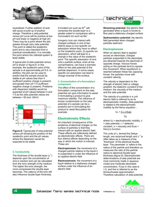
Zeta Potential 
An Introduction in 30 Minutes 
Introduction 
Zeta potential is a physical property 
which is exhibited by any particle in 
suspension. It can be used to 
optimize the formulations of 
suspensions and emulsions. 
Knowledge of the zeta potential can 
reduce the time needed to produce 
trial formulations. It is also an aid in 
predicting long-term stability. 
Colloid Science 
Three of the fundamental states of 
matter are solids, liquids and gases. If 
one of these states is finely dispersed 
in another then we have a ‘colloidal 
system’. These materials have special 
properties that are of great practical 
importance. 
There are various examples of 
colloidal systems that include 
aerosols, emulsions, colloidal 
suspensions and association colloids. 
In certain circumstances, the 
particles in a dispersion may adhere 
to one another and form aggregates 
of successively increasing size, which 
may settle out under the influence of 
gravity. An initially formed aggregate 
is called a floc and the process of its 
formation flocculation. The floc may or 
may not sediment or phase separate. 
If the aggregate changes to a much 
denser form, it is said to undergo 
coagulation. An aggregate usually 
separates out either by sedimentation 
(if it is more dense than the medium) 
or by creaming (if it less dense than 
the medium). The terms flocculation 
and coagulation have often been used 
interchangeably. Usually coagulation 
is irreversible whereas flocculation 
can be reversed by the process of 
deflocculation. Figure 1 schematically 
represents some of these processes. 
Figure 1: Schematic diagram 
showing various mechanisms where 
stability may be lost in a colloidal 
dispersion 
Colloidal Stability and 
DVLO Theory 
The scientists Derjaguin, Verwey, 
Landau and Overbeek developed a 
theory in the 1940s which dealt with 
the stability of colloidal systems. 
DVLO theory suggests that the 
stability of a particle in solution is 
dependent upon its total potential 
energy function VT. This theory 
recognizes that VT is the balance of 
several competing contributions: 
VT = VA + VR + VS 
VS is the potential energy due to the 
solvent, it usually only makes a 
marginal contribution to the total 
potential energy over the last few 
nanometers of separation. Much more 
important is the balance between VA 
and VR, these are the attractive and 
repulsive contributions. They 
potentially are much larger and 
operate over a much larger distance 
VA = -A/(12 π D2) 
where A is the Hamaker constant and 
D is the particle separation. The 
repulsive potential VR is a far more 
complex function. 
VR = 2 π ε a ζ2 exp(-κD) 
where a is the particle radius, π is the 
solvent permeability, κ is a function of 
the ionic composition and ζ is the zeta 
potential. 
Figure 2(a): Schematic diagram of the 
variation of free energy with particle 
separation according to DVLO theory. 
DVLO theory suggests that the 
stability of a colloidal system is 
determined by the sum of these van 
der Waals attractive (VA) and 
electrical double layer repulsive (VR) 
forces that exist between particles as 
they approach each other due to the 
Brownian motion they are undergoing. 
This theory proposes that an energy 
barrier resulting from the repulsive 
force prevents two particles 
approaching one another and 
adhering together (figure 2 (a)). But if 
the particles collide with sufficient 
energy to overcome that barrier, the 
1 Zetasizer Nano series technical note MRK654-01
attractive force will pull them into 
contact where they adhere strongly 
and irreversibly together. 
Therefore if the particles have a 
sufficiently high repulsion, the 
dispersion will resist flocculation and 
the colloidal system will be stable. 
However if a repulsion mechanism 
does not exist then flocculation or 
coagulation will eventually take place. 
Figure 2(b): Schematic diagram of the 
variation of free energy with particle 
separation at higher salt concentrations 
showing the possibility of a secondary 
minimum. 
If the zeta potential is reduced (e.g. in 
high salt concentrations), there is a 
possibility of a “secondary minimum” 
being created, where a much weaker 
and potentially reversible adhesion 
between particles exists (figure 2 (b)). 
These weak flocs are sufficiently 
stable not to be broken up by 
Brownian motion, but may disperse 
under an externally applied force such 
as vigorous agitation. 
Therefore to maintain the stability of 
the colloidal system, the repulsive 
forces must be dominant. How can 
colloidal stability be achieved? There 
are two fundamental mechanisms that 
affect dispersion stability (figure 3): 
• Steric repulsion - this involves 
polymers added to the system 
adsorbing onto the particle 
surface and preventing the 
particle surfaces coming into 
close contact. If enough polymer 
adsorbs, the thickness of the 
coating is sufficient to keep 
particles separated by steric 
repulsions between the polymer 
layers, and at those separations 
the van der Waals forces are too 
weak to cause the particles to 
adhere. 
• Electrostatic or charge 
stabilization - this is the effect on 
particle interaction due to the 
distribution of charged species in 
the system. 
Each mechanism has its benefits for 
particular systems. Steric stabilization 
is simple, requiring just the addition of 
a suitable polymer. However it can be 
difficult to subsequently flocculate the 
system if this is required, the polymer 
can be expensive and in some cases 
the polymer is undesirable e.g. when 
Figure 3: Steric and electrostatic 
stabilization mechanisms of 
colloidal dispersions 
a ceramic slip is cast and sintered, the 
polymer has to be ‘burnt out’. This 
causes shrinkage and can lead to 
defects. 
Electrostatic or charge stabilization 
has the benefits of stabilizing or 
flocculating a system by simply 
altering the concentration of ions in 
the system. This is a reversible 
process and is potentially 
inexpensive. 
It has long been recognised that the 
zeta potential is a very good index of 
the magnitude of the interaction 
between colloidal particles and 
measurements of zeta potential are 
commonly used to assess the stability 
of colloidal systems. 
Origins of Surface Charge 
Most colloidal dispersions in aqueous 
media carry an electric charge. There 
are many origins of this surface 
charge depending upon the nature of 
the particle and it’s surrounding 
medium but we will consider the more 
important mechanisms. 
Ionisation of Surface Groups 
Dissociation of acidic groups on the 
surface of a particle will give rise to a 
negatively charged surface. 
Conversely, a basic surface will take 
on a positive charge (figure 4). In both 
cases, the magnitude of the surface 
charge depends on the acidic or basic 
strengths of the surface groups and 
on the pH of the solution. The surface 
charge can be reduced to zero by 
suppressing the surface ionisation by 
decreasing the pH in case of 
negatively charged particles (figure 
4(a)) or by increasing the pH in the 
case of positively charged particles 
(figure 4(b)). 
Figure 4(a): Origin of surface 
charge by ionisation of acidic 
groups to give a negatively 
charged surface 
Figure 4(b): Origin of surface 
charge by ionisation of basic 
groups to give a positively charged 
surface 
2 Zetasizer Nano series technical note MRK654-01
Differential loss of ions from 
the crystal lattice 
As an example, consider a crystal of 
silver iodide placed in water. Solution 
of ions occurs. If equal amounts of 
Ag+ and I- ions were to dissolve, the 
surface would be uncharged. In fact 
silver ions dissolve preferentially, 
leaving a negatively charged surface 
(figure 5). If Ag+ ions are now added 
the charge falls to zero. Further 
addition leads to a positively charged 
surface. 
Figure 5: Origin of surface charge 
by differential solution of silver 
ions from a AgI surface 
Adsorption of charged species 
(ions and ionic surfactants) 
Surfactant ions may be specifically 
adsorbed on the surface of a particle, 
leading, in the case of cationic 
surfactants, to a positively charged 
surface (figure 6(a)) and, in the case 
of anionic surfactants, to a negatively 
charged surface (figure 6(b)). 
Figure 6(b): Origin of surface 
charge by specific adsorption 
of an anonic surfactant. R = 
hydrocarbon chain 
The Electrical 
Double Layer 
The development of a nett charge at 
the particle surface affects the 
distribution of ions in the surrounding 
interfacial region, resulting in an 
increased concentration of counter 
ions, ions of opposite charge to that of 
the particle, close to the surface. Thus 
an electrical double layer exists round 
each particle. 
Zeta Potential 
The liquid layer surrounding the 
particle exists as two parts; an inner 
region (Stern layer) where the ions 
are strongly bound and an outer 
(diffuse) region where they are less 
firmly associated. Within the diffuse 
layer there is a notional boundary 
inside which the ions and particles 
form a stable entity. When a particle 
moves (e.g. due to gravity), ions 
within the boundary move it. Those 
ions beyond the boundary stay with 
the bulk dispersant. The potential at 
this boundary (surface of 
hydrodynamic shear) is the zeta 
potential (figure 7). 
The magnitude of the zeta potential 
gives an indication of the potential 
stability of the colloidal system. If all 
the particles in suspension have a 
large negative or positive zeta 
potential then they will tend to repel 
each other and there will be no 
tendency for the particles to come 
together. However, if the particles 
have low zeta potential values then 
there will be no force to prevent the 
Figure 7: Schematic representation of 
zeta potential 
particles coming together and 
flocculating. 
The general dividing line between 
stable and unstable suspensions is 
generally taken at either +30 or -30 
mV. Particles with zeta potentials 
more positive than +30 mV or more 
negative than -30 mV are normally 
considered stable. However, if the 
particles have a density different form 
the dispersant, they will eventually 
sediment forming a close packed bed 
(i.e. a hard cake). 
Factors Affecting Zeta Potential 
(1) pH 
In aqueous media, the pH of the 
sample is one of the most important 
factors that affects its zeta potential. A 
zeta potential value on its own without 
defining the solution conditions is a 
virtually meaningless number. 
Imagine a particle in suspension with 
a negative zeta potential. If more 
alkali is added to this suspension then 
the particles tend to acquire more 
negative charge. If acid is added to 
this suspension then a point will be 
reached where the charge will be 
Figure 6(a): Origin of surface 
charge by specific adsorption 
of a cationic surfactant. R = 
hydrocarbon chain 
3 Zetasizer Nano series technical note MRK654-01
neutralised. Further addition of acid 
will cause a build up of positive 
charge. Therefore a zeta potential 
versus pH curve will be positive at low 
pH and lower or negative at high pH. 
There may be a point where the plot 
passes through zero zeta potential. 
This point is called the isoelectric 
point and is very important from a 
practical consideration. It is normally 
the point where the colloidal system is 
least stable. 
A typical plot of zeta potential versus 
pH is shown in figure 8. In this 
example, the isoelectric point of the 
sample is at approximately pH 5.5. In 
addition, the plot can be used to 
predict that the sample should be 
stable at pH values less than 4 
(sufficient positive charge is present) 
and greater than pH 7.5 (sufficient 
negative charge is present). Problems 
with dispersion stability would be 
expected at pH values between 4 and 
7.5 as the zeta potential values are 
between +30 and -30mV. 
2. Conductivity 
The thickness of the double layer (κ-1) 
depends upon the concentration of 
ions in solution and can be calculated 
from the ionic strength of the medium. 
The higher the ionic strength, the 
more compressed the double layer 
becomes. The valency of the ions will 
also influence double layer thickness. 
A trivalent ion such as Al3+ will 
compress the double layer to a 
greater extent in comparison with a 
monovalent ion such as Na+. 
Inorganic ions can interact with 
charged surfaces in one of two 
distinct ways (i) non-specific ion 
adsorption where they have no effect 
on the isoelectric point. (ii) specific ion 
adsorption, which will lead to a 
change in the value of the isoelectric 
point. The specific adsorption of ions 
onto a particle surface, even at low 
concentrations, can have a dramatic 
effect on the zeta potential of the 
particle dispersion. In some cases, 
specific ion adsorption can lead to 
charge reversal of the surface. 
3. Concentration of a formulation 
component 
The effect of the concentration of a 
formulation component on the zeta 
potential can give information to assist 
in formulating a product to give 
maximum stability. The influence of 
known contaminants on the zeta 
potential of a sample can be a 
powerful tool in formulating the 
product to resist flocculation for 
example. 
Electrokinetic Effects 
An important consequence of the 
existence of electrical charges on the 
surface of particles is that they 
interact with an applied electric field. 
These effects are collectively defined 
as electrokinetic effects. There are 
four distinct effects depending on the 
way in which the motion is induced. 
These are: 
Electrophoresis: the movement of a 
charged particle relative to the liquid it 
is suspended in under the influence of 
an applied electric field 
Electroosmosis: the movement of a 
liquid relative to a stationary charged 
surface under the influence of an 
electric field 
Streaming potential: the electric field 
generated when a liquid is forced to 
flow past a stationary charged surface 
Sedimentation potential: the electric 
field generated when charged 
particles sediment 
Electrophoresis 
When an electric field is applied 
across an electrolyte, charged 
particles suspended in the electrolyte 
are attracted towards the electrode of 
opposite charge. Viscous forces 
acting on the particles tend to oppose 
this movement. When equilibrium is 
reached between these two opposing 
forces, the particles move with 
constant velocity. 
The velocity is dependent on the 
strength of electric field or voltage 
gradient, the dielectric constant of the 
medium, the viscosity of the medium 
and the zeta potential. 
The velocity of a particle in a unit 
electric field is referred to as its 
electrophoretic mobility. Zeta potential 
is related to the electrophoretic 
mobility by the Henry equation:- 
4 Zetasizer Nano series technical note MRK654-01 
UE = 2 ε z f(κa) 
3η 
where UE = electrophoretic mobility, z 
= zeta potential, ε = dielectric 
constant, η = viscosity and f(κa) = 
Henry’s function. 
The units of κ, termed the Debye 
length, are reciprocal length and κ-1 is 
often taken as a measure of the 
“thickness” of the electrical double 
layer. The parameter ‘a’ refers to the 
radius of the particle and therefore κa 
measures the ratio of the particle 
radius to electrical double layer 
thickness (figure 9). Electrophoretic 
determinations of zeta potential are 
most commonly made in aqueous 
media and moderate electrolyte 
concentration. F(κa) in this case is 
1.5, and this is referred to as the 
Smoluchowski approximation. 
Therefore calculation of zeta potential 
Figure 8: Typical plot of zeta potential 
versus pH showing the position of the 
isoelectric point and the pH values 
where the dispersion would be 
expected to be stable
from the mobility is straightforward for 
systems that fit the Smoluchowski 
model, i.e. particles larger than about 
0.2 microns dispersed in electrolytes 
containing more that 10-3 molar salt. 
For small particles in low dielectric 
constant media (eg non-aqueous 
media), f(κa) becomes 1.0 and allows 
an equally simple calculation. This is 
referred to as the Huckel 
approximation. 
Figure 9: Schematic illustrating 
Huckel and Smoluchowski's 
approximations used for the 
conversion of electrophoretic mobility 
into zeta potential 
Measuring Electrophoretic 
Mobility 
The essence of a classical micro-electrophoresis 
system is a capillary 
cell with electrodes at either end to 
which a potential is applied. Particles 
move towards the electrode, their 
velocity is measured and expressed in 
unit field strength as their mobility. 
Early methods involved the process of 
directly observing individual particles 
using ultra-microscope techniques 
and manually tracking their progress 
over a measured distance. This 
procedure, although still being used 
by many groups world wide, suffers 
from several disadvantages, not least 
that of the strenuous effort required to 
make a measurement, particularly 
with small or poorly scattering 
particles. The technique used in 
Malvern’s Zetasizer Nano range of 
instruments is laser Doppler 
electrophoresis in combination with 
M3-PALS. 
The M3-PALS Technique 
The Zetasizer Nano Series uses a 
combination of laser Doppler 
velocimetry and phase analysis light 
scattering (PALS) in a patented 
technique called M3-PALS to 
measure particle electrophoretic 
mobility. Implementation of M3-PALS 
enables even samples of very low 
mobility to be analysed and their 
mobility distributions calculated. 
PALS can give an increase in 
performance of greater than 100 
times that associated with standard 
measurement techniques. This allows 
the measurement of high conductivity 
samples, plus the ability to accurately 
measure samples that have low 
particle mobilities, such as samples 
dispersed in non-aqueous solvents. 
Low applied voltages can now be 
used to avoid any risk of sample 
effects due to Joule heating. 
Further information discussing the 
techniques of laser Doppler 
electrophoresis and M3-PALS can be 
found in various articles available on 
the Malvern Instruments web-site 
Optical Configuration of a 
Zeta Potential Instrument 
A zeta potential measurement system 
comprises of six main components 
(figure 10). Firstly, a laser 1 is used 
to provides a light source to illuminate 
the particles within the sample. For 
zeta potential measurements, this 
light source is split to provide an 
incident and reference beam. The 
incident laser beam passes through 
the centre of the sample cell 2, and 
the scattered light at an angle of 
about 13o is detected 3. When an 
electric field is applied to the cell, any 
particles moving through the 
measurement volume will cause the 
intensity of light detected to fluctuate 
with a frequency proportional to the 
particle speed and this information is 
passed to a digital signal processor 4 
and then to a computer 5. The 
Zetasizer Nano software produces a 
frequency spectrum from which the 
electrophoretic mobility and hence 
zeta potential is calculated. The 
intensity of the detected, scattered 
light must be within a specific range 
for the detector to successfully 
measure it. This is achieved using an 
attenuator 6, which adjusts the 
intensity of the light reaching the 
sample and hence the intensity of the 
scattering. To correct for any 
differences in the cell wall thickness 
and dispersant refraction, 
compensation optics 7 are installed 
to maintain optimum alignment. 
Figure 10: Optical configuration of 
the Zetasizer Nano series for zeta 
potential measurements 
References 
Derjaguin, B.V. and Landau, L. (1941) 
Acta Physiochim. URSS, 14, 633. 
Verway, E.J.W. and Overbeek, J. Th. 
G. (1948) Theory of the Stability of 
Lyophobic Colloids, Elsevier, 
Amsterdam. 
Hunter, R.J. (1988) Zeta Potential In 
Colloid Science: Principles And 
Applications, Academic Press, UK. 
5 Zetasizer Nano series technical note MRK654-01
Shaw, D.J. (1992) Introduction To 
Colloid And Surface Chemistry, 
Butterworth Heinemann, UK. 
Everett, D.H. (1994) Basic Principles 
Of Colloid Science, The Royal Society 
of Chemistry, UK. 
Ross, S. and Morrison, I.D. (1988) 
Colloidal Systems and Interfaces, 
John Wiley and Sons, USA. 
Lyklema, J. (2000) Fundamentals of 
Interface and Colloid Science: Volume 
1 (Fundamentals), Academic Press, 
UK. 
Measuring Zeta Potential: Laser 
Doppler Electrophoresis, Technical 
Note available from 
www.malvern.co.uk 
Measuring Zeta Potential Using 
Phase Analysis Light Scattering 
(PALS), Technical Note available from 
www.malvern.co.uk 
Measuring Zeta Potential: A New 
Technique, Technical Note available 
from www.malvern.co.uk 
Simplifying the Measurement of Zeta 
Potential Using M3-PALS, Technical 
Note available from 
www.malvern.co.uk 
Malvern Instruments Ltd 
Enigma Business Park • Grovewood Road • Malvern • Worcestershire • UK • WR14 1XZ 
Tel: +44 (0)1684 892456 • Fax: +44 (0)1684 892789 
Malvern Instruments Worldwide 
Sales and service centers in over 50 countries for details visit www.malvern.co.uk/contact 
more information at www.malvern.co.uk 
6 Zetasizer Nano series technical note MRK654-01

Zeta

  • 1.
    Zeta Potential AnIntroduction in 30 Minutes Introduction Zeta potential is a physical property which is exhibited by any particle in suspension. It can be used to optimize the formulations of suspensions and emulsions. Knowledge of the zeta potential can reduce the time needed to produce trial formulations. It is also an aid in predicting long-term stability. Colloid Science Three of the fundamental states of matter are solids, liquids and gases. If one of these states is finely dispersed in another then we have a ‘colloidal system’. These materials have special properties that are of great practical importance. There are various examples of colloidal systems that include aerosols, emulsions, colloidal suspensions and association colloids. In certain circumstances, the particles in a dispersion may adhere to one another and form aggregates of successively increasing size, which may settle out under the influence of gravity. An initially formed aggregate is called a floc and the process of its formation flocculation. The floc may or may not sediment or phase separate. If the aggregate changes to a much denser form, it is said to undergo coagulation. An aggregate usually separates out either by sedimentation (if it is more dense than the medium) or by creaming (if it less dense than the medium). The terms flocculation and coagulation have often been used interchangeably. Usually coagulation is irreversible whereas flocculation can be reversed by the process of deflocculation. Figure 1 schematically represents some of these processes. Figure 1: Schematic diagram showing various mechanisms where stability may be lost in a colloidal dispersion Colloidal Stability and DVLO Theory The scientists Derjaguin, Verwey, Landau and Overbeek developed a theory in the 1940s which dealt with the stability of colloidal systems. DVLO theory suggests that the stability of a particle in solution is dependent upon its total potential energy function VT. This theory recognizes that VT is the balance of several competing contributions: VT = VA + VR + VS VS is the potential energy due to the solvent, it usually only makes a marginal contribution to the total potential energy over the last few nanometers of separation. Much more important is the balance between VA and VR, these are the attractive and repulsive contributions. They potentially are much larger and operate over a much larger distance VA = -A/(12 π D2) where A is the Hamaker constant and D is the particle separation. The repulsive potential VR is a far more complex function. VR = 2 π ε a ζ2 exp(-κD) where a is the particle radius, π is the solvent permeability, κ is a function of the ionic composition and ζ is the zeta potential. Figure 2(a): Schematic diagram of the variation of free energy with particle separation according to DVLO theory. DVLO theory suggests that the stability of a colloidal system is determined by the sum of these van der Waals attractive (VA) and electrical double layer repulsive (VR) forces that exist between particles as they approach each other due to the Brownian motion they are undergoing. This theory proposes that an energy barrier resulting from the repulsive force prevents two particles approaching one another and adhering together (figure 2 (a)). But if the particles collide with sufficient energy to overcome that barrier, the 1 Zetasizer Nano series technical note MRK654-01
  • 2.
    attractive force willpull them into contact where they adhere strongly and irreversibly together. Therefore if the particles have a sufficiently high repulsion, the dispersion will resist flocculation and the colloidal system will be stable. However if a repulsion mechanism does not exist then flocculation or coagulation will eventually take place. Figure 2(b): Schematic diagram of the variation of free energy with particle separation at higher salt concentrations showing the possibility of a secondary minimum. If the zeta potential is reduced (e.g. in high salt concentrations), there is a possibility of a “secondary minimum” being created, where a much weaker and potentially reversible adhesion between particles exists (figure 2 (b)). These weak flocs are sufficiently stable not to be broken up by Brownian motion, but may disperse under an externally applied force such as vigorous agitation. Therefore to maintain the stability of the colloidal system, the repulsive forces must be dominant. How can colloidal stability be achieved? There are two fundamental mechanisms that affect dispersion stability (figure 3): • Steric repulsion - this involves polymers added to the system adsorbing onto the particle surface and preventing the particle surfaces coming into close contact. If enough polymer adsorbs, the thickness of the coating is sufficient to keep particles separated by steric repulsions between the polymer layers, and at those separations the van der Waals forces are too weak to cause the particles to adhere. • Electrostatic or charge stabilization - this is the effect on particle interaction due to the distribution of charged species in the system. Each mechanism has its benefits for particular systems. Steric stabilization is simple, requiring just the addition of a suitable polymer. However it can be difficult to subsequently flocculate the system if this is required, the polymer can be expensive and in some cases the polymer is undesirable e.g. when Figure 3: Steric and electrostatic stabilization mechanisms of colloidal dispersions a ceramic slip is cast and sintered, the polymer has to be ‘burnt out’. This causes shrinkage and can lead to defects. Electrostatic or charge stabilization has the benefits of stabilizing or flocculating a system by simply altering the concentration of ions in the system. This is a reversible process and is potentially inexpensive. It has long been recognised that the zeta potential is a very good index of the magnitude of the interaction between colloidal particles and measurements of zeta potential are commonly used to assess the stability of colloidal systems. Origins of Surface Charge Most colloidal dispersions in aqueous media carry an electric charge. There are many origins of this surface charge depending upon the nature of the particle and it’s surrounding medium but we will consider the more important mechanisms. Ionisation of Surface Groups Dissociation of acidic groups on the surface of a particle will give rise to a negatively charged surface. Conversely, a basic surface will take on a positive charge (figure 4). In both cases, the magnitude of the surface charge depends on the acidic or basic strengths of the surface groups and on the pH of the solution. The surface charge can be reduced to zero by suppressing the surface ionisation by decreasing the pH in case of negatively charged particles (figure 4(a)) or by increasing the pH in the case of positively charged particles (figure 4(b)). Figure 4(a): Origin of surface charge by ionisation of acidic groups to give a negatively charged surface Figure 4(b): Origin of surface charge by ionisation of basic groups to give a positively charged surface 2 Zetasizer Nano series technical note MRK654-01
  • 3.
    Differential loss ofions from the crystal lattice As an example, consider a crystal of silver iodide placed in water. Solution of ions occurs. If equal amounts of Ag+ and I- ions were to dissolve, the surface would be uncharged. In fact silver ions dissolve preferentially, leaving a negatively charged surface (figure 5). If Ag+ ions are now added the charge falls to zero. Further addition leads to a positively charged surface. Figure 5: Origin of surface charge by differential solution of silver ions from a AgI surface Adsorption of charged species (ions and ionic surfactants) Surfactant ions may be specifically adsorbed on the surface of a particle, leading, in the case of cationic surfactants, to a positively charged surface (figure 6(a)) and, in the case of anionic surfactants, to a negatively charged surface (figure 6(b)). Figure 6(b): Origin of surface charge by specific adsorption of an anonic surfactant. R = hydrocarbon chain The Electrical Double Layer The development of a nett charge at the particle surface affects the distribution of ions in the surrounding interfacial region, resulting in an increased concentration of counter ions, ions of opposite charge to that of the particle, close to the surface. Thus an electrical double layer exists round each particle. Zeta Potential The liquid layer surrounding the particle exists as two parts; an inner region (Stern layer) where the ions are strongly bound and an outer (diffuse) region where they are less firmly associated. Within the diffuse layer there is a notional boundary inside which the ions and particles form a stable entity. When a particle moves (e.g. due to gravity), ions within the boundary move it. Those ions beyond the boundary stay with the bulk dispersant. The potential at this boundary (surface of hydrodynamic shear) is the zeta potential (figure 7). The magnitude of the zeta potential gives an indication of the potential stability of the colloidal system. If all the particles in suspension have a large negative or positive zeta potential then they will tend to repel each other and there will be no tendency for the particles to come together. However, if the particles have low zeta potential values then there will be no force to prevent the Figure 7: Schematic representation of zeta potential particles coming together and flocculating. The general dividing line between stable and unstable suspensions is generally taken at either +30 or -30 mV. Particles with zeta potentials more positive than +30 mV or more negative than -30 mV are normally considered stable. However, if the particles have a density different form the dispersant, they will eventually sediment forming a close packed bed (i.e. a hard cake). Factors Affecting Zeta Potential (1) pH In aqueous media, the pH of the sample is one of the most important factors that affects its zeta potential. A zeta potential value on its own without defining the solution conditions is a virtually meaningless number. Imagine a particle in suspension with a negative zeta potential. If more alkali is added to this suspension then the particles tend to acquire more negative charge. If acid is added to this suspension then a point will be reached where the charge will be Figure 6(a): Origin of surface charge by specific adsorption of a cationic surfactant. R = hydrocarbon chain 3 Zetasizer Nano series technical note MRK654-01
  • 4.
    neutralised. Further additionof acid will cause a build up of positive charge. Therefore a zeta potential versus pH curve will be positive at low pH and lower or negative at high pH. There may be a point where the plot passes through zero zeta potential. This point is called the isoelectric point and is very important from a practical consideration. It is normally the point where the colloidal system is least stable. A typical plot of zeta potential versus pH is shown in figure 8. In this example, the isoelectric point of the sample is at approximately pH 5.5. In addition, the plot can be used to predict that the sample should be stable at pH values less than 4 (sufficient positive charge is present) and greater than pH 7.5 (sufficient negative charge is present). Problems with dispersion stability would be expected at pH values between 4 and 7.5 as the zeta potential values are between +30 and -30mV. 2. Conductivity The thickness of the double layer (κ-1) depends upon the concentration of ions in solution and can be calculated from the ionic strength of the medium. The higher the ionic strength, the more compressed the double layer becomes. The valency of the ions will also influence double layer thickness. A trivalent ion such as Al3+ will compress the double layer to a greater extent in comparison with a monovalent ion such as Na+. Inorganic ions can interact with charged surfaces in one of two distinct ways (i) non-specific ion adsorption where they have no effect on the isoelectric point. (ii) specific ion adsorption, which will lead to a change in the value of the isoelectric point. The specific adsorption of ions onto a particle surface, even at low concentrations, can have a dramatic effect on the zeta potential of the particle dispersion. In some cases, specific ion adsorption can lead to charge reversal of the surface. 3. Concentration of a formulation component The effect of the concentration of a formulation component on the zeta potential can give information to assist in formulating a product to give maximum stability. The influence of known contaminants on the zeta potential of a sample can be a powerful tool in formulating the product to resist flocculation for example. Electrokinetic Effects An important consequence of the existence of electrical charges on the surface of particles is that they interact with an applied electric field. These effects are collectively defined as electrokinetic effects. There are four distinct effects depending on the way in which the motion is induced. These are: Electrophoresis: the movement of a charged particle relative to the liquid it is suspended in under the influence of an applied electric field Electroosmosis: the movement of a liquid relative to a stationary charged surface under the influence of an electric field Streaming potential: the electric field generated when a liquid is forced to flow past a stationary charged surface Sedimentation potential: the electric field generated when charged particles sediment Electrophoresis When an electric field is applied across an electrolyte, charged particles suspended in the electrolyte are attracted towards the electrode of opposite charge. Viscous forces acting on the particles tend to oppose this movement. When equilibrium is reached between these two opposing forces, the particles move with constant velocity. The velocity is dependent on the strength of electric field or voltage gradient, the dielectric constant of the medium, the viscosity of the medium and the zeta potential. The velocity of a particle in a unit electric field is referred to as its electrophoretic mobility. Zeta potential is related to the electrophoretic mobility by the Henry equation:- 4 Zetasizer Nano series technical note MRK654-01 UE = 2 ε z f(κa) 3η where UE = electrophoretic mobility, z = zeta potential, ε = dielectric constant, η = viscosity and f(κa) = Henry’s function. The units of κ, termed the Debye length, are reciprocal length and κ-1 is often taken as a measure of the “thickness” of the electrical double layer. The parameter ‘a’ refers to the radius of the particle and therefore κa measures the ratio of the particle radius to electrical double layer thickness (figure 9). Electrophoretic determinations of zeta potential are most commonly made in aqueous media and moderate electrolyte concentration. F(κa) in this case is 1.5, and this is referred to as the Smoluchowski approximation. Therefore calculation of zeta potential Figure 8: Typical plot of zeta potential versus pH showing the position of the isoelectric point and the pH values where the dispersion would be expected to be stable
  • 5.
    from the mobilityis straightforward for systems that fit the Smoluchowski model, i.e. particles larger than about 0.2 microns dispersed in electrolytes containing more that 10-3 molar salt. For small particles in low dielectric constant media (eg non-aqueous media), f(κa) becomes 1.0 and allows an equally simple calculation. This is referred to as the Huckel approximation. Figure 9: Schematic illustrating Huckel and Smoluchowski's approximations used for the conversion of electrophoretic mobility into zeta potential Measuring Electrophoretic Mobility The essence of a classical micro-electrophoresis system is a capillary cell with electrodes at either end to which a potential is applied. Particles move towards the electrode, their velocity is measured and expressed in unit field strength as their mobility. Early methods involved the process of directly observing individual particles using ultra-microscope techniques and manually tracking their progress over a measured distance. This procedure, although still being used by many groups world wide, suffers from several disadvantages, not least that of the strenuous effort required to make a measurement, particularly with small or poorly scattering particles. The technique used in Malvern’s Zetasizer Nano range of instruments is laser Doppler electrophoresis in combination with M3-PALS. The M3-PALS Technique The Zetasizer Nano Series uses a combination of laser Doppler velocimetry and phase analysis light scattering (PALS) in a patented technique called M3-PALS to measure particle electrophoretic mobility. Implementation of M3-PALS enables even samples of very low mobility to be analysed and their mobility distributions calculated. PALS can give an increase in performance of greater than 100 times that associated with standard measurement techniques. This allows the measurement of high conductivity samples, plus the ability to accurately measure samples that have low particle mobilities, such as samples dispersed in non-aqueous solvents. Low applied voltages can now be used to avoid any risk of sample effects due to Joule heating. Further information discussing the techniques of laser Doppler electrophoresis and M3-PALS can be found in various articles available on the Malvern Instruments web-site Optical Configuration of a Zeta Potential Instrument A zeta potential measurement system comprises of six main components (figure 10). Firstly, a laser 1 is used to provides a light source to illuminate the particles within the sample. For zeta potential measurements, this light source is split to provide an incident and reference beam. The incident laser beam passes through the centre of the sample cell 2, and the scattered light at an angle of about 13o is detected 3. When an electric field is applied to the cell, any particles moving through the measurement volume will cause the intensity of light detected to fluctuate with a frequency proportional to the particle speed and this information is passed to a digital signal processor 4 and then to a computer 5. The Zetasizer Nano software produces a frequency spectrum from which the electrophoretic mobility and hence zeta potential is calculated. The intensity of the detected, scattered light must be within a specific range for the detector to successfully measure it. This is achieved using an attenuator 6, which adjusts the intensity of the light reaching the sample and hence the intensity of the scattering. To correct for any differences in the cell wall thickness and dispersant refraction, compensation optics 7 are installed to maintain optimum alignment. Figure 10: Optical configuration of the Zetasizer Nano series for zeta potential measurements References Derjaguin, B.V. and Landau, L. (1941) Acta Physiochim. URSS, 14, 633. Verway, E.J.W. and Overbeek, J. Th. G. (1948) Theory of the Stability of Lyophobic Colloids, Elsevier, Amsterdam. Hunter, R.J. (1988) Zeta Potential In Colloid Science: Principles And Applications, Academic Press, UK. 5 Zetasizer Nano series technical note MRK654-01
  • 6.
    Shaw, D.J. (1992)Introduction To Colloid And Surface Chemistry, Butterworth Heinemann, UK. Everett, D.H. (1994) Basic Principles Of Colloid Science, The Royal Society of Chemistry, UK. Ross, S. and Morrison, I.D. (1988) Colloidal Systems and Interfaces, John Wiley and Sons, USA. Lyklema, J. (2000) Fundamentals of Interface and Colloid Science: Volume 1 (Fundamentals), Academic Press, UK. Measuring Zeta Potential: Laser Doppler Electrophoresis, Technical Note available from www.malvern.co.uk Measuring Zeta Potential Using Phase Analysis Light Scattering (PALS), Technical Note available from www.malvern.co.uk Measuring Zeta Potential: A New Technique, Technical Note available from www.malvern.co.uk Simplifying the Measurement of Zeta Potential Using M3-PALS, Technical Note available from www.malvern.co.uk Malvern Instruments Ltd Enigma Business Park • Grovewood Road • Malvern • Worcestershire • UK • WR14 1XZ Tel: +44 (0)1684 892456 • Fax: +44 (0)1684 892789 Malvern Instruments Worldwide Sales and service centers in over 50 countries for details visit www.malvern.co.uk/contact more information at www.malvern.co.uk 6 Zetasizer Nano series technical note MRK654-01