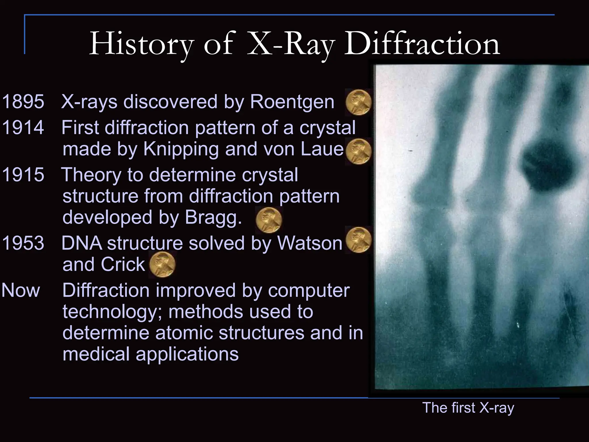
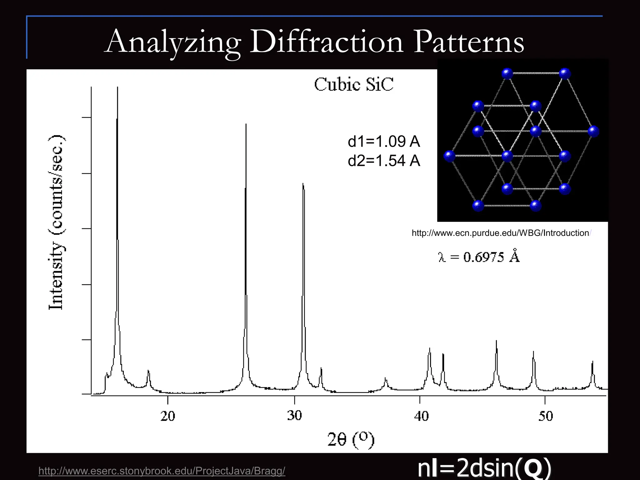
X-Ray Diffraction
Emily Day and Sage Ross
Advanced Lab 1
Spring 2004
Outline
 Introduction
 History
 How Diffraction Works
 Demonstration
 Analyzing Diffraction Patterns
 Solving DNA
 Applications
 Summary and Conclusions
Introduction
Motivation:
• X-ray diffraction is used to obtain structural
information about crystalline solids.
• Useful in biochemistry to solve the 3D structures of
complex biomolecules.
• Bridge the gaps between physics, chemistry, and
biology.
X-ray diffraction is important for:
• Solid-state physics
• Biophysics
• Medical physics
• Chemistry and Biochemistry
X-ray Diffractometer
History of X-Ray Diffraction
1895 X-rays discovered by Roentgen
1914 First diffraction pattern of a crystal
made by Knipping and von Laue
1915 Theory to determine crystal
structure from diffraction pattern
developed by Bragg.
1953 DNA structure solved by Watson
and Crick
Now Diffraction improved by computer
technology; methods used to
determine atomic structures and in
medical applications
The first X-ray
How Diffraction Works
 Wave Interacting with a Single Particle
 Incident beams scattered uniformly in all directions
 Wave Interacting with a Solid
 Scattered beams interfere constructively in some
directions, producing diffracted beams
 Random arrangements cause beams to randomly
interfere and no distinctive pattern is produced
 Crystalline Material
 Regular pattern of crystalline atoms produces
regular diffraction pattern.
 Diffraction pattern gives information on crystal
structure
NaCl
nl=2dsin(Q)
• Similar principle to multiple slit experiments
• Constructive and destructive interference patterns depend on
lattice spacing (d) and wavelength of radiation (l)
• By varying wavelength and observing diffraction patterns,
information about lattice spacing is obtained
How Diffraction Works: Bragg’s Law
d
Q Q
Q
X-rays of
wavelength l
l
How Diffraction Works: Schematic
http://mrsec.wisc.edu/edetc/modules/xray/X-raystm.pdf
NaCl
How Diffraction Works: Schematic
http://mrsec.wisc.edu/edetc/modules/xray/X-raystm.pdf
NaCl
Demonstration
Array A versus Array B
•Dots in A are closer together than in B
•Diffraction pattern A has spots farther
apart than pattern B
Array E
•Hexagonal arrangement
Array F
•Pattern created from the word “NANO”
written repeatedly
•Any repeating arrangement produces a
characteristic diffraction pattern
Array G versus Array H
•G represents one line of the chains of
atoms of DNA (a single helix)
•H represents a double helix
•Distinct patterns for single and double
helices
Credit: Exploring the Nanoworld
A
C
E
G
B
D
F
H
Analyzing Diffraction Patterns
 Data is taken from a full range of angles
 For simple crystal structures, diffraction
patterns are easily recognizable
 Phase Problem
 Only intensities of diffracted beams are measured
 Phase info is lost and must be inferred from data
 For complicated structures, diffraction
patterns at each angle can be used to
produce a 3-D electron density map
Analyzing Diffraction Patterns
http://www.eserc.stonybrook.edu/ProjectJava/Bragg/
http://www.ecn.purdue.edu/WBG/Introduction/
d1=1.09 A
d2=1.54 A
nl=2dsin(Q)
Solving the Structure of DNA:
History
 Rosalind Franklin- physical chemist
and x-ray crystallographer who first
crystallized and photographed BDNA
 Maurice Wilkins- collaborator of
Franklin
 Watson & Crick- chemists who
combined the information from Photo
51 with molecular modeling to solve
the structure of DNA in 1953
Rosalind Franklin
Solving the Structure of DNA
 Photo 51 Analysis
 “X” pattern characteristic
of helix
 Diamond shapes
indicate long, extended
molecules
 Smear spacing reveals
distance between
repeating structures
 Missing smears indicate
interference from second
helix
Photo 51- The x-ray diffraction image
that allowed Watson and Crick to solve
the structure of DNA
www.pbs.org/wgbh/nova/photo51
Solving the Structure of DNA
Photo 51- The x-ray diffraction image
that allowed Watson and Crick to solve
the structure of DNA
 Photo 51 Analysis
 “X” pattern characteristic
of helix
 Diamond shapes
indicate long, extended
molecules
 Smear spacing reveals
distance between
repeating structures
 Missing smears indicate
interference from second
helix
www.pbs.org/wgbh/nova/photo51
 Information Gained from Photo 51
 Double Helix
 Radius: 10 angstroms
 Distance between bases: 3.4 angstroms
 Distance per turn: 34 angstroms
 Combining Data with Other Information
 DNA made from:
sugar
phosphates
4 nucleotides (A,C,G,T)
 Chargaff’s Rules
 %A=%T
 %G=%C
 Molecular Modeling
Solving the Structure of DNA
Watson and Crick’s model
Applications of X-Ray Diffraction
 Find structure to determine function of proteins
 Convenient three letter acronym: XRD
 Distinguish between different crystal structures with
identical compositions
 Study crystal deformation and stress properties
 Study of rapid biological and chemical processes
 …and much more!
Summary and Conclusions
 X-ray diffraction is a technique for analyzing
structures of biological molecules
 X-ray beam hits a crystal, scattering the beam in a
manner characterized by the atomic structure
 Even complex structures can be analyzed by x-ray
diffraction, such as DNA and proteins
 This will provide useful in the future for combining
knowledge from physics, chemistry, and biology
Questions?
References
www.matter.org.uk/diffraction
www.embo.or/projects/scisoc/download/TW02weiss.pdf
www.branta.connectfree.co.uk/x-ray_diffraction.htm
www.xraydiffrac.com/xrd.htm
www.samford.edu/~gekeller/casey.html
neon.mems.cmu.edu/xray/Introduction.html
www.omega.dawsoncollege.qc.ca/ray/dna/franklin.htm
mrsec.wisc.edu/edetc/modules/xray/X-raystm.pdf
Exploring the Nanoworld
www.eserc.stonybrook.edu/ProjectJava/Bragg/
www.pbs.org/wgbh/nova/photo51

X-Ray Diffraction-Instrumentation and application

  • 1.
    X-Ray Diffraction Emily Dayand Sage Ross Advanced Lab 1 Spring 2004
  • 2.
    Outline  Introduction  History How Diffraction Works  Demonstration  Analyzing Diffraction Patterns  Solving DNA  Applications  Summary and Conclusions
  • 3.
    Introduction Motivation: • X-ray diffractionis used to obtain structural information about crystalline solids. • Useful in biochemistry to solve the 3D structures of complex biomolecules. • Bridge the gaps between physics, chemistry, and biology. X-ray diffraction is important for: • Solid-state physics • Biophysics • Medical physics • Chemistry and Biochemistry X-ray Diffractometer
  • 4.
    History of X-RayDiffraction 1895 X-rays discovered by Roentgen 1914 First diffraction pattern of a crystal made by Knipping and von Laue 1915 Theory to determine crystal structure from diffraction pattern developed by Bragg. 1953 DNA structure solved by Watson and Crick Now Diffraction improved by computer technology; methods used to determine atomic structures and in medical applications The first X-ray
  • 5.
    How Diffraction Works Wave Interacting with a Single Particle  Incident beams scattered uniformly in all directions  Wave Interacting with a Solid  Scattered beams interfere constructively in some directions, producing diffracted beams  Random arrangements cause beams to randomly interfere and no distinctive pattern is produced  Crystalline Material  Regular pattern of crystalline atoms produces regular diffraction pattern.  Diffraction pattern gives information on crystal structure NaCl
  • 6.
    nl=2dsin(Q) • Similar principleto multiple slit experiments • Constructive and destructive interference patterns depend on lattice spacing (d) and wavelength of radiation (l) • By varying wavelength and observing diffraction patterns, information about lattice spacing is obtained How Diffraction Works: Bragg’s Law d Q Q Q X-rays of wavelength l l
  • 7.
    How Diffraction Works:Schematic http://mrsec.wisc.edu/edetc/modules/xray/X-raystm.pdf NaCl
  • 8.
    How Diffraction Works:Schematic http://mrsec.wisc.edu/edetc/modules/xray/X-raystm.pdf NaCl
  • 9.
    Demonstration Array A versusArray B •Dots in A are closer together than in B •Diffraction pattern A has spots farther apart than pattern B Array E •Hexagonal arrangement Array F •Pattern created from the word “NANO” written repeatedly •Any repeating arrangement produces a characteristic diffraction pattern Array G versus Array H •G represents one line of the chains of atoms of DNA (a single helix) •H represents a double helix •Distinct patterns for single and double helices Credit: Exploring the Nanoworld A C E G B D F H
  • 10.
    Analyzing Diffraction Patterns Data is taken from a full range of angles  For simple crystal structures, diffraction patterns are easily recognizable  Phase Problem  Only intensities of diffracted beams are measured  Phase info is lost and must be inferred from data  For complicated structures, diffraction patterns at each angle can be used to produce a 3-D electron density map
  • 11.
  • 12.
    Solving the Structureof DNA: History  Rosalind Franklin- physical chemist and x-ray crystallographer who first crystallized and photographed BDNA  Maurice Wilkins- collaborator of Franklin  Watson & Crick- chemists who combined the information from Photo 51 with molecular modeling to solve the structure of DNA in 1953 Rosalind Franklin
  • 13.
    Solving the Structureof DNA  Photo 51 Analysis  “X” pattern characteristic of helix  Diamond shapes indicate long, extended molecules  Smear spacing reveals distance between repeating structures  Missing smears indicate interference from second helix Photo 51- The x-ray diffraction image that allowed Watson and Crick to solve the structure of DNA www.pbs.org/wgbh/nova/photo51
  • 14.
    Solving the Structureof DNA Photo 51- The x-ray diffraction image that allowed Watson and Crick to solve the structure of DNA  Photo 51 Analysis  “X” pattern characteristic of helix  Diamond shapes indicate long, extended molecules  Smear spacing reveals distance between repeating structures  Missing smears indicate interference from second helix www.pbs.org/wgbh/nova/photo51
  • 15.
    Solving the Structureof DNA Photo 51- The x-ray diffraction image that allowed Watson and Crick to solve the structure of DNA  Photo 51 Analysis  “X” pattern characteristic of helix  Diamond shapes indicate long, extended molecules  Smear spacing reveals distance between repeating structures  Missing smears indicate interference from second helix www.pbs.org/wgbh/nova/photo51
  • 16.
    Solving the Structureof DNA Photo 51- The x-ray diffraction image that allowed Watson and Crick to solve the structure of DNA  Photo 51 Analysis  “X” pattern characteristic of helix  Diamond shapes indicate long, extended molecules  Smear spacing reveals distance between repeating structures  Missing smears indicate interference from second helix www.pbs.org/wgbh/nova/photo51
  • 17.
    Solving the Structureof DNA Photo 51- The x-ray diffraction image that allowed Watson and Crick to solve the structure of DNA  Photo 51 Analysis  “X” pattern characteristic of helix  Diamond shapes indicate long, extended molecules  Smear spacing reveals distance between repeating structures  Missing smears indicate interference from second helix www.pbs.org/wgbh/nova/photo51
  • 18.
     Information Gainedfrom Photo 51  Double Helix  Radius: 10 angstroms  Distance between bases: 3.4 angstroms  Distance per turn: 34 angstroms  Combining Data with Other Information  DNA made from: sugar phosphates 4 nucleotides (A,C,G,T)  Chargaff’s Rules  %A=%T  %G=%C  Molecular Modeling Solving the Structure of DNA Watson and Crick’s model
  • 19.
    Applications of X-RayDiffraction  Find structure to determine function of proteins  Convenient three letter acronym: XRD  Distinguish between different crystal structures with identical compositions  Study crystal deformation and stress properties  Study of rapid biological and chemical processes  …and much more!
  • 20.
    Summary and Conclusions X-ray diffraction is a technique for analyzing structures of biological molecules  X-ray beam hits a crystal, scattering the beam in a manner characterized by the atomic structure  Even complex structures can be analyzed by x-ray diffraction, such as DNA and proteins  This will provide useful in the future for combining knowledge from physics, chemistry, and biology
  • 21.
  • 22.