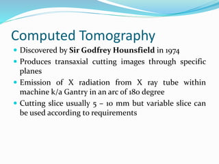
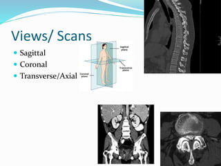
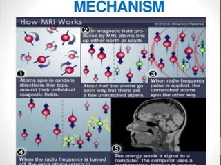
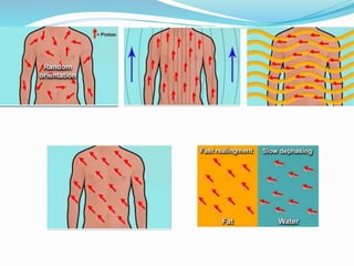
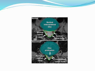
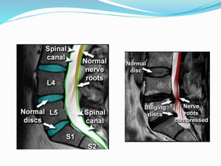
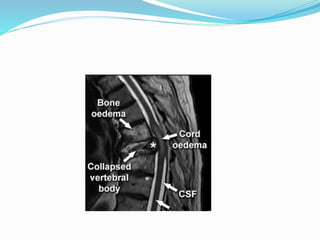
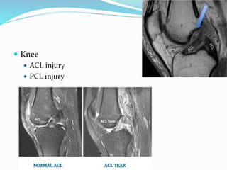
Presenter: Dr. Bijay Mehta
Moderator: Dr. Kuldip Mangal Joshi
INTRODUCTION
 IMAGING MODALITIES REMAINS A KEYSTONE
IN DIAGNOSING ORTHOPAEDIC CONDITIONS
 HISTORY DATES BACK TO 1895 –DISCOVERY OF
XRAY
 USEFUL IN DIAGNOSIS, STAGING AS WELL AS
FOR MANAGEMENT PURPOSES
IMAGING MODALITIES
1. Xray(Plain Radiograph)
2. Ultrasonography
3. Computed Tomography
4. Magnetic Resonance Imaging
5. Bone scan / scintigraphy
6. Single Photon Emission Computed Tomography(SPECT)
7. Positron Emission Tomography
8. Bone Mineral Densitometry
X Ray(Plain Film Radiography)
 High energy electromagnetic radiation
 wavelength ranging from 0.01 to 10 nanometers
 Penetrates the tissue producing images based on the
density/absorption of tissue
 Produced by Xray tube
 Absorbed by film after passing through a medium
 Denser the tissue, greater xray attenuation and brighter
the image
 Discovered by WK Roentgen in 1895
 Most commonly used as 1st line investigation
 Produce 2 D images
 “One view is no view”
 Generally at least 2 views are used
 Inexpensive
Rule of 2
 2 views
 2 joints
 2 sides
 2 occasions
 2 Sites
 Most commonly used views- Anteroposterior and
Lateral Views
Special views
Scapula Stryker Notch view, Scapular Y view
Shoulder Y view, Axillary View, Hill Sachs View,
Elbow Coyle view
Wrist Carpal tunnel view
Hand Ball catcher view
Pelvis Inlet/Outlet View
Acetabulum Judet – iliac/ obturator oblique view
Ankle Mortise view
Calcaneum Harris axial view
Radiographic Interpretation
 Should be done in a sequence- preferably- the patient-
the bone- the soft tissues- the joint
 The patient- confirm name,age and history
 The bone- shape, change in density, fracture or
localized change
 The soft tissues- Any soft tissue shadows
 The Joint-the joint space, shape , Erosions
Uses in orthopedics
 Fracture / dislocation/ subluxation
 Soft tissue shadows
 Evaluation of implants
 Guiding image in cases of closed reduction
 Identifying infections/Tumors
Fracture /Dislocations
Union vs Consolidation
Union Consolidation
Incomplete Repair Complete Repair
Ensheathing callus is calcified Callus is ossified
Attempted angulation is painful painless
Xray- fracture line still visible
with callus around it
Fracture line obliterated and
crossed by bony trabeculae
Infections
 During 1st week- No xray
abnormalities
 2nd week- Periosteal Reactions
 Later- patchy rarefaction of
metaphysis and bone
destruction
 Chronic osteomyelitis-
Sequestrum, Involucrum may
be seen
Fluoroscopy
 Uses X-rays to obtain real-time moving images
Uses
1. Arthrography (visualization of a joint or joints)
2. To asses joint stability
3. During Intraop Imaging
4. Biopsy guidance
Computed Tomography
 Discovered by Sir Godfrey Hounsfield in 1974
 Produces transaxial cutting images through specific
planes
 Emission of X radiation from X ray tube within
machine k/a Gantry in an arc of 180 degree
 Cutting slice usually 5 – 10 mm but variable slice can
be used according to requirements
Views/ Scans
 Sagittal
 Coronal
 Transverse/Axial
Advantages
 Cross sectional image
 No hindrances by overlying soft tissues
 Superior contrast resolution
 Detection of even early calcification
 3D reconstruction
Disadvantages
 Radiation exposure
 Soft tissues aren’t delineated properly
 Cost
Recent advances
 3 dimensional CT
 Real time multiplanar reconstruction
 High resolution CT
 Dynamic CT/ contrast enhanced
Uses
 Fractures/subluxation/dislocations requiring
transaxial/ 3D reconstruct like carpal/ tarsal/ tibial
plateau/calcaneal/pelvic
Spinal trauma
Spinal canal stenosis
CT guided biopsy
Magnetic Resonance Imaging
 Felix Bloch and EM Purcell discovered
 Paul C Lauterbur and Peter Mansfield won Nobel prize
for 3D MRI
Mechanism
TR and TE
 The repetition time (TR) is the length of time between
corresponding consecutive points on a repeating series
of pulses and echoes
 The echo time (TE) represents the time from the
center of the RF-pulse to the center of the echo
 T1 weighted image
 Longitudinal relaxation time
 Short TR and short TE
 return of protons back to equilibrium after application
and removal of the RF pulse
 Good anatomic detail- sharp / well defined
 Fat- bright ; Fluid – dark
 Contrast Enhanced MRI are T1 weighted
 T2 weighted image
 Transverse relaxation time
 Long TR and long TE
 Associated loss of coherence or phase b/w individual
protons immediately after application of RF pulse
 Evaluation of pathological process/ sensitively detect
edema
 Fat – bright ( fatty tissue/ bone marrow)
 Fluid – brighter ( CSF/ fluid)
T1 Vs T2 images
STIR (Short Tau Inversion Recovery)
 Highly water sensitive
 Timing of pulse sequence to supress signal from fatty
tissue
 Used to null the signal from fats
FLAIR ( Fluid Attenuated Inverse Recovery)
 Used to null the signal from water/ CSF
Utilities
 Spine
 PIVD
 Cord compression
 Knee
 ACL injury
 PCL injury
 Hip
 AVN
 Occult Fractures
 Shoulder
 Ankle
Advantages
 No risk of radiation
 Clear soft tissue visualisation
Disadvantages
 Expensive
 Patient must lie still
 Claustrophobic
Ultrasonography
 Sound frequency more than 20 KHz
 Uses based on intensity of USG
 High intensities(1-2 W/cm2)-for therapy/ operative
 Low intensities( 50mW/cm2)- diagnostics
Uses
 Deep seated cystic lesions
 DDH
 Rotator cuff injuries
 Soft tissue masses
 Advantages
 No radiation
 Cost effective
Positron Emission Tomography(PET)
 Advanced Nuclear Medicine technique that allows
functional imaging of disease process
 Uses positron emitting isotopes with short half lives
 Most commonly used is 18-fluoro-2-deoxy –D-
glucose(18FDG)
 Measures the consumption rate of glucose by different
tissues
 Useful in oncology to identify occult malignant tumors
and stage them
Bone Mineral Densitometry
 Used in identifying osteoporosis and osteopenia
 Techniques used- Radiographic Absorptiometry(RA),
Quantitative CT(QCT), Quantitaive US(QUS), DEXA
 Most commonly used technique-Dual Energy Xray
Absorptiometry(DEXA scan)
DEXA scan
 Uses low dose xray beams of two different beams to
distinguish density of soft tissue and bones
 Measures xray absorption by bones (esp hip and spine
) and compares with age and sex matched
population(Z score) and also to peak adult bone mass(
T score)
 According to WHO- T score <-1.5- osteopenia and T-
score <- 2.5 -osteoporosis
References
 Clinical Orthopedics Diagnosis 3rd edition
 Apley and Solomon’s System of Orthopaedics and
Trauma, 10th Edition
 Radiopedia
 Radiography masterclass
Thank You

Diagnostic imaging in orthopaedics

  • 1.
    Presenter: Dr. BijayMehta Moderator: Dr. Kuldip Mangal Joshi
  • 2.
    INTRODUCTION  IMAGING MODALITIESREMAINS A KEYSTONE IN DIAGNOSING ORTHOPAEDIC CONDITIONS  HISTORY DATES BACK TO 1895 –DISCOVERY OF XRAY  USEFUL IN DIAGNOSIS, STAGING AS WELL AS FOR MANAGEMENT PURPOSES
  • 3.
    IMAGING MODALITIES 1. Xray(PlainRadiograph) 2. Ultrasonography 3. Computed Tomography 4. Magnetic Resonance Imaging 5. Bone scan / scintigraphy 6. Single Photon Emission Computed Tomography(SPECT) 7. Positron Emission Tomography 8. Bone Mineral Densitometry
  • 4.
    X Ray(Plain FilmRadiography)  High energy electromagnetic radiation  wavelength ranging from 0.01 to 10 nanometers  Penetrates the tissue producing images based on the density/absorption of tissue  Produced by Xray tube  Absorbed by film after passing through a medium
  • 5.
     Denser thetissue, greater xray attenuation and brighter the image  Discovered by WK Roentgen in 1895  Most commonly used as 1st line investigation  Produce 2 D images  “One view is no view”  Generally at least 2 views are used  Inexpensive
  • 6.
    Rule of 2 2 views  2 joints  2 sides  2 occasions  2 Sites  Most commonly used views- Anteroposterior and Lateral Views
  • 7.
    Special views Scapula StrykerNotch view, Scapular Y view Shoulder Y view, Axillary View, Hill Sachs View, Elbow Coyle view Wrist Carpal tunnel view Hand Ball catcher view Pelvis Inlet/Outlet View Acetabulum Judet – iliac/ obturator oblique view Ankle Mortise view Calcaneum Harris axial view
  • 8.
    Radiographic Interpretation  Shouldbe done in a sequence- preferably- the patient- the bone- the soft tissues- the joint  The patient- confirm name,age and history  The bone- shape, change in density, fracture or localized change  The soft tissues- Any soft tissue shadows  The Joint-the joint space, shape , Erosions
  • 9.
    Uses in orthopedics Fracture / dislocation/ subluxation  Soft tissue shadows  Evaluation of implants  Guiding image in cases of closed reduction  Identifying infections/Tumors
  • 10.
  • 11.
    Union vs Consolidation UnionConsolidation Incomplete Repair Complete Repair Ensheathing callus is calcified Callus is ossified Attempted angulation is painful painless Xray- fracture line still visible with callus around it Fracture line obliterated and crossed by bony trabeculae
  • 12.
    Infections  During 1stweek- No xray abnormalities  2nd week- Periosteal Reactions  Later- patchy rarefaction of metaphysis and bone destruction  Chronic osteomyelitis- Sequestrum, Involucrum may be seen
  • 13.
    Fluoroscopy  Uses X-raysto obtain real-time moving images
  • 14.
    Uses 1. Arthrography (visualizationof a joint or joints) 2. To asses joint stability 3. During Intraop Imaging 4. Biopsy guidance
  • 15.
    Computed Tomography  Discoveredby Sir Godfrey Hounsfield in 1974  Produces transaxial cutting images through specific planes  Emission of X radiation from X ray tube within machine k/a Gantry in an arc of 180 degree  Cutting slice usually 5 – 10 mm but variable slice can be used according to requirements
  • 16.
    Views/ Scans  Sagittal Coronal  Transverse/Axial
  • 17.
    Advantages  Cross sectionalimage  No hindrances by overlying soft tissues  Superior contrast resolution  Detection of even early calcification  3D reconstruction
  • 18.
    Disadvantages  Radiation exposure Soft tissues aren’t delineated properly  Cost
  • 19.
    Recent advances  3dimensional CT  Real time multiplanar reconstruction  High resolution CT  Dynamic CT/ contrast enhanced
  • 20.
    Uses  Fractures/subluxation/dislocations requiring transaxial/3D reconstruct like carpal/ tarsal/ tibial plateau/calcaneal/pelvic
  • 22.
  • 23.
  • 24.
  • 25.
    Magnetic Resonance Imaging Felix Bloch and EM Purcell discovered  Paul C Lauterbur and Peter Mansfield won Nobel prize for 3D MRI
  • 26.
  • 28.
    TR and TE The repetition time (TR) is the length of time between corresponding consecutive points on a repeating series of pulses and echoes  The echo time (TE) represents the time from the center of the RF-pulse to the center of the echo
  • 29.
     T1 weightedimage  Longitudinal relaxation time  Short TR and short TE  return of protons back to equilibrium after application and removal of the RF pulse  Good anatomic detail- sharp / well defined  Fat- bright ; Fluid – dark  Contrast Enhanced MRI are T1 weighted
  • 30.
     T2 weightedimage  Transverse relaxation time  Long TR and long TE  Associated loss of coherence or phase b/w individual protons immediately after application of RF pulse  Evaluation of pathological process/ sensitively detect edema  Fat – bright ( fatty tissue/ bone marrow)  Fluid – brighter ( CSF/ fluid)
  • 31.
    T1 Vs T2images
  • 32.
    STIR (Short TauInversion Recovery)  Highly water sensitive  Timing of pulse sequence to supress signal from fatty tissue  Used to null the signal from fats
  • 33.
    FLAIR ( FluidAttenuated Inverse Recovery)  Used to null the signal from water/ CSF
  • 34.
  • 38.
     Knee  ACLinjury  PCL injury
  • 40.
     Hip  AVN Occult Fractures
  • 41.
  • 42.
  • 43.
    Advantages  No riskof radiation  Clear soft tissue visualisation
  • 44.
    Disadvantages  Expensive  Patientmust lie still  Claustrophobic
  • 45.
    Ultrasonography  Sound frequencymore than 20 KHz  Uses based on intensity of USG  High intensities(1-2 W/cm2)-for therapy/ operative  Low intensities( 50mW/cm2)- diagnostics
  • 46.
    Uses  Deep seatedcystic lesions  DDH  Rotator cuff injuries  Soft tissue masses
  • 47.
     Advantages  Noradiation  Cost effective
  • 48.
    Positron Emission Tomography(PET) Advanced Nuclear Medicine technique that allows functional imaging of disease process  Uses positron emitting isotopes with short half lives  Most commonly used is 18-fluoro-2-deoxy –D- glucose(18FDG)  Measures the consumption rate of glucose by different tissues  Useful in oncology to identify occult malignant tumors and stage them
  • 49.
    Bone Mineral Densitometry Used in identifying osteoporosis and osteopenia  Techniques used- Radiographic Absorptiometry(RA), Quantitative CT(QCT), Quantitaive US(QUS), DEXA  Most commonly used technique-Dual Energy Xray Absorptiometry(DEXA scan)
  • 50.
    DEXA scan  Useslow dose xray beams of two different beams to distinguish density of soft tissue and bones  Measures xray absorption by bones (esp hip and spine ) and compares with age and sex matched population(Z score) and also to peak adult bone mass( T score)  According to WHO- T score <-1.5- osteopenia and T- score <- 2.5 -osteoporosis
  • 51.
    References  Clinical OrthopedicsDiagnosis 3rd edition  Apley and Solomon’s System of Orthopaedics and Trauma, 10th Edition  Radiopedia  Radiography masterclass
  • 52.