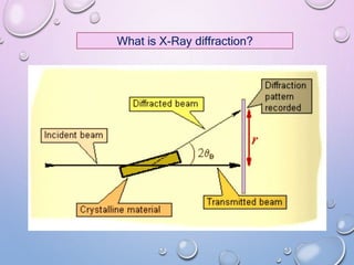
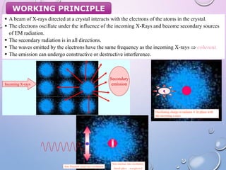
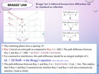
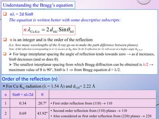
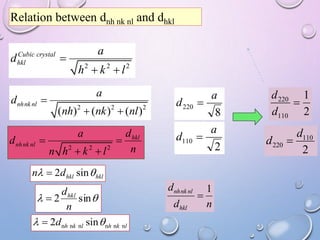
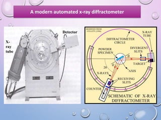
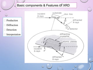
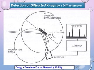
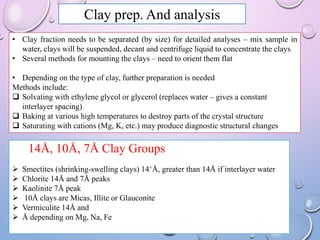
SlideShare a Scribd company logo
X-RAY
DIFFRACTION(XRD)
A PRESENTATION
ON
Submitted to-
Dr. Sonal Tripathi
Associate Professor
Dept. of Soil Science
N.M.C.A., NAU, Navsari
Submitted by-
Srikumar Debasis Swain
1st year M.Sc. (Agri)
2nd semester
Dept.- Agronomy
Regd. No.- 2010117110
INTRODUCTION
• It is a novel and non-destructive method of chemical analysis
meaning the chemical substance is not affected by the
analysis.
• The atomic planes of a crystal cause an incident beam of X-
rays to interfere with one another as they leave the crystal.
The phenomenon is called X-ray diffraction.
• Every crystalline substance gives a pattern; the same
substance always gives the same pattern; and in a mixture of
substances each produces its pattern independently of the
others.
• The X-ray diffraction pattern of a pure substance is,
therefore, like a fingerprint of the substance. It is based on
the scattering of x-rays by crystals.
WHAT IS X-RAY ?
• X-rays are electromagnetic radiation of wavelength about 1 Å (10-10m)which is
about the same size as an atom.
• They occur in that portion of the electromagnetic spectrum between gamma
rays and the ultraviolet.
• The discovery of X-rays in 1895 enabled scientists to probe crystalline structure
at the atomic level.
If we use a white light we cannot look
at objects smaller than the wavelength
of light, which is about 10 -6 m. Since
the atom has dimensions of about 10-10
m we cannot image an atom with a
photon of white light. X-rays, on the
other hand, have a wavelength of about
10 -10 m and are suitable for imaging
objects at the atomic scale.
WHY X-RAY IS
USED
GENERATION OF X-RAYS
 X-rays can be generated by decelerating electrons.
 Hence, X-rays are generated by bombarding a target (say Cu) with an electron beam.
 The resultant spectrum of X-rays generated (i.e. X-rays versus Intensity plot) is shown
in the next slide. The pattern shows intense peaks on a ‘broad’ background.
 The intense peaks can be ‘thought of’ as monochromatic radiation and be used for
X-ray diffraction studies.
Beam of electrons Target X-rays
An accelerating (or
decelerating) charge
radiates
electromagnetic
radiation
What is X-Ray diffraction?
WHY XRD ?
 Measure the average spacing's between layers or rows of atoms
 Determine the orientation of a single crystal or grain
 Find the crystal structure of an unknown material
 Measure the size, shape and internal stress of small crystalline regions
X-RAY
SOURCES
the sealed-
tube
the rotating
anode
Sealed tube
The sealed tube is simply a glass or ceramic tube where a tungsten cathode has been
placed above a metallic stationary anode. The tube is then evacuated and current is
applied to the cathode and the anode
Rotating anode
A rotating anode is similar to the sealed tube instrument except for the fact that the metallic
anode is now spinning. The spinning anode spreads the heat of the electron bombardment
over a wider area. This allows for higher wattages, which produces a higher X-ray flux.
For diffraction experiments the X-rays should be monochromatic.
The crystal
monochromator
produces more
monochromatic X-rays
at the expense of X-ray
flux.
The metallic filter is
normally used with
powder diffraction
and results in high
X-ray flux with poor
monochromation.
To do this we employ either a crystal monochromator or a metallic filter.
The anode is also rectangular which allows for a line focus (which is broad but
has low flux and a point focus, which is intense but has a narrow illumination
area. In practice the line focus is used with powder diffraction so as to
illuminate more sample and the point focus is used in single crystal and
small angle x-ray scattering instruments for higher flux for small samples.
The X-rays that are generated are of two types
1) Characteristic (ejection of electrons from the atom in the anode
2) White Radiation (synchrotron effect)
Electron strikes the
target and ejects an
electron. The cascade
of electrons from
higher orbitals
generates X-ray
M
Characteristic
X-rays
Kalpha Kbeta
Electron reaccelerate
when entering the metal
and "bend" their
trajectory path. Loss of
momentum results in
generation of X-rays.
M
White Radiation
“Bremsstrahlung”
or breaking
radiation
The energy of the X-ray is determined from the observed wavelength and is
given by the formula :
Energy (KeV) = 1.2398 / λ (nm)
Intensity
Wavelength ()
0.2 0.6 1.0 1.4
White
radiation
Characteristic radiation →
due to energy transitions
in the atom
K
K
Intense peak, nearly
monochromatic
Energy for K alpha (for Mo) = 17.28 KeV
Mo Target impacted by electrons
accelerated by a 35 kV potential shows
the emission spectrum as in the
figure(schematic)
X-ray sources with different 
for doing XRD studies
Target
Metal
 Of K
radiation (Å)
Mo 0.71
Cu 1.54
Co 1.79
Fe 1.94
Cr 2.29
X-ray sources with different  for doing XRD studies
Elements (KV)  Of K1
radiation
(Å)
 Of K2
radiation (Å)
 Of Kβ
radiation (Å)
Kβ-Filter
(mm)
Ag 25.52 0.55941 0.5638 0.49707 Pd
0.0461
Mo 20 0.7093 0.71359 0.63229 Zr
0.0678
Cu 8.98 1.540598 1.54439 1.39222 Ni
0.017
Ni 8.33 1.65791 1.66175 1.50014 Co
0.0158
Co 7.71 1.78897 1.79285 1.62079 Fe
0.0166
Fe 7.11 1.93604 1.93998 1.75661 Mn
0.0168
Cr 5.99 2.2897 2.29361 2.08487 V
0.169
WORKING PRINCIPLE
 A beam of X-rays directed at a crystal interacts with the electrons of the atoms in the crystal.
 The electrons oscillate under the influence of the incoming X-Rays and become secondary sources
of EM radiation.
 The secondary radiation is in all directions.
 The waves emitted by the electrons have the same frequency as the incoming X-rays  coherent.
 The emission can undergo constructive or destructive interference.
Incoming X-rays
Secondary
emission
Oscillating charge re-radiates  In phase with
the incoming x-rays
Sets Electron cloud into oscillation
Sets nucleus into oscillation
Small effect  neglected
BRAGGS’ LAW Braggs’ law is followed because here diffraction can
be visualised as reflection
 The scattering planes have a spacing ‘d’.
 Ray-2 travels an extra path as compared to Ray-1 (= ABC). The path difference between
Ray-1 and Ray-2 = ABC = (d Sin + d Sin) = (2d.Sin).
 For constructive interference, this path difference should be an integral multiple of :
n = 2d Sin  the Bragg’s equation. (More about this sooner).
 The path difference between Ray-1 and Ray-3 is = 2(2d.Sin) = 2n = 2n. This implies
that if Ray-1 and Ray-2 constructively interfere Ray-1 and Ray-3 will also constructively
interfere. (And so forth).
Understanding the Bragg’s equation
 n = 2d Sin
The equation is written better with some descriptive subscripts:
 n is an integer and is the order of the reflection
(i.e. how many wavelengths of the X-ray go on to make the path difference between planes).
Note: if hkl reflection (corresponding to n=1) occurs at hkl then 2h 2k 2l reflection (n=2) will occur at a higher angle 2h 2k 2l.
  For large interplanar spacing the angle of reflection tends towards zero → as d increases,
Sin decreases (and so does ).
 The smallest interplanar spacing from which Bragg diffraction can be obtained is /2 →
maximum value of  is 90, Sin is 1  from Bragg equation d = /2.
2 SinCu K hkl hkln d 
Order of the reflection (n)
 For Cu K radiation ( = 1.54 Å) and d110= 2.22 Å
n Sin = n/2d 
1 0.34 20.7º • First order reflection from (110)  110
2 0.69 43.92º
• Second order reflection from (110) planes  110
• Also considered as first order reflection from (220) planes  220
Relation between dnh nk nl and dhkl
2 2 2
Cubic crystal
hkl
a
d
h k l

 
8
220
a
d 
2
110
a
d 
2
1
110
220

d
d
2 2 2
( ) ( ) ( )
nhnk nl
a
d
nh nk nl

 
2 2 2
hkl
nhnk nl
da
d
nn h k l
 
 
110
220
2
d
d 
2 sinhkl hkln d 
 sin2
n
dhkl

n n n n n n2 sinh k l h k ld 
1nhnk nl
hkl
d
d n

A modern automated x-ray diffractometer
X-
ray
tube
Detector
Basic components & Features of XRD
Production
Diffraction
Detection
Interpretation
Detection of Diffracted X-rays by a Diffractometer
Bragg - Brentano Focus Geometry, Cullity
Clay prep. And analysis
• Clay fraction needs to be separated (by size) for detailed analyses – mix sample in
water, clays will be suspended, decant and centrifuge liquid to concentrate the clays
• Several methods for mounting the clays – need to orient them flat
• Depending on the type of clay, further preparation is needed
Methods include:
 Solvating with ethylene glycol or glycerol (replaces water – gives a constant
interlayer spacing)
 Baking at various high temperatures to destroy parts of the crystal structure
 Saturating with cations (Mg, K, etc.) may produce diagnostic structural changes
14Å, 10Å, 7Å Clay Groups
 Smectites (shrinking-swelling clays) 14+Å, greater than 14Å if interlayer water
 Chlorite 14Å and 7Å peaks
 Kaolinite 7Å peak
 10Å clays are Micas, Illite or Glauconite
 Vermiculite 14Å and
 Å depending on Mg, Na, Fe
X ray diffraction(xrd) principle and use

More Related Content

What's hot

X ray diffraction
X ray diffractionX ray diffraction
X ray diffraction
Faraz Khan
 
X ray diffraction
X ray diffractionX ray diffraction
X ray diffraction
hdghcfgfgftf
 
X-ray Photoelectron Spectrocopy (XPS)
X-ray Photoelectron Spectrocopy (XPS)X-ray Photoelectron Spectrocopy (XPS)
X-ray Photoelectron Spectrocopy (XPS)
CNPEM
 
Xrd nanomaterials course_s2015_bates
Xrd nanomaterials course_s2015_batesXrd nanomaterials course_s2015_bates
Xrd nanomaterials course_s2015_bates
Michael Bates
 
X Ray Diffraction Spectroscopy
X Ray Diffraction SpectroscopyX Ray Diffraction Spectroscopy
X Ray Diffraction Spectroscopy
hephz
 
Xrd (X-ray Diffraction) presentation
Xrd (X-ray Diffraction) presentationXrd (X-ray Diffraction) presentation
Xrd (X-ray Diffraction) presentation
Kishan Kasundra
 
Xrd
XrdXrd
X ray diffraction
X ray diffractionX ray diffraction
X ray diffraction
Arvind Singh Heer
 
X ray photoelectron spectroscopy
X ray photoelectron spectroscopyX ray photoelectron spectroscopy
X ray photoelectron spectroscopy
Zubair Aslam
 
X-ray Photoelecctron Spectroscopy (XPS)
X-ray Photoelecctron Spectroscopy (XPS)X-ray Photoelecctron Spectroscopy (XPS)
X-ray Photoelecctron Spectroscopy (XPS)
faheem maqsood
 
Transmission electron microscope (TEM)
Transmission electron microscope (TEM)Transmission electron microscope (TEM)
Transmission electron microscope (TEM)
Nano Encryption
 
X ray diffraction ppt
X ray diffraction pptX ray diffraction ppt
X ray diffraction ppt
VanithaVaniN1
 
Applications of raman spectroscopy
Applications of raman spectroscopyApplications of raman spectroscopy
Applications of raman spectroscopy
kaavyabalachandran
 
X ray diffraction
X ray diffractionX ray diffraction
X ray diffraction
Priyanka Jaiswal
 
Raman spectroscopy
Raman spectroscopy Raman spectroscopy
Raman spectroscopy
Aravind AB
 
XRD- X-Ray diffraction
XRD- X-Ray diffractionXRD- X-Ray diffraction
XRD- X-Ray diffraction
BASANTKUMAR123
 
X ray Photoelectron Spectroscopy (XPS)
X ray Photoelectron Spectroscopy (XPS)X ray Photoelectron Spectroscopy (XPS)
X ray Photoelectron Spectroscopy (XPS)
Nano Encryption
 
Basics of-xrd
Basics of-xrdBasics of-xrd
Basics of-xrd
ceutics1315
 
Energy Dispersive Spectroscopy
Energy Dispersive SpectroscopyEnergy Dispersive Spectroscopy
Energy Dispersive Spectroscopy
Saad Shaukat
 
Scanning tunneling microscopy
Scanning tunneling microscopyScanning tunneling microscopy
Scanning tunneling microscopy
Azhar Ansari
 

What's hot (20)

X ray diffraction
X ray diffractionX ray diffraction
X ray diffraction
 
X ray diffraction
X ray diffractionX ray diffraction
X ray diffraction
 
X-ray Photoelectron Spectrocopy (XPS)
X-ray Photoelectron Spectrocopy (XPS)X-ray Photoelectron Spectrocopy (XPS)
X-ray Photoelectron Spectrocopy (XPS)
 
Xrd nanomaterials course_s2015_bates
Xrd nanomaterials course_s2015_batesXrd nanomaterials course_s2015_bates
Xrd nanomaterials course_s2015_bates
 
X Ray Diffraction Spectroscopy
X Ray Diffraction SpectroscopyX Ray Diffraction Spectroscopy
X Ray Diffraction Spectroscopy
 
Xrd (X-ray Diffraction) presentation
Xrd (X-ray Diffraction) presentationXrd (X-ray Diffraction) presentation
Xrd (X-ray Diffraction) presentation
 
Xrd
XrdXrd
Xrd
 
X ray diffraction
X ray diffractionX ray diffraction
X ray diffraction
 
X ray photoelectron spectroscopy
X ray photoelectron spectroscopyX ray photoelectron spectroscopy
X ray photoelectron spectroscopy
 
X-ray Photoelecctron Spectroscopy (XPS)
X-ray Photoelecctron Spectroscopy (XPS)X-ray Photoelecctron Spectroscopy (XPS)
X-ray Photoelecctron Spectroscopy (XPS)
 
Transmission electron microscope (TEM)
Transmission electron microscope (TEM)Transmission electron microscope (TEM)
Transmission electron microscope (TEM)
 
X ray diffraction ppt
X ray diffraction pptX ray diffraction ppt
X ray diffraction ppt
 
Applications of raman spectroscopy
Applications of raman spectroscopyApplications of raman spectroscopy
Applications of raman spectroscopy
 
X ray diffraction
X ray diffractionX ray diffraction
X ray diffraction
 
Raman spectroscopy
Raman spectroscopy Raman spectroscopy
Raman spectroscopy
 
XRD- X-Ray diffraction
XRD- X-Ray diffractionXRD- X-Ray diffraction
XRD- X-Ray diffraction
 
X ray Photoelectron Spectroscopy (XPS)
X ray Photoelectron Spectroscopy (XPS)X ray Photoelectron Spectroscopy (XPS)
X ray Photoelectron Spectroscopy (XPS)
 
Basics of-xrd
Basics of-xrdBasics of-xrd
Basics of-xrd
 
Energy Dispersive Spectroscopy
Energy Dispersive SpectroscopyEnergy Dispersive Spectroscopy
Energy Dispersive Spectroscopy
 
Scanning tunneling microscopy
Scanning tunneling microscopyScanning tunneling microscopy
Scanning tunneling microscopy
 

Similar to X ray diffraction(xrd) principle and use

263 4.pdf
263 4.pdf263 4.pdf
263 4.pdf
AbitiEthiopia
 
263 4.pdf
263 4.pdf263 4.pdf
Crystal structure analysis
Crystal structure analysisCrystal structure analysis
Crystal structure analysis
zoelfalia
 
X ray crystallography for mpharm
X ray crystallography for mpharm X ray crystallography for mpharm
X ray crystallography for mpharm
Martin Jacob
 
Chapter 6 x ray
Chapter 6 x rayChapter 6 x ray
Chapter 6 x ray
Miza Kamaruzzaman
 
Chapter 6 x ray
Chapter 6 x rayChapter 6 x ray
Chapter 6 x ray
Miza Kamaruzzaman
 
X ray crystallography
X ray crystallographyX ray crystallography
X ray crystallography
Sayeda Salma S.A.
 
B.Tech sem I Engineering Physics U-IV Chapter 2-X-Rays
B.Tech sem I Engineering Physics U-IV Chapter 2-X-RaysB.Tech sem I Engineering Physics U-IV Chapter 2-X-Rays
B.Tech sem I Engineering Physics U-IV Chapter 2-X-Rays
Abhi Hirpara
 
Ph 101-1
Ph 101-1Ph 101-1
Ph 101-1
Chandan Singh
 
X ray crystallography
X ray crystallographyX ray crystallography
X ray crystallography
Rajput1998
 
Ir spectroscopy by Bhavana P Khobragade
Ir spectroscopy by Bhavana P KhobragadeIr spectroscopy by Bhavana P Khobragade
Ir spectroscopy by Bhavana P Khobragade
BhavanaKhobragade
 
B.tech sem i engineering physics u iv chapter 2-x-rays
B.tech sem i engineering physics u iv chapter 2-x-raysB.tech sem i engineering physics u iv chapter 2-x-rays
B.tech sem i engineering physics u iv chapter 2-x-rays
Rai University
 
Atomic Plane Resolution Electron Magnetic Circular Dichroism
Atomic Plane Resolution Electron Magnetic Circular DichroismAtomic Plane Resolution Electron Magnetic Circular Dichroism
Atomic Plane Resolution Electron Magnetic Circular Dichroism
Riccardo Di Stefano
 
XRD.pptx
XRD.pptxXRD.pptx
XRD.pptx
MajedAmini2
 
Optics
OpticsOptics
PH 101 Optics
PH 101 OpticsPH 101 Optics
PH 101 Optics
Chandan Singh
 
Chapter_3c_X_Ray_Diffraction.ppt
Chapter_3c_X_Ray_Diffraction.pptChapter_3c_X_Ray_Diffraction.ppt
Chapter_3c_X_Ray_Diffraction.ppt
MithunSarkar33
 
Interference and the Wave Nature of Light
Interference and the Wave Nature of LightInterference and the Wave Nature of Light
Interference and the Wave Nature of Light
Taimoor Muzaffar Gondal
 
X – ray diffraction by iswar hazarika
X – ray diffraction by iswar hazarikaX – ray diffraction by iswar hazarika
X –ray diffraction
X –ray diffractionX –ray diffraction
X –ray diffraction
Rajinapp
 

Similar to X ray diffraction(xrd) principle and use (20)

263 4.pdf
263 4.pdf263 4.pdf
263 4.pdf
 
263 4.pdf
263 4.pdf263 4.pdf
263 4.pdf
 
Crystal structure analysis
Crystal structure analysisCrystal structure analysis
Crystal structure analysis
 
X ray crystallography for mpharm
X ray crystallography for mpharm X ray crystallography for mpharm
X ray crystallography for mpharm
 
Chapter 6 x ray
Chapter 6 x rayChapter 6 x ray
Chapter 6 x ray
 
Chapter 6 x ray
Chapter 6 x rayChapter 6 x ray
Chapter 6 x ray
 
X ray crystallography
X ray crystallographyX ray crystallography
X ray crystallography
 
B.Tech sem I Engineering Physics U-IV Chapter 2-X-Rays
B.Tech sem I Engineering Physics U-IV Chapter 2-X-RaysB.Tech sem I Engineering Physics U-IV Chapter 2-X-Rays
B.Tech sem I Engineering Physics U-IV Chapter 2-X-Rays
 
Ph 101-1
Ph 101-1Ph 101-1
Ph 101-1
 
X ray crystallography
X ray crystallographyX ray crystallography
X ray crystallography
 
Ir spectroscopy by Bhavana P Khobragade
Ir spectroscopy by Bhavana P KhobragadeIr spectroscopy by Bhavana P Khobragade
Ir spectroscopy by Bhavana P Khobragade
 
B.tech sem i engineering physics u iv chapter 2-x-rays
B.tech sem i engineering physics u iv chapter 2-x-raysB.tech sem i engineering physics u iv chapter 2-x-rays
B.tech sem i engineering physics u iv chapter 2-x-rays
 
Atomic Plane Resolution Electron Magnetic Circular Dichroism
Atomic Plane Resolution Electron Magnetic Circular DichroismAtomic Plane Resolution Electron Magnetic Circular Dichroism
Atomic Plane Resolution Electron Magnetic Circular Dichroism
 
XRD.pptx
XRD.pptxXRD.pptx
XRD.pptx
 
Optics
OpticsOptics
Optics
 
PH 101 Optics
PH 101 OpticsPH 101 Optics
PH 101 Optics
 
Chapter_3c_X_Ray_Diffraction.ppt
Chapter_3c_X_Ray_Diffraction.pptChapter_3c_X_Ray_Diffraction.ppt
Chapter_3c_X_Ray_Diffraction.ppt
 
Interference and the Wave Nature of Light
Interference and the Wave Nature of LightInterference and the Wave Nature of Light
Interference and the Wave Nature of Light
 
X – ray diffraction by iswar hazarika
X – ray diffraction by iswar hazarikaX – ray diffraction by iswar hazarika
X – ray diffraction by iswar hazarika
 
X –ray diffraction
X –ray diffractionX –ray diffraction
X –ray diffraction
 

Recently uploaded

Phenomics assisted breeding in crop improvement
Phenomics assisted breeding in crop improvementPhenomics assisted breeding in crop improvement
Phenomics assisted breeding in crop improvement
IshaGoswami9
 
The debris of the ‘last major merger’ is dynamically young
The debris of the ‘last major merger’ is dynamically youngThe debris of the ‘last major merger’ is dynamically young
The debris of the ‘last major merger’ is dynamically young
Sérgio Sacani
 
ANAMOLOUS SECONDARY GROWTH IN DICOT ROOTS.pptx
ANAMOLOUS SECONDARY GROWTH IN DICOT ROOTS.pptxANAMOLOUS SECONDARY GROWTH IN DICOT ROOTS.pptx
ANAMOLOUS SECONDARY GROWTH IN DICOT ROOTS.pptx
RASHMI M G
 
Micronuclei test.M.sc.zoology.fisheries.
Micronuclei test.M.sc.zoology.fisheries.Micronuclei test.M.sc.zoology.fisheries.
Micronuclei test.M.sc.zoology.fisheries.
Aditi Bajpai
 
SAR of Medicinal Chemistry 1st by dk.pdf
SAR of Medicinal Chemistry 1st by dk.pdfSAR of Medicinal Chemistry 1st by dk.pdf
SAR of Medicinal Chemistry 1st by dk.pdf
KrushnaDarade1
 
8.Isolation of pure cultures and preservation of cultures.pdf
8.Isolation of pure cultures and preservation of cultures.pdf8.Isolation of pure cultures and preservation of cultures.pdf
8.Isolation of pure cultures and preservation of cultures.pdf
by6843629
 
Oedema_types_causes_pathophysiology.pptx
Oedema_types_causes_pathophysiology.pptxOedema_types_causes_pathophysiology.pptx
Oedema_types_causes_pathophysiology.pptx
muralinath2
 
NuGOweek 2024 Ghent programme overview flyer
NuGOweek 2024 Ghent programme overview flyerNuGOweek 2024 Ghent programme overview flyer
NuGOweek 2024 Ghent programme overview flyer
pablovgd
 
Equivariant neural networks and representation theory
Equivariant neural networks and representation theoryEquivariant neural networks and representation theory
Equivariant neural networks and representation theory
Daniel Tubbenhauer
 
bordetella pertussis.................................ppt
bordetella pertussis.................................pptbordetella pertussis.................................ppt
bordetella pertussis.................................ppt
kejapriya1
 
aziz sancar nobel prize winner: from mardin to nobel
aziz sancar nobel prize winner: from mardin to nobelaziz sancar nobel prize winner: from mardin to nobel
aziz sancar nobel prize winner: from mardin to nobel
İsa Badur
 
What is greenhouse gasses and how many gasses are there to affect the Earth.
What is greenhouse gasses and how many gasses are there to affect the Earth.What is greenhouse gasses and how many gasses are there to affect the Earth.
What is greenhouse gasses and how many gasses are there to affect the Earth.
moosaasad1975
 
Chapter 12 - climate change and the energy crisis
Chapter 12 - climate change and the energy crisisChapter 12 - climate change and the energy crisis
Chapter 12 - climate change and the energy crisis
tonzsalvador2222
 
Deep Software Variability and Frictionless Reproducibility
Deep Software Variability and Frictionless ReproducibilityDeep Software Variability and Frictionless Reproducibility
Deep Software Variability and Frictionless Reproducibility
University of Rennes, INSA Rennes, Inria/IRISA, CNRS
 
EWOCS-I: The catalog of X-ray sources in Westerlund 1 from the Extended Weste...
EWOCS-I: The catalog of X-ray sources in Westerlund 1 from the Extended Weste...EWOCS-I: The catalog of X-ray sources in Westerlund 1 from the Extended Weste...
EWOCS-I: The catalog of X-ray sources in Westerlund 1 from the Extended Weste...
Sérgio Sacani
 
Eukaryotic Transcription Presentation.pptx
Eukaryotic Transcription Presentation.pptxEukaryotic Transcription Presentation.pptx
Eukaryotic Transcription Presentation.pptx
RitabrataSarkar3
 
Travis Hills' Endeavors in Minnesota: Fostering Environmental and Economic Pr...
Travis Hills' Endeavors in Minnesota: Fostering Environmental and Economic Pr...Travis Hills' Endeavors in Minnesota: Fostering Environmental and Economic Pr...
Travis Hills' Endeavors in Minnesota: Fostering Environmental and Economic Pr...
Travis Hills MN
 
Nucleic Acid-its structural and functional complexity.
Nucleic Acid-its structural and functional complexity.Nucleic Acid-its structural and functional complexity.
Nucleic Acid-its structural and functional complexity.
Nistarini College, Purulia (W.B) India
 
The binding of cosmological structures by massless topological defects
The binding of cosmological structures by massless topological defectsThe binding of cosmological structures by massless topological defects
The binding of cosmological structures by massless topological defects
Sérgio Sacani
 
Randomised Optimisation Algorithms in DAPHNE
Randomised Optimisation Algorithms in DAPHNERandomised Optimisation Algorithms in DAPHNE
Randomised Optimisation Algorithms in DAPHNE
University of Maribor
 

Recently uploaded (20)

Phenomics assisted breeding in crop improvement
Phenomics assisted breeding in crop improvementPhenomics assisted breeding in crop improvement
Phenomics assisted breeding in crop improvement
 
The debris of the ‘last major merger’ is dynamically young
The debris of the ‘last major merger’ is dynamically youngThe debris of the ‘last major merger’ is dynamically young
The debris of the ‘last major merger’ is dynamically young
 
ANAMOLOUS SECONDARY GROWTH IN DICOT ROOTS.pptx
ANAMOLOUS SECONDARY GROWTH IN DICOT ROOTS.pptxANAMOLOUS SECONDARY GROWTH IN DICOT ROOTS.pptx
ANAMOLOUS SECONDARY GROWTH IN DICOT ROOTS.pptx
 
Micronuclei test.M.sc.zoology.fisheries.
Micronuclei test.M.sc.zoology.fisheries.Micronuclei test.M.sc.zoology.fisheries.
Micronuclei test.M.sc.zoology.fisheries.
 
SAR of Medicinal Chemistry 1st by dk.pdf
SAR of Medicinal Chemistry 1st by dk.pdfSAR of Medicinal Chemistry 1st by dk.pdf
SAR of Medicinal Chemistry 1st by dk.pdf
 
8.Isolation of pure cultures and preservation of cultures.pdf
8.Isolation of pure cultures and preservation of cultures.pdf8.Isolation of pure cultures and preservation of cultures.pdf
8.Isolation of pure cultures and preservation of cultures.pdf
 
Oedema_types_causes_pathophysiology.pptx
Oedema_types_causes_pathophysiology.pptxOedema_types_causes_pathophysiology.pptx
Oedema_types_causes_pathophysiology.pptx
 
NuGOweek 2024 Ghent programme overview flyer
NuGOweek 2024 Ghent programme overview flyerNuGOweek 2024 Ghent programme overview flyer
NuGOweek 2024 Ghent programme overview flyer
 
Equivariant neural networks and representation theory
Equivariant neural networks and representation theoryEquivariant neural networks and representation theory
Equivariant neural networks and representation theory
 
bordetella pertussis.................................ppt
bordetella pertussis.................................pptbordetella pertussis.................................ppt
bordetella pertussis.................................ppt
 
aziz sancar nobel prize winner: from mardin to nobel
aziz sancar nobel prize winner: from mardin to nobelaziz sancar nobel prize winner: from mardin to nobel
aziz sancar nobel prize winner: from mardin to nobel
 
What is greenhouse gasses and how many gasses are there to affect the Earth.
What is greenhouse gasses and how many gasses are there to affect the Earth.What is greenhouse gasses and how many gasses are there to affect the Earth.
What is greenhouse gasses and how many gasses are there to affect the Earth.
 
Chapter 12 - climate change and the energy crisis
Chapter 12 - climate change and the energy crisisChapter 12 - climate change and the energy crisis
Chapter 12 - climate change and the energy crisis
 
Deep Software Variability and Frictionless Reproducibility
Deep Software Variability and Frictionless ReproducibilityDeep Software Variability and Frictionless Reproducibility
Deep Software Variability and Frictionless Reproducibility
 
EWOCS-I: The catalog of X-ray sources in Westerlund 1 from the Extended Weste...
EWOCS-I: The catalog of X-ray sources in Westerlund 1 from the Extended Weste...EWOCS-I: The catalog of X-ray sources in Westerlund 1 from the Extended Weste...
EWOCS-I: The catalog of X-ray sources in Westerlund 1 from the Extended Weste...
 
Eukaryotic Transcription Presentation.pptx
Eukaryotic Transcription Presentation.pptxEukaryotic Transcription Presentation.pptx
Eukaryotic Transcription Presentation.pptx
 
Travis Hills' Endeavors in Minnesota: Fostering Environmental and Economic Pr...
Travis Hills' Endeavors in Minnesota: Fostering Environmental and Economic Pr...Travis Hills' Endeavors in Minnesota: Fostering Environmental and Economic Pr...
Travis Hills' Endeavors in Minnesota: Fostering Environmental and Economic Pr...
 
Nucleic Acid-its structural and functional complexity.
Nucleic Acid-its structural and functional complexity.Nucleic Acid-its structural and functional complexity.
Nucleic Acid-its structural and functional complexity.
 
The binding of cosmological structures by massless topological defects
The binding of cosmological structures by massless topological defectsThe binding of cosmological structures by massless topological defects
The binding of cosmological structures by massless topological defects
 
Randomised Optimisation Algorithms in DAPHNE
Randomised Optimisation Algorithms in DAPHNERandomised Optimisation Algorithms in DAPHNE
Randomised Optimisation Algorithms in DAPHNE
 

X ray diffraction(xrd) principle and use

  • 1. X-RAY DIFFRACTION(XRD) A PRESENTATION ON Submitted to- Dr. Sonal Tripathi Associate Professor Dept. of Soil Science N.M.C.A., NAU, Navsari Submitted by- Srikumar Debasis Swain 1st year M.Sc. (Agri) 2nd semester Dept.- Agronomy Regd. No.- 2010117110
  • 2. INTRODUCTION • It is a novel and non-destructive method of chemical analysis meaning the chemical substance is not affected by the analysis. • The atomic planes of a crystal cause an incident beam of X- rays to interfere with one another as they leave the crystal. The phenomenon is called X-ray diffraction. • Every crystalline substance gives a pattern; the same substance always gives the same pattern; and in a mixture of substances each produces its pattern independently of the others. • The X-ray diffraction pattern of a pure substance is, therefore, like a fingerprint of the substance. It is based on the scattering of x-rays by crystals.
  • 3. WHAT IS X-RAY ? • X-rays are electromagnetic radiation of wavelength about 1 Å (10-10m)which is about the same size as an atom. • They occur in that portion of the electromagnetic spectrum between gamma rays and the ultraviolet. • The discovery of X-rays in 1895 enabled scientists to probe crystalline structure at the atomic level. If we use a white light we cannot look at objects smaller than the wavelength of light, which is about 10 -6 m. Since the atom has dimensions of about 10-10 m we cannot image an atom with a photon of white light. X-rays, on the other hand, have a wavelength of about 10 -10 m and are suitable for imaging objects at the atomic scale. WHY X-RAY IS USED
  • 4. GENERATION OF X-RAYS  X-rays can be generated by decelerating electrons.  Hence, X-rays are generated by bombarding a target (say Cu) with an electron beam.  The resultant spectrum of X-rays generated (i.e. X-rays versus Intensity plot) is shown in the next slide. The pattern shows intense peaks on a ‘broad’ background.  The intense peaks can be ‘thought of’ as monochromatic radiation and be used for X-ray diffraction studies. Beam of electrons Target X-rays An accelerating (or decelerating) charge radiates electromagnetic radiation
  • 5. What is X-Ray diffraction?
  • 6. WHY XRD ?  Measure the average spacing's between layers or rows of atoms  Determine the orientation of a single crystal or grain  Find the crystal structure of an unknown material  Measure the size, shape and internal stress of small crystalline regions X-RAY SOURCES the sealed- tube the rotating anode
  • 7. Sealed tube The sealed tube is simply a glass or ceramic tube where a tungsten cathode has been placed above a metallic stationary anode. The tube is then evacuated and current is applied to the cathode and the anode Rotating anode A rotating anode is similar to the sealed tube instrument except for the fact that the metallic anode is now spinning. The spinning anode spreads the heat of the electron bombardment over a wider area. This allows for higher wattages, which produces a higher X-ray flux. For diffraction experiments the X-rays should be monochromatic. The crystal monochromator produces more monochromatic X-rays at the expense of X-ray flux. The metallic filter is normally used with powder diffraction and results in high X-ray flux with poor monochromation. To do this we employ either a crystal monochromator or a metallic filter.
  • 8. The anode is also rectangular which allows for a line focus (which is broad but has low flux and a point focus, which is intense but has a narrow illumination area. In practice the line focus is used with powder diffraction so as to illuminate more sample and the point focus is used in single crystal and small angle x-ray scattering instruments for higher flux for small samples.
  • 9.
  • 10. The X-rays that are generated are of two types 1) Characteristic (ejection of electrons from the atom in the anode 2) White Radiation (synchrotron effect) Electron strikes the target and ejects an electron. The cascade of electrons from higher orbitals generates X-ray M Characteristic X-rays Kalpha Kbeta Electron reaccelerate when entering the metal and "bend" their trajectory path. Loss of momentum results in generation of X-rays. M White Radiation “Bremsstrahlung” or breaking radiation
  • 11. The energy of the X-ray is determined from the observed wavelength and is given by the formula : Energy (KeV) = 1.2398 / λ (nm) Intensity Wavelength () 0.2 0.6 1.0 1.4 White radiation Characteristic radiation → due to energy transitions in the atom K K Intense peak, nearly monochromatic Energy for K alpha (for Mo) = 17.28 KeV Mo Target impacted by electrons accelerated by a 35 kV potential shows the emission spectrum as in the figure(schematic) X-ray sources with different  for doing XRD studies Target Metal  Of K radiation (Å) Mo 0.71 Cu 1.54 Co 1.79 Fe 1.94 Cr 2.29
  • 12. X-ray sources with different  for doing XRD studies Elements (KV)  Of K1 radiation (Å)  Of K2 radiation (Å)  Of Kβ radiation (Å) Kβ-Filter (mm) Ag 25.52 0.55941 0.5638 0.49707 Pd 0.0461 Mo 20 0.7093 0.71359 0.63229 Zr 0.0678 Cu 8.98 1.540598 1.54439 1.39222 Ni 0.017 Ni 8.33 1.65791 1.66175 1.50014 Co 0.0158 Co 7.71 1.78897 1.79285 1.62079 Fe 0.0166 Fe 7.11 1.93604 1.93998 1.75661 Mn 0.0168 Cr 5.99 2.2897 2.29361 2.08487 V 0.169
  • 13. WORKING PRINCIPLE  A beam of X-rays directed at a crystal interacts with the electrons of the atoms in the crystal.  The electrons oscillate under the influence of the incoming X-Rays and become secondary sources of EM radiation.  The secondary radiation is in all directions.  The waves emitted by the electrons have the same frequency as the incoming X-rays  coherent.  The emission can undergo constructive or destructive interference. Incoming X-rays Secondary emission Oscillating charge re-radiates  In phase with the incoming x-rays Sets Electron cloud into oscillation Sets nucleus into oscillation Small effect  neglected
  • 14. BRAGGS’ LAW Braggs’ law is followed because here diffraction can be visualised as reflection  The scattering planes have a spacing ‘d’.  Ray-2 travels an extra path as compared to Ray-1 (= ABC). The path difference between Ray-1 and Ray-2 = ABC = (d Sin + d Sin) = (2d.Sin).  For constructive interference, this path difference should be an integral multiple of : n = 2d Sin  the Bragg’s equation. (More about this sooner).  The path difference between Ray-1 and Ray-3 is = 2(2d.Sin) = 2n = 2n. This implies that if Ray-1 and Ray-2 constructively interfere Ray-1 and Ray-3 will also constructively interfere. (And so forth).
  • 15. Understanding the Bragg’s equation  n = 2d Sin The equation is written better with some descriptive subscripts:  n is an integer and is the order of the reflection (i.e. how many wavelengths of the X-ray go on to make the path difference between planes). Note: if hkl reflection (corresponding to n=1) occurs at hkl then 2h 2k 2l reflection (n=2) will occur at a higher angle 2h 2k 2l.   For large interplanar spacing the angle of reflection tends towards zero → as d increases, Sin decreases (and so does ).  The smallest interplanar spacing from which Bragg diffraction can be obtained is /2 → maximum value of  is 90, Sin is 1  from Bragg equation d = /2. 2 SinCu K hkl hkln d  Order of the reflection (n)  For Cu K radiation ( = 1.54 Å) and d110= 2.22 Å n Sin = n/2d  1 0.34 20.7º • First order reflection from (110)  110 2 0.69 43.92º • Second order reflection from (110) planes  110 • Also considered as first order reflection from (220) planes  220
  • 16. Relation between dnh nk nl and dhkl 2 2 2 Cubic crystal hkl a d h k l    8 220 a d  2 110 a d  2 1 110 220  d d 2 2 2 ( ) ( ) ( ) nhnk nl a d nh nk nl    2 2 2 hkl nhnk nl da d nn h k l     110 220 2 d d  2 sinhkl hkln d   sin2 n dhkl  n n n n n n2 sinh k l h k ld  1nhnk nl hkl d d n 
  • 17. A modern automated x-ray diffractometer X- ray tube Detector
  • 18. Basic components & Features of XRD Production Diffraction Detection Interpretation
  • 19. Detection of Diffracted X-rays by a Diffractometer Bragg - Brentano Focus Geometry, Cullity
  • 20. Clay prep. And analysis • Clay fraction needs to be separated (by size) for detailed analyses – mix sample in water, clays will be suspended, decant and centrifuge liquid to concentrate the clays • Several methods for mounting the clays – need to orient them flat • Depending on the type of clay, further preparation is needed Methods include:  Solvating with ethylene glycol or glycerol (replaces water – gives a constant interlayer spacing)  Baking at various high temperatures to destroy parts of the crystal structure  Saturating with cations (Mg, K, etc.) may produce diagnostic structural changes 14Å, 10Å, 7Å Clay Groups  Smectites (shrinking-swelling clays) 14+Å, greater than 14Å if interlayer water  Chlorite 14Å and 7Å peaks  Kaolinite 7Å peak  10Å clays are Micas, Illite or Glauconite  Vermiculite 14Å and  Å depending on Mg, Na, Fe