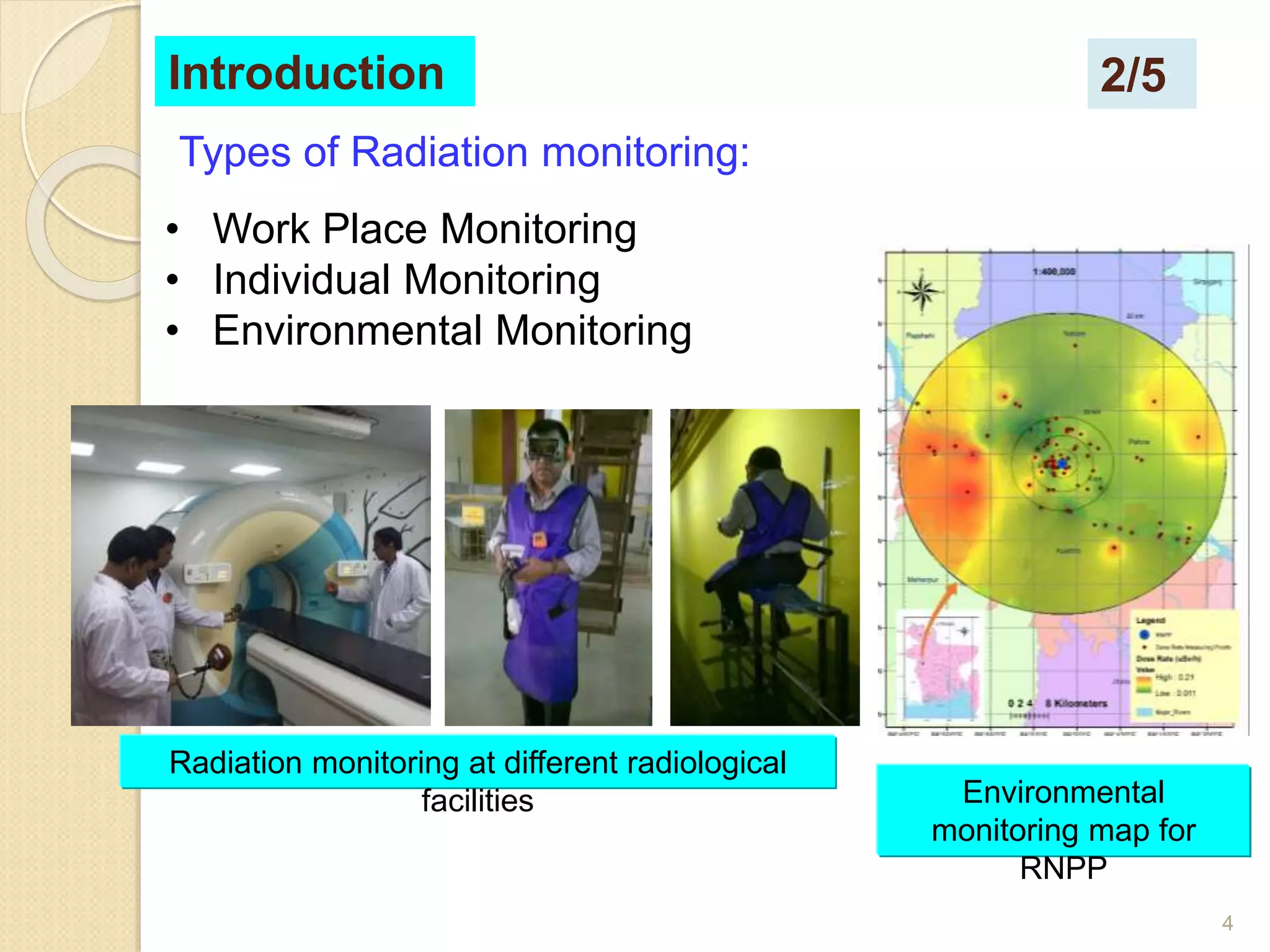
Radiation monitoring involves measuring radiation levels in workplaces, areas, and the environment. There are several types of radiation monitoring:
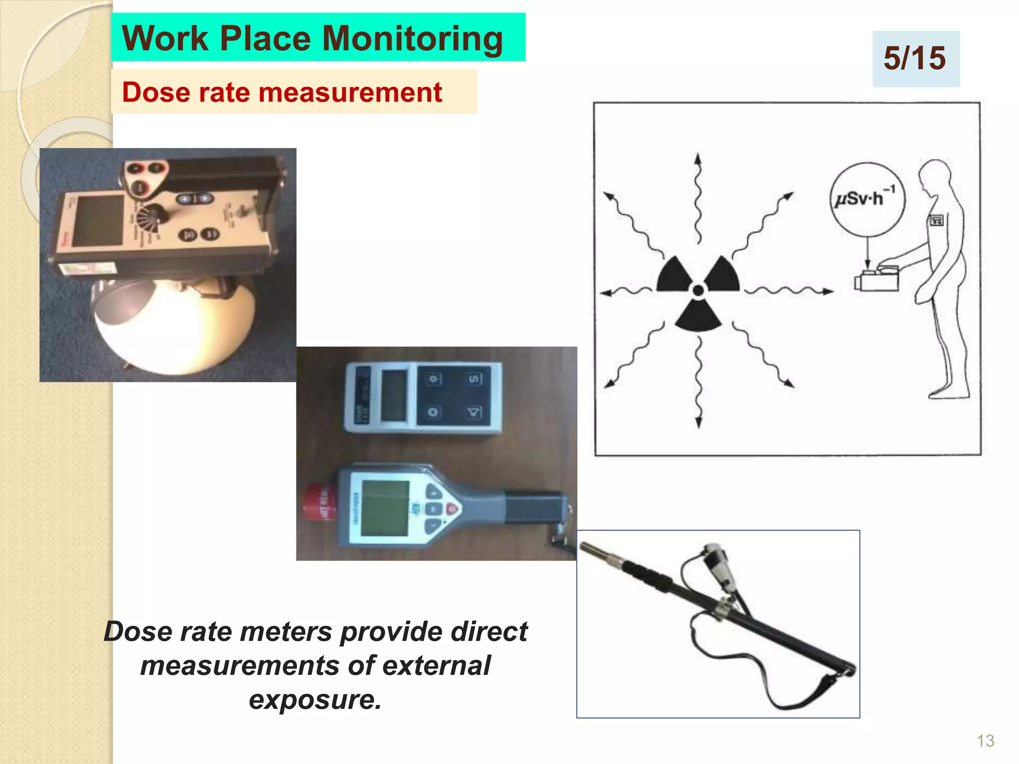
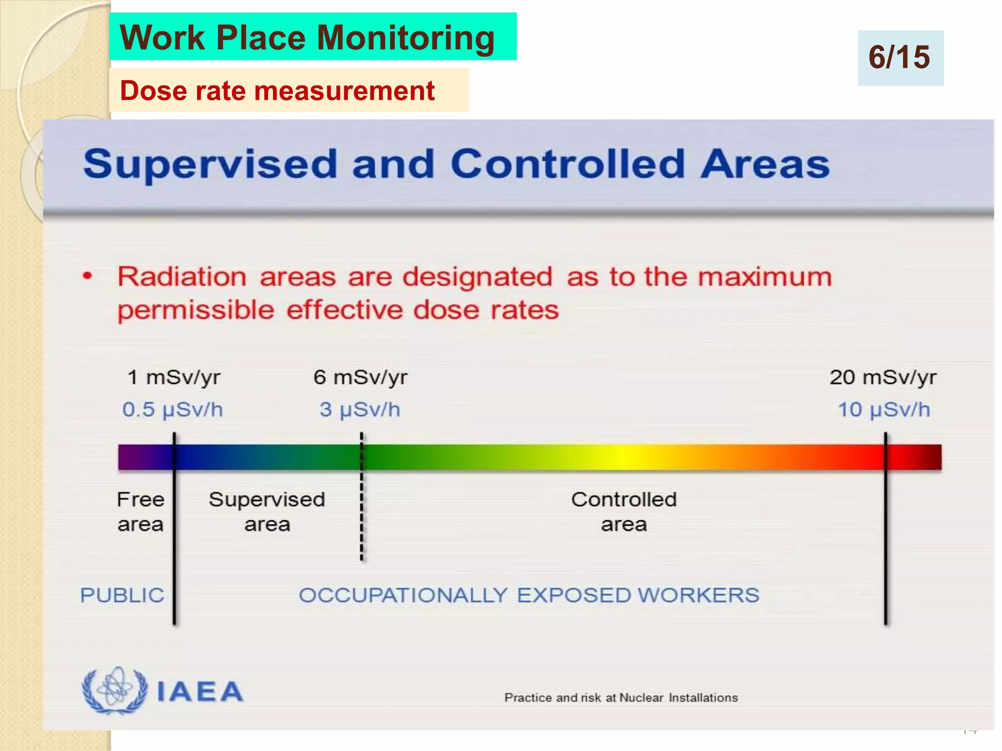
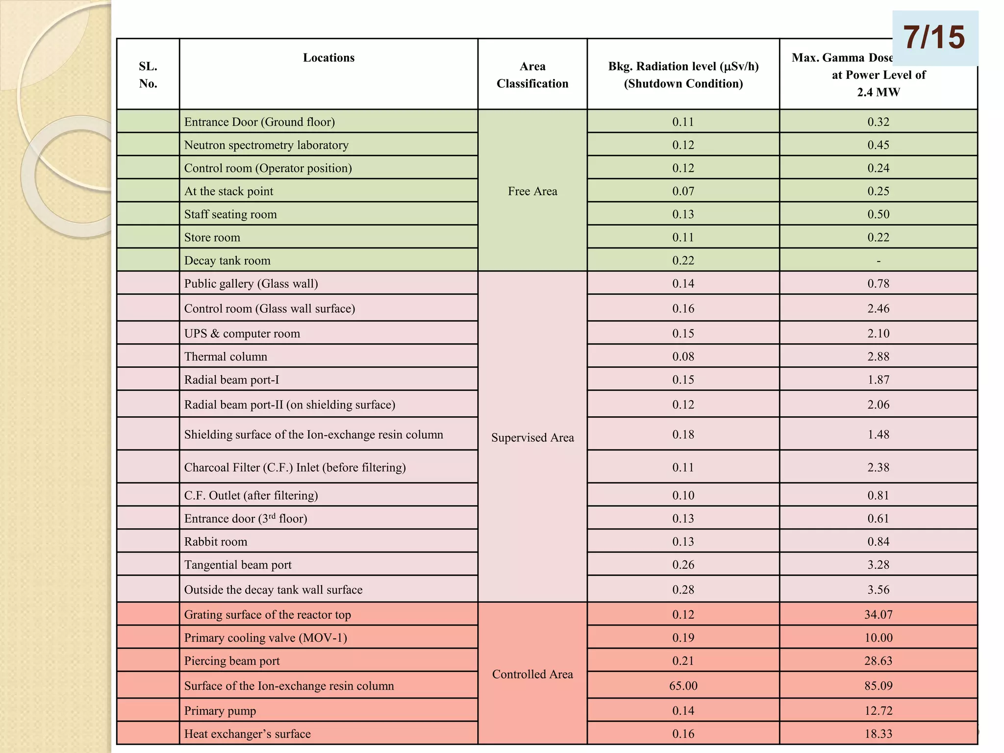
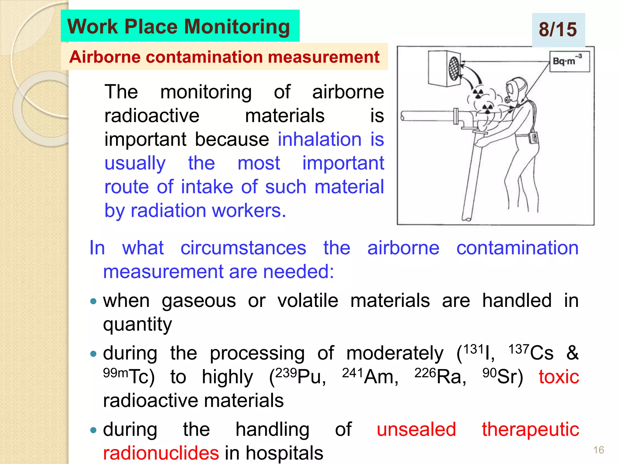
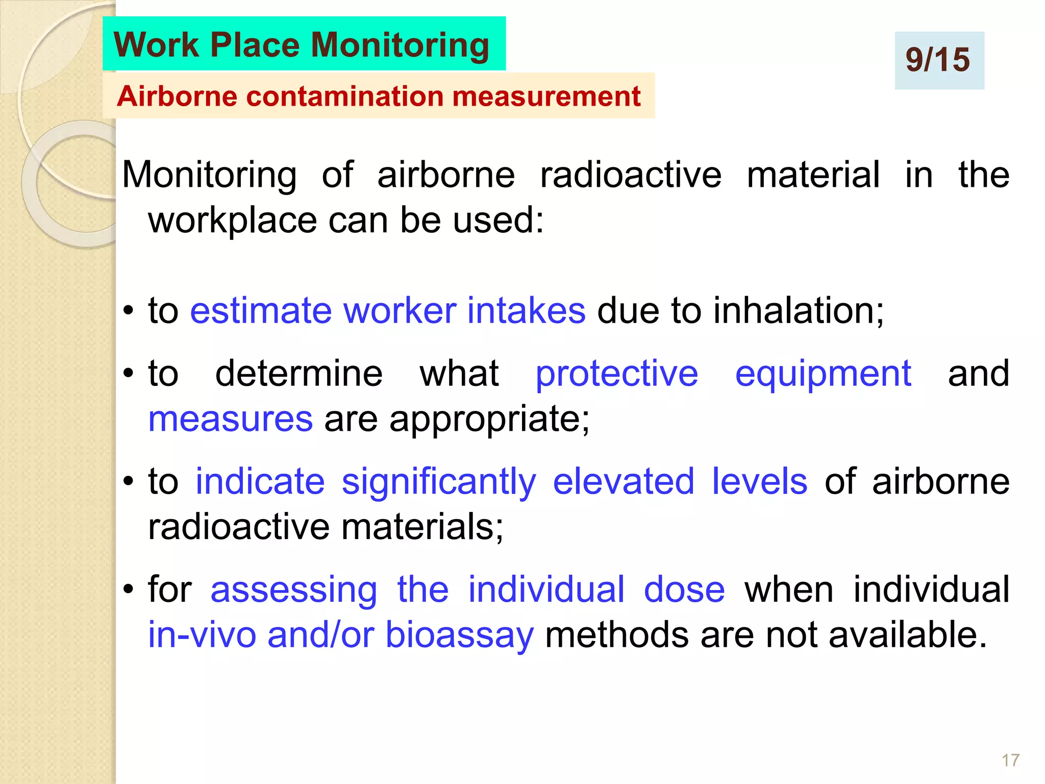
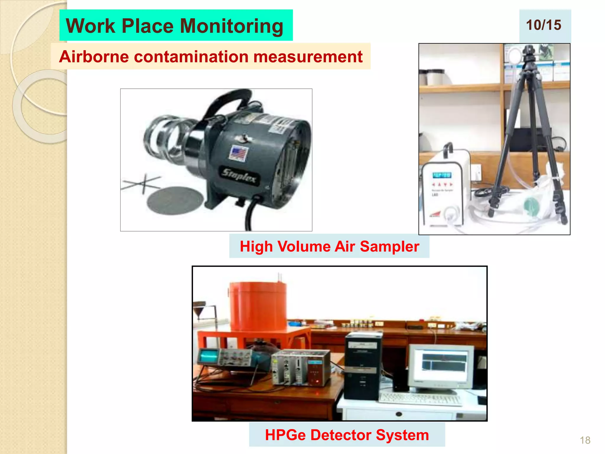
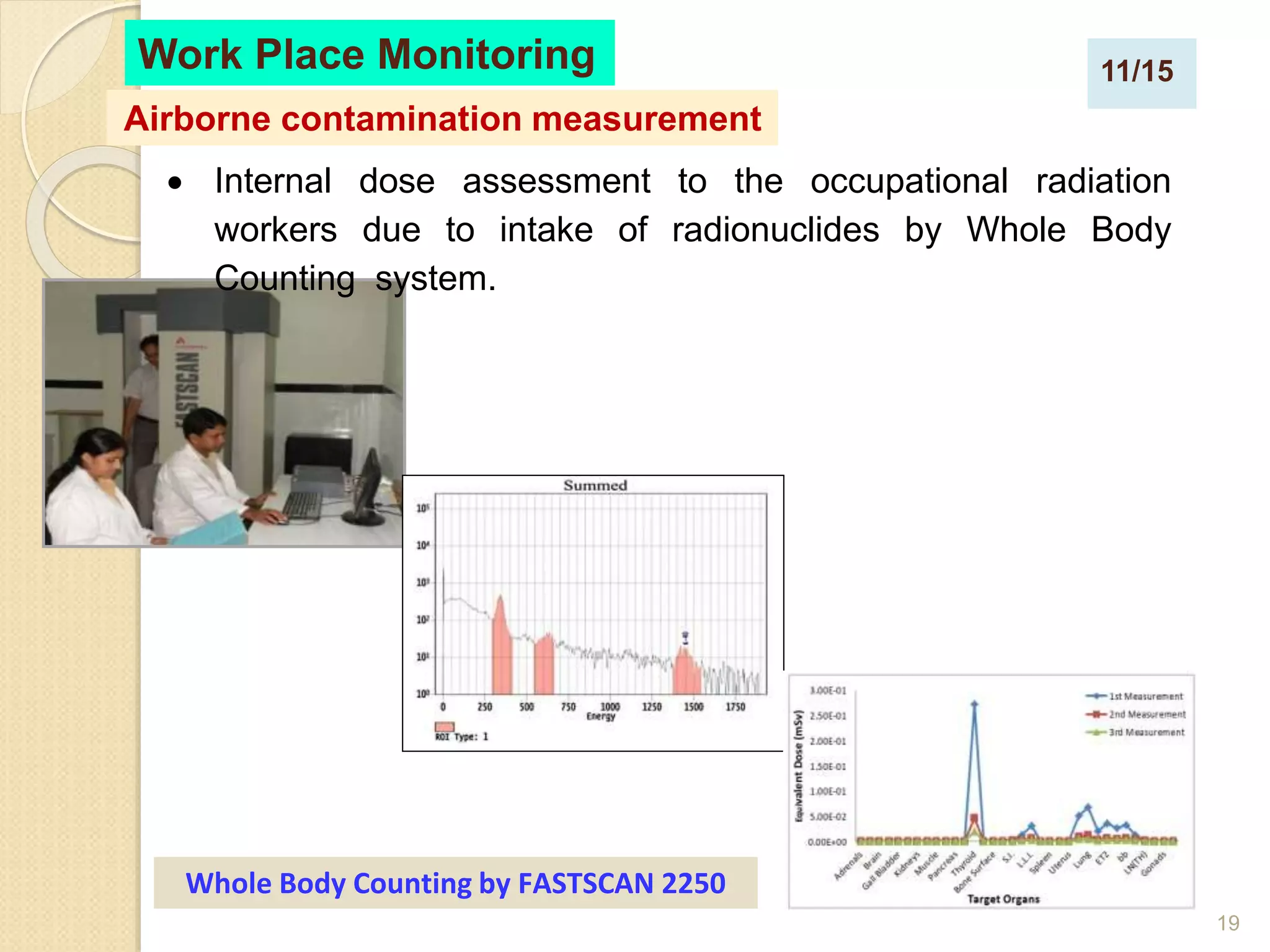
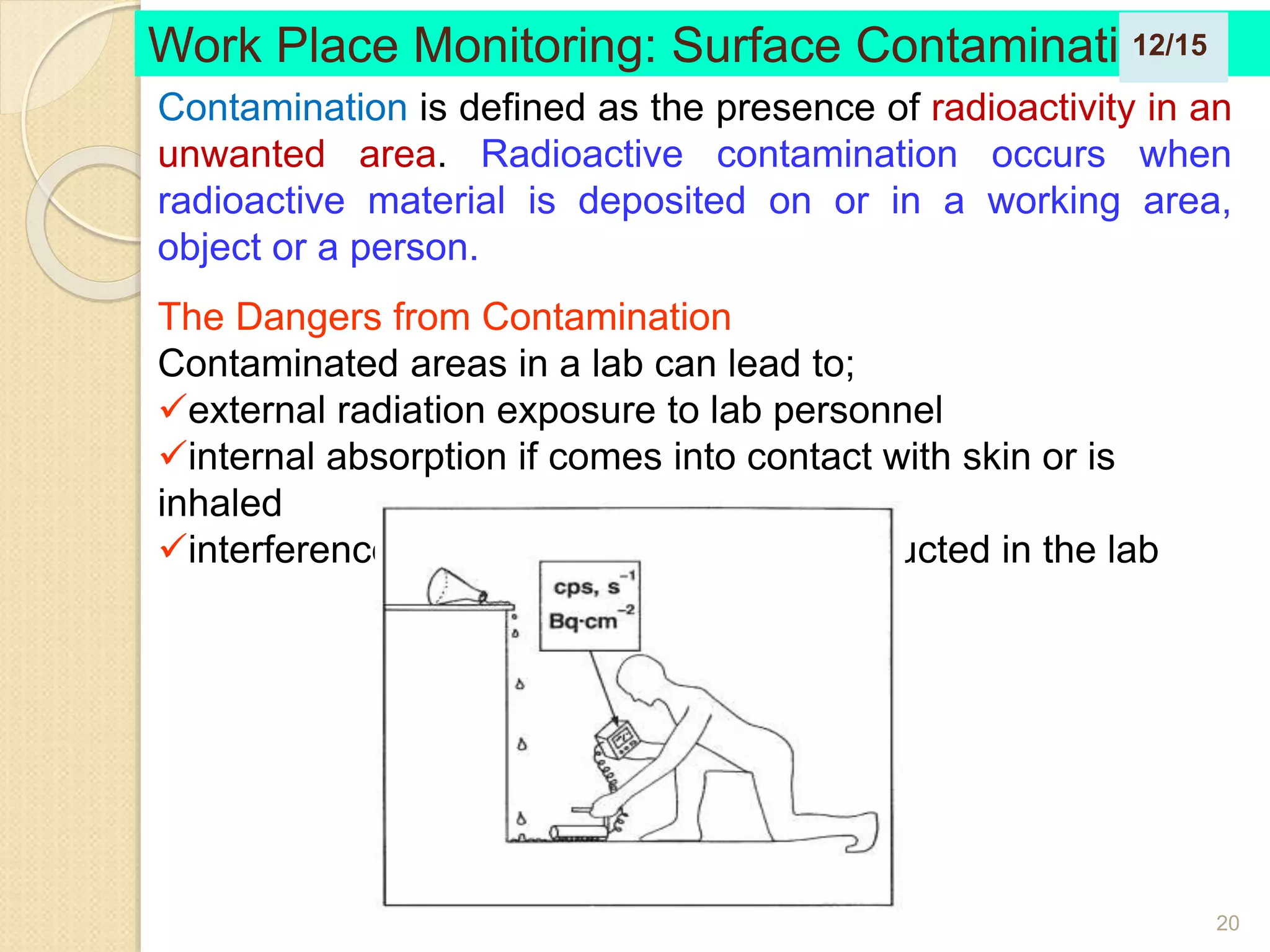
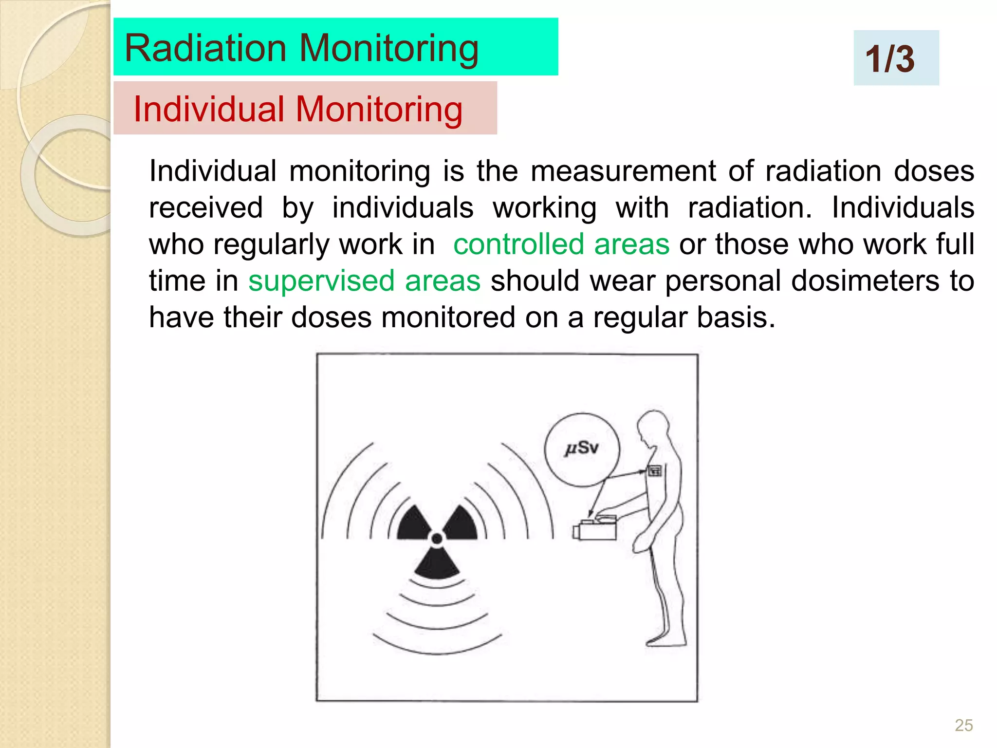
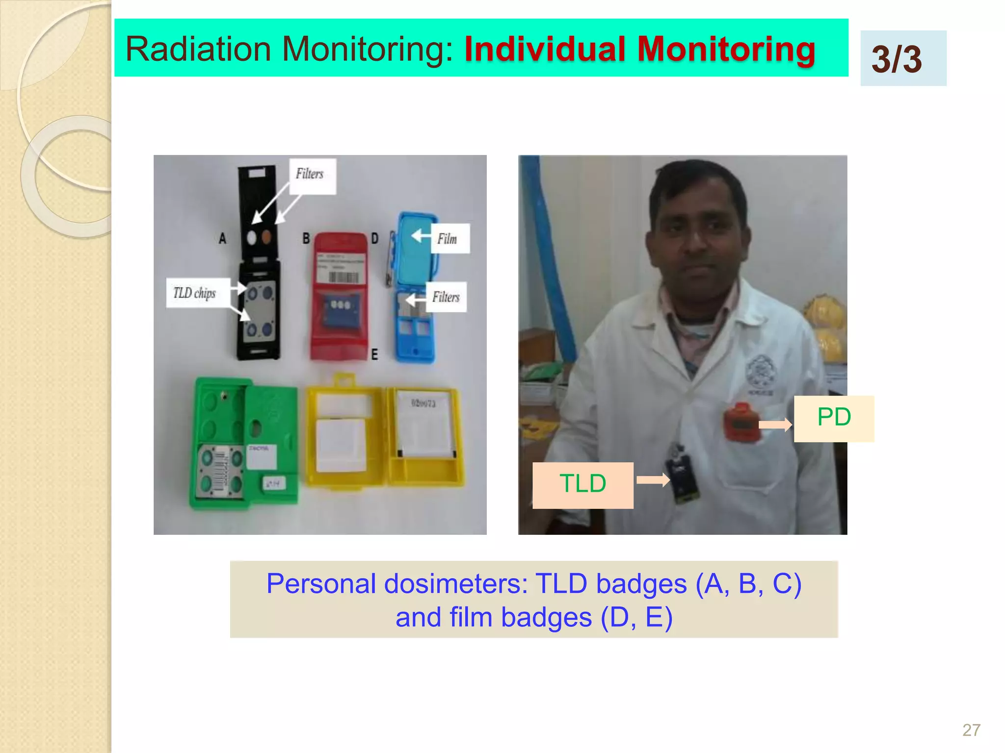
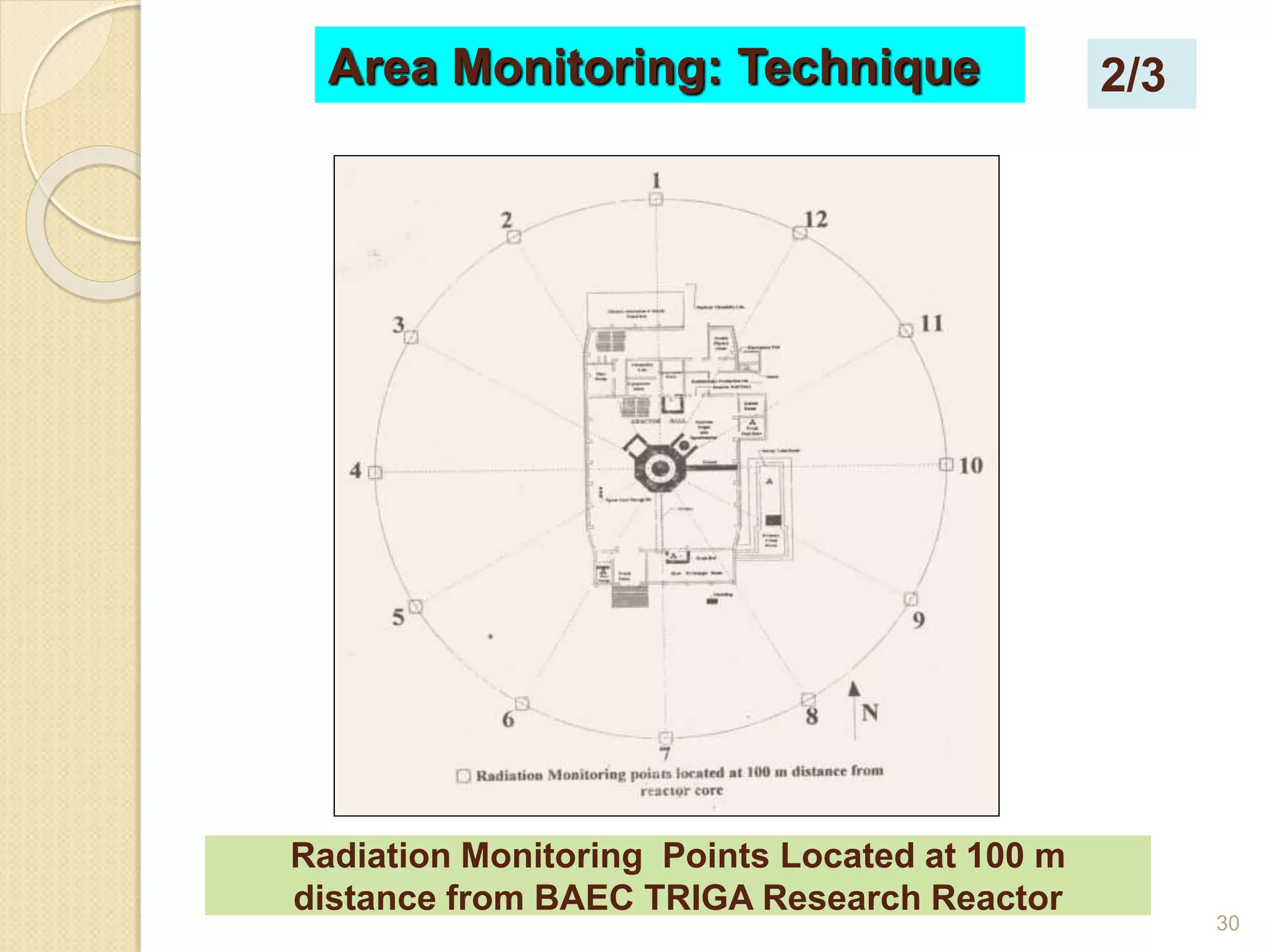
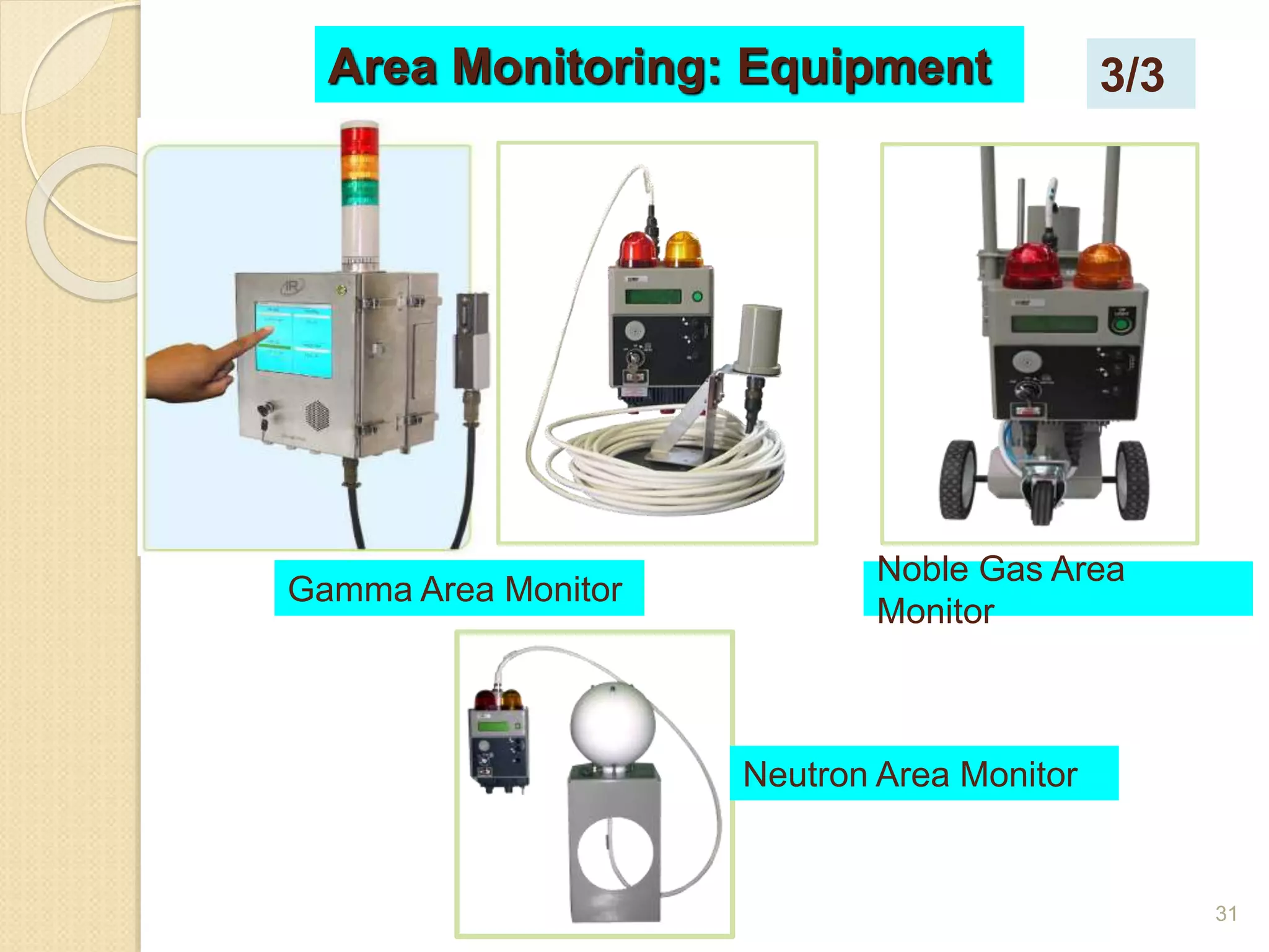
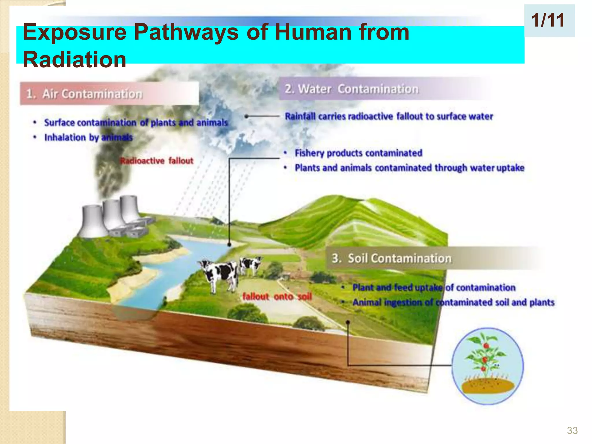
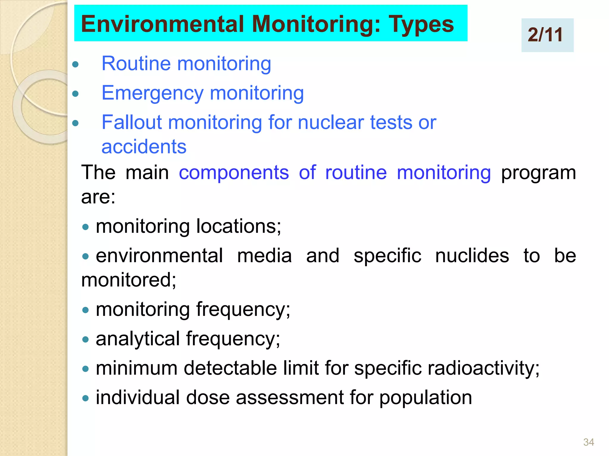
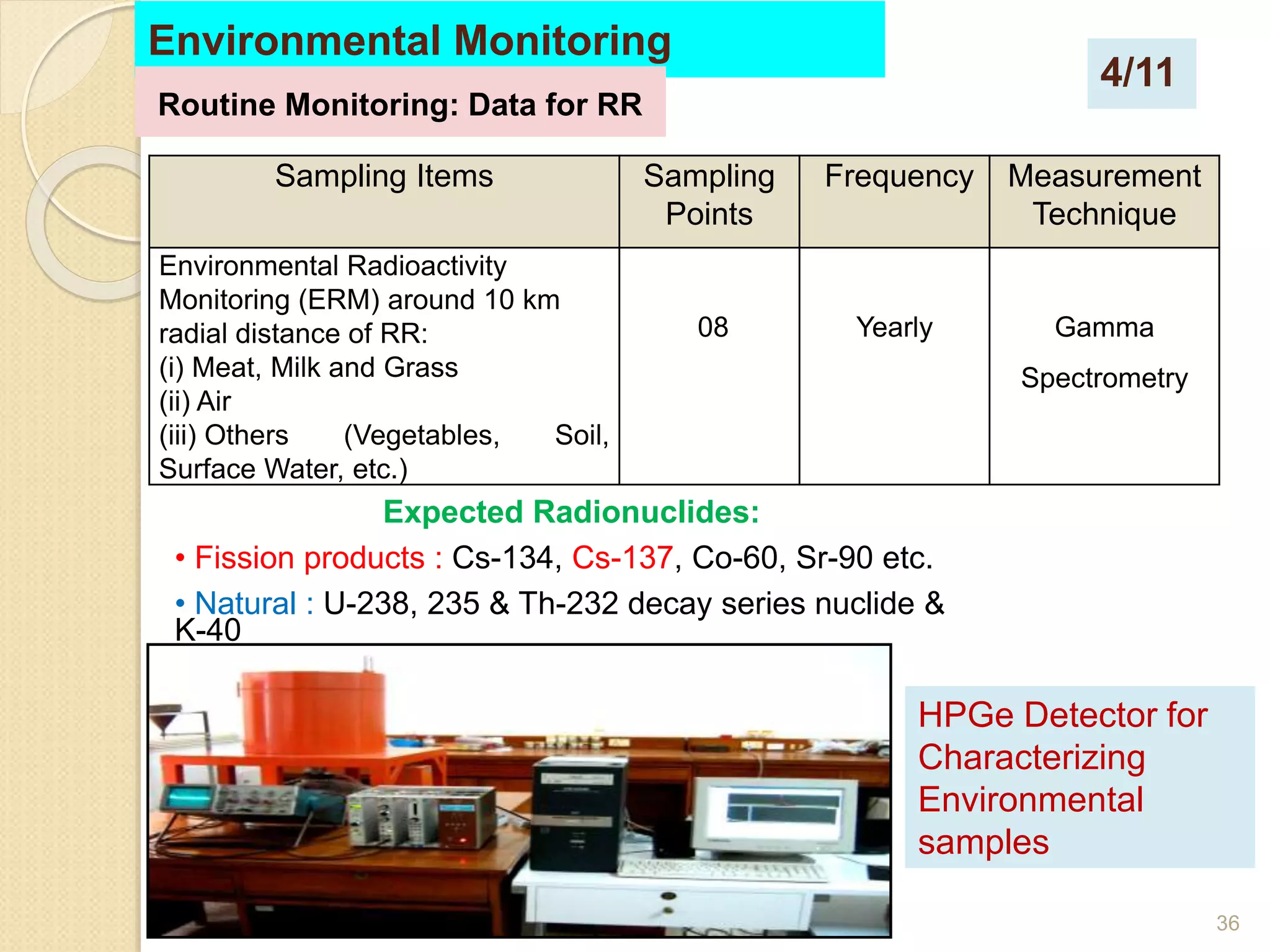
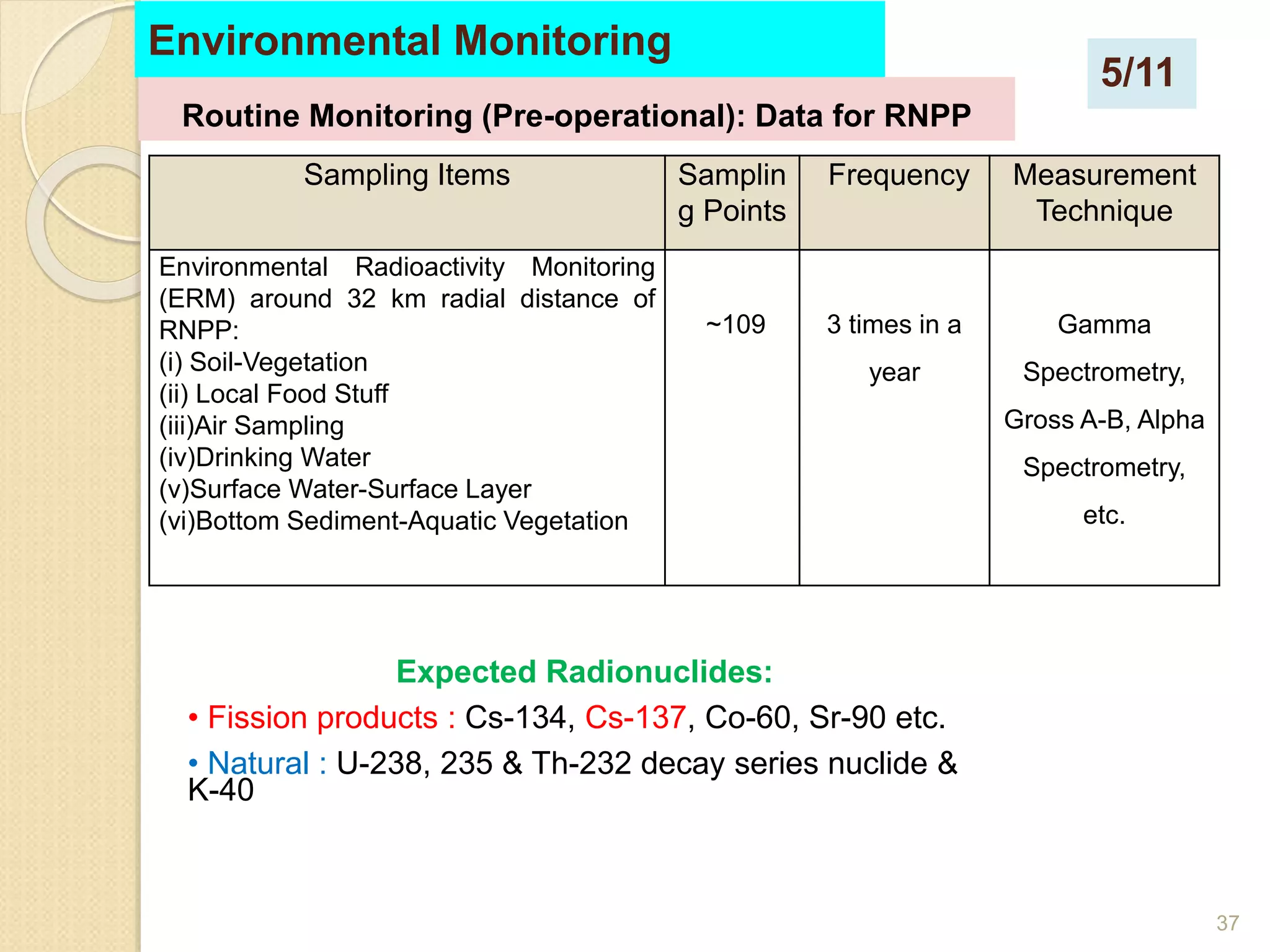
Workplace monitoring measures radiation dose rates, surface contamination, and airborne radioactivity where radiation sources are used. Individual monitoring tracks radiation doses received by workers through personal dosimeters. Area monitoring measures radiation levels at predefined locations around facilities to ensure safety. Environmental monitoring routinely samples media like food, water and air near facilities to measure radiation levels and ensure public safety.
Occupational radiation safety in Radiotherapy, Timothy Peace Sohscmcvellore
This document discusses occupational radiation safety in radiotherapy. It outlines potential radiation hazards from teletherapy equipment like telecobalt units and linear accelerators, as well as brachytherapy sources. Case studies of accidents are presented to illustrate hazards that can occur from equipment malfunctions, improper safety procedures, and lack of regulatory oversight. The document recommends strict adherence to safety guidelines and regulatory standards to minimize risks and ensure occupational exposures are kept as low as reasonably achievable. Regular equipment maintenance, staff training, and quality assurance are emphasized.
The document discusses recommendations from ICRP 60 & 103 regarding radiation protection. It begins with background on natural and artificial radiation sources and their effects. It then summarizes the evolution of ICRP recommendations over time, from early annual dose limits of 1000 mSv reduced gradually to current limits. Key concepts discussed include justification of practices, optimization of protection, and application of dose limits. Occupational, public, and medical exposure dose limits are provided. ICRP 103 introduced changes like new tissue weighting factors and computational phantoms.
Electron beam therapy uses accelerated electrons to treat superficial tumors. Electrons interact with matter through inelastic collisions that cause ionization and excitation, and elastic collisions that scatter the electrons. This gives electron beams a characteristically sharp dose drop-off beyond the tumor depth. Key applications of electron beams include treatment of skin cancers, chest wall irradiation for breast cancer, and boost doses to lymph nodes.
The document discusses the International Commission on Radiological Protection (ICRP), which sets standards for radiation protection. The ICRP relies on the linear no-threshold model to establish dose limits for workers and the public. This model assumes that any amount of radiation exposure increases cancer risk proportionally. The ICRP cites data from studies of atomic bomb survivors and other exposed groups to determine that radiation carries a 5% increased risk of cancer per sievert of lifetime dose. Using this risk factor, the ICRP calculates annual dose limits of 20 millisieverts for occupational workers and 1 millisievert for members of the public. Though other models question the linear no-threshold model, the ICRP maintains it is a
Radiation protection in nuclear medicine.ppt 2Rad Tech
This document provides guidance on radiation protection procedures for radionuclide therapy, including administration of therapy, management of radioactive patients, and optimization of protection for medical staff, visitors, and the hospitalized patient. Key points addressed include justifying therapy based on clinical benefits, ensuring proper training and responsibilities of medical personnel, constraining doses to comforters and visitors, providing instructions to hospitalized patients, and surveying rooms prior to releasing patients or decommissioning areas.
Radiation is energy that is given off by particular materials and devices.
Radiation protection, also known as radiological protection, is defined by the International Atomic Energy Agency (IAEA) as "The protection of people from harmful effects of exposure to ionizing radiation, and the means for achieving this". Exposure can be from a source of radiation external to the human body or due to internal irradiation caused by the ingestion of radioactive contamination
Radiation safety in diagnostic nuclear medicineSGPGIMS
1. Radiation is a form of energy emitted by atoms in the form of electromagnetic waves or particles. Ionizing radiation can eject electrons from atoms and produce ions, while non-ionizing radiation excites electrons.
2. People are exposed to ionizing radiation from natural and man-made sources. Naturally occurring sources include terrestrial radiation, cosmic radiation, and internal radiation. Medical procedures such as CT scans, nuclear medicine exams, and fluoroscopy account for over 90% of man-made radiation exposure.
3. Radiation protection aims to take advantage of the benefits of radiation use while preventing deterministic effects and limiting stochastic effects to acceptable levels. Occupational dose limits are higher than public limits, and some populations like
This document summarizes key aspects of acceptance testing and commissioning for a new radiation therapy machine. It describes the necessary measurement equipment, including radiation survey meters, ionization chambers, and phantoms. Acceptance tests and commissioning involve measuring various beam properties to ensure the machine meets specifications and performs reliably before clinical use. This process establishes the machine's baseline performance values which are then monitored ongoing through periodic quality assurance tests.
The document discusses quality assurance in nuclear medicine, outlining general principles and procedures for ensuring high quality patient care and radiation safety. It covers organizing a quality assurance program, administrative routines like requesting exams and generating reports, monitoring occupational and medical exposure, maintaining instrumentation, and educating staff. The overall goal is continual improvement in diagnostic accuracy, effective use of resources, and optimization of radiation dose for patients and workers.
1. This report discusses radiation protection in clinical applications like radiology, radiation therapy, and nuclear medicine. It focuses on establishing radiation protection areas like monitoring areas, controlled areas, and exclusion areas based on radiation dose levels.
2. Experiments were conducted to test diffuse radiation, inverse square law, and shielding verification of a CT scanner. Measurements showed radiation dose decreased with distance from the source and that shielding blocked radiation effectively.
3. The results indicate good clinical practice and shielding can decrease radiation dose to patients and workers, helping to prevent deterministic effects and reduce stochastic effect probabilities in accordance with international safety standards.
Radiation safety precautions (General Principles, Power Plant Safety, Radionu...Sabir Rasheed
Radiation safety precaution. General Principles of Radiation Safety.
Aspects of shielding in diagnostic radiology.
Nuclear Power Plant Safety.
Specific Handling Precautions For Various Radionuclides.
Evolution of gynaecological brachytherapyRitam Joarder
This document provides a historical overview of brachytherapy and the evolution of radiation sources used. It discusses some of the early discoveries in x-rays and radioactivity in the 1890s. It then describes some of the early uses of radium to treat skin lesions and cervical cancer in the early 1900s. The document outlines several early brachytherapy systems developed between 1913-1953, including the Stockholm, Paris, Manchester, and Paterson-Parker systems. It also discusses the introduction of the Quimby system using radium needles. The document notes the evolution of brachytherapy sources over time from radium to cesium-137 to iridium-192 to improve dosimetry, specific activity,
This document discusses key considerations for designing radiation shielding in diagnostic radiology facilities. It outlines parameters to calculate shielding needs such as workload, occupancy, beam direction and tube leakage. Common shielding materials like lead, concrete and gypsum are described. The importance of continuity, integrity and quality control of the shielding installation is emphasized through inspection and record keeping.
This document discusses electron beam therapy. It begins by explaining that electron beams allow for uniform dose delivery to within approximately 6 cm of the surface, sparing deeper tissues. Electrons are useful for treating various cancers of the skin, head and neck, breast, and other sites. It then covers topics like electron beam production, interactions in tissue, depth dose curves, effects of field size and obliquity, tissue heterogeneities, treatment planning, and future directions like intensity-modulated electron therapy.
Radiation safety involves understanding radiation sources and their biological effects, and implementing principles of protection. Ionizing radiation can damage tissue through stochastic effects like cancer. Dose limits regulate worker and public exposure, and the ALARA principle aims to keep doses as low as reasonably achievable. Proper signage, dosimetry, shielding, security, and training help ensure radiation is used safely in medicine.
This document discusses the history and development of radiotherapy machines. It describes early machines that used X-rays and radium to treat cancers from the late 19th century up to the 1950s. The development of cobalt-60 teletherapy units in the 1950s provided a more powerful and practical radiation source. The document focuses on describing the Theratron 780C cobalt-60 teletherapy machine, including its parts, radiation modes, source, controls, specifications and safety features. It also discusses concepts like isocenter, penumbra and the advantages cobalt-60 provided over earlier radiation sources.
This document provides an overview of nuclear medicine and the technologies used. It discusses radiopharmaceuticals, which consist of a chemical molecule and radionuclide, and are used in nuclear medicine to provide information about organ function. Gamma cameras are described as detecting radiation emitted from radiopharmaceuticals and producing images, while SPECT involves a gamma camera rotating around the patient to generate 3D tomographic images. The key components of gamma cameras and their operation are also summarized.
The document discusses the history of radiation protection, including early pioneers who discovered radiation hazards and effects. It describes some key events like the establishment of the ICRP and AERB, and definitions of key radiation terms. It also outlines the biological effects of radiation exposure, distinguishing between deterministic and stochastic effects. The three principles of radiation protection - justification, optimization and dose limitation - are explained.
Radioactive Contamination and Procedures of Decontaminationmahbubul hassan
Training Course on Radiation Protection for Radiation Workers and RCOs of BAEC, Medical Facilities and Industries, TI, AERE, BAEC Savar, 27 October 2021
Radiation Introduction, Hazards and Measuring Equipment used in Radiation Pro...Sabir Rasheed
Introduction of radiation, hazards and Measuring Equipment used in Radiation Protection.
Biology Effects.
Nuclear effects.
Different Radiation Measuring instruments.
1.Types of personnel monitoring devices
2.Instruments for measuring external Exposure.
The document provides information about radiation safety at Wayne State University. It introduces the Office of Environmental Health and Safety (OEHS) and its roles in protecting health and safety regarding hazardous materials use. It also provides contact information for the radiation safety and hazardous waste staff. Basic radiation safety training requirements and rights of radiation workers are outlined.
The document discusses various factors that affect image quality in nuclear medicine imaging, including spatial resolution, contrast, and noise. It describes methods for evaluating spatial resolution such as using bar phantoms or line spread functions. Modulation transfer functions can also be used to characterize spatial resolution and compare different imaging systems. Image contrast and noise are affected by factors like radiopharmaceutical uptake, scatter radiation, and count rates. Quality assurance tests are important for ensuring optimal system performance and image quality.
This document discusses radiation protection and dosimetry concepts. It defines key terms like absorbed dose, equivalent dose, effective dose and their calculations. It describes stochastic and deterministic effects and the objectives of radiation protection to limit both. The ALARA principle and its application are explained. Various radiation measurement instruments like survey meters, dosimeters and their uses are outlined. The document also discusses radiation shielding calculations and definitions of controlled, supervised and uncontrolled areas.
Radiation protection involves protecting people from harmful effects of ionizing radiation. There are three types of radiation: primary radiation which is most intense; scattered radiation resulting from the Compton effect; and leakage radiation emitted from x-ray equipment. The three cardinal principles of radiation protection are time, distance, and shielding. The system of radiation protection justifies practices where benefits outweigh risks, uses ALARA to keep doses as low as reasonably achievable, and limits doses to individuals. Radiation can cause stochastic or non-stochastic effects depending on dose thresholds. Exposure includes medical exposure to patients, occupational exposure to workers, and public exposure. Radiation is monitored through personnel and workplace monitoring devices. Radiation facilities use controlled and
This document discusses emergency response and preparedness in a radiation department. It defines a radiation emergency and classifies emergencies by whether they affect equipment, an individual patient, or many patients. Potential sources of error leading to emergencies in radiotherapy, nuclear medicine, brachytherapy, and diagnostic radiology are described. Regulations regarding reporting and investigating emergencies are summarized. Steps for handling common emergency situations like source stucks are outlined. The responsibilities of licensees and radiation safety officers in emergency planning and response are also covered.
This document discusses radiation protection and provides definitions, types of radiation effects, sources of radiation exposure, units of measurement, dose limits, and techniques to reduce radiation exposure in medical imaging. It defines radiation protection as protecting people from harmful effects of ionizing radiation. It describes stochastic and deterministic effects and lists examples of radiation anomalies. It also outlines regulatory bodies, dose limits for occupational workers and the public, and principles of radiation safety including time, distance, shielding and reducing exposure.
The document outlines the key components of a radiation protection program for industrial radiography and irradiator facilities. It discusses organizational responsibilities, radiation protection responsibilities, area classification, radiation monitoring, quality assurance, emergency response plans, training and health surveillance of workers, and record keeping. Safety working procedures are also described for industrial practices, including personnel monitoring, radiation surveys, warning signals, operating exposure rooms, and radiographic work procedures.
The document discusses sources of radiation, natural and man-made, and outlines safety procedures for radiography operations. It notes that radiation comes from cosmic, terrestrial, and internal sources. For radiography work, safety equipment like dosimeters, barricades, signs, and certified radiography workers are required to limit exposure and ensure safe operation. Regular monitoring and training are also needed to protect radiography workers.
Individual and area monitoring are important aspects of radiation protection. Individual monitoring involves measuring radiation doses received by individuals working with radiation, typically using dosimeters like TLD badges worn by workers in controlled and supervised areas. Area monitoring involves taking radiation measurements at different points in a workplace to assess conditions and ensure safe radiological conditions. The results of monitoring are recorded and investigated if exposure limits are exceeded.
This ppt is all about dosimetry used in radiology department.
it also consist of history of dosimetry ,conventional dosimeters like Film badge,TLD , OSLD ,Pocket dosimetry.
Further it is all about the latest advancements in dosimetry mailny by MIRION technologies.
This document provides guidelines for the manufacture of sterile medicinal products in the European Union. It outlines classification grades (A-D) for clean rooms based on airborne particle limits, with Grade A being the highest standard for filling zones. It recommends environmental monitoring for viable and non-viable particles in grades A and B. Guidelines are provided for terminally sterilized products and aseptically prepared products, specifying the appropriate grade for different manufacturing steps. Personnel requirements include training in hygiene and microbiology and limits on those handling non-sterile materials.
This document provides guidelines for the manufacture of sterile medicinal products in the European Union. It outlines classification grades (A-D) for clean rooms based on airborne particle limits, with Grade A being the highest standard for filling zones. It recommends environmental monitoring for viable and non-viable particles in grades A and B. Guidelines are provided for terminally sterilized products and aseptically prepared products, specifying the appropriate grade for different manufacturing steps. Personnel requirements include training in hygiene and microbiology and maintaining high standards of personal cleanliness.
This document provides information on detection and measurement of ionizing radiation. It discusses different types of radiation monitors used for source monitoring, environmental monitoring, and individual monitoring. These include dose rate meters, contamination monitors, Geiger counters, scintillation counters, film badges, and thermoluminescent dosimeters (TLDs). It also covers topics like instrument ranges, surface contamination limits, and control standards for radiation exposure. The goal of radiation measurement is to evaluate radiation conditions, assess potential exposures, and review classification of controlled areas.
1) The document discusses good practices for dispensing and sampling of raw materials in pharmaceutical manufacturing. It emphasizes carefully following standard operating procedures to prevent contamination.
2) Key steps in dispensing include cleaning the room, checking that the right chemical and amount are issued, weighing while being double checked, and immediately cleaning up any dust.
3) Tips to prevent contamination include sampling one material at a time in a segregated booth, cleaning all containers before storage, having appropriate air control systems, and avoiding simultaneous charging of raw materials for different batches.
The document discusses various aspects of naturally occurring radioactive material (NORM) in the oil and gas industry, including:
1) Methods for characterizing radionuclides in produced water and NORM waste through gamma spectrometry analysis.
2) Exposure control measures needed for NORM, including personal protective equipment, area monitoring, and record keeping.
3) Regulatory options for controlling NORM, ranging from exemption to licensing depending on exposure levels.
4) Guidelines for handling, storing, and disposing of NORM-contaminated equipment to minimize radiation exposure.
This document evaluates quality assurance and radiation safety procedures for ruthenium-106 plaque brachytherapy treatment of eye tumors. Leakage tests found radiation levels from plaques to be below limits. Radiation surveys found levels around the facility to be slightly above background. During procedures, dose rates to surgeons were 3.5 mR/hr at the wrist and 55 μR/hr at eye level. The maximum leakage near autoclaves used to sterilize plaques was 400 μR/hr at 5 cm, dropping to 80 μR/hr at 1 meter. Proper shielding, monitoring, and safety protocols were followed to ensure protection of patients, staff and the public.
The document discusses environmental monitoring programs in clean rooms and aseptic processing areas. It describes the purpose of monitoring to control microbial and particle contamination and prevent release of contaminated products. Key aspects covered include viable and non-viable monitoring of air, surfaces, personnel and drains using methods like air sampling, surface swabbing and particle counting. It provides classification standards and action limits for contamination and validation procedures for HVAC systems. Personnel are identified as the main challenge to control in aseptic processing.
This document discusses air pollution monitoring and different monitoring methods. It describes the need for well-planned monitoring to make rational decisions about environmental protection programs. Discrete sampling using manual methods are presented as a practical alternative for most monitoring objectives in India. High volume air samplers and respirable dust samplers are discussed as methods for ambient and workplace air quality monitoring. Continuous automatic instruments are better for surveillance but discrete sampling is more versatile and cost-effective for monitoring multiple locations and parameters.
The document discusses good radiopharmaceutical practices (GRP) which provide guidelines for safely handling radiopharmaceuticals. GRP focuses on personnel, premises, equipment, preparation, quality control, and documentation. Personnel must be trained and premises must limit public access while allowing for sterile production. Quality control testing ensures product safety and sterility. Documentation of all production and testing is required to maintain standards. GRP aims to protect workers, patients, and the public from radiation hazards during radiopharmaceutical development and use.
This document outlines the key elements of a radiation protection program for facilities using nuclear gauges and well logging tools. It discusses the responsibilities of management, radiation protection officers, and workers. It describes the classification of controlled and supervised areas and requirements for area and individual monitoring, record keeping, training, emergency planning, and auditing the radiation protection program. The overall goal is to ensure radiation exposures are adequately controlled and safety measures are followed.
This document discusses environmental microbial monitoring (EMM) in cleanrooms and pharmaceutical facilities. It provides an overview of EMM purposes and regulations, who performs EMM, what areas are monitored, sampling plans and methods used. Key points covered include:
- EMM determines microbial and particulate levels to ensure cleanroom quality and identify contamination sources.
- Quality Control and Assurance departments perform EMM to demonstrate safety and ensure GMP compliance.
- Non-viable air, viable air and surface samples are monitored from areas like personnel, equipment and facilities.
- Sampling frequency, sites and methods like air samplers, settle plates, contact plates and swabbing are discussed in accordance with regulations like USP 39
The document provides information about an internship program conducted at Vimta Labs Ltd in Coimbatore, India. It discusses the company profile and services provided by Vimta related to environmental assessments, monitoring, and chemistry. It also outlines the objectives of the internship which were to monitor ambient air quality in an industrial estate, compare results to national standards, and calculate the air pollution index. The methodology section describes the various instruments and parameters used to monitor ambient air, stack emissions, water quality, noise, and light levels.
This document discusses occupational hygiene and its role in protecting worker health and safety through preventing or reducing risks from chemical, physical, and biological hazards in the workplace. Occupational hygiene applies scientific and managerial principles to control exposures to harmful substances like dusts, gases, vapors, noise, vibration and biological agents. Proper ventilation, atmospheric monitoring, use of personal protective equipment, and hazard assessments are important controls to consider.
The document discusses the qualification of analytical equipment. It describes the types of qualification which include design qualification, installation qualification, operational qualification, and performance qualification. It provides details on qualifying various analytical instruments such as FTIR, GC, and HPLC. Specific parameters to be tested during qualification are discussed for components like the inlet, oven, detector, autosampler, and others, along with typical acceptance criteria. The document emphasizes the importance of qualifying analytical equipment to ensure it is properly installed, operates correctly, and provides expected results.
Occupational hygiene plays a vital role in the university's occupational health and safety program by protecting worker health through preventing or reducing risks from chemical, physical, and biological hazards. Such hazards can include dusts, gases, vapors, noise, vibration, lighting issues, and biological exposures. Proper controls and monitoring help ensure a safe workplace.
This document provides information about radiation detection and measurement instruments. It discusses various types of gas-filled detectors like ionization chambers, proportional counters, and Geiger-Muller counters. It also describes semiconductor detectors, scintillation counters, and instruments used for personnel dosimetry and measuring dose rates and contamination levels. The key purpose of these various instruments is to detect and measure different types of ionizing radiation like alpha, beta, gamma, and neutrons by converting radiation interactions into electrical pulses or light flashes that can be analyzed. Regular calibration of radiation monitors is emphasized to ensure accurate measurements for different radionuclides and radiation energies.
Basic concept of radiation, radioactivity, radiation dosemahbubul hassan
This document provides information about a radiation protection training course taking place from October 24-28, 2021 in Dhaka, Bangladesh. It covers basic concepts in radiation, radioactivity, radiation dose units, types of radiation including alpha, beta, gamma, x-rays, and neutrons. It also discusses units of radioactivity, absorbed dose, dose equivalent, radiation weighting factors, and tissue weighting factors which are important concepts in radiation protection.
6th Training Course on Radiation Protection for Radiation Workers and RCOs of BAEC, Medical Facilities & Industries
Training Institute, AERE, Savar, BAEC
24 - 29 October 2021
FUNDAMENTALS OF RADIATION PROTECTION – EXTERNAL & INTERNAL mahbubul hassan
Training Course on Radiation Protection for Radiation Workers
and RCOs of BAEC, Medical Facilities & Industries
24 - 28 October 2021
Training Institute
Atomic Energy Research Establishment, Savar, Dhaka
BASIC CONCEPT OF RADIATION SHIELDING AND ITS CALCULATION TECHNIQUES mahbubul hassan
Training Course on Radiation Protection for Radiation Workers
and RCOs of BAEC, Medical Facilities & Industries
24 - 28 October 2021
Training Institute
Atomic Energy Research Establishment, Savar, Dhaka
6th Training Course on Radiation Protection for Radiation Workers and RCOs of BAEC, Medical Facilities & Industries
Training Institute, AERE, Savar, BAEC
24 - 29 October 2021
Ionizing radiation can cause biological damage through direct or indirect action on cells. The effects of radiation exposure depend on factors like total dose, dose rate, and part of body exposed. There are stochastic effects like cancer which occur randomly with no safe threshold, and deterministic effects like skin burns which have a threshold below which no effect occurs. Early effects appear within days of exposure while late effects can take years. Acute exposure involves a high dose over a short time compared to chronic low dose exposure. The principles of radiation protection are justification, optimization and dose limitation to reduce risk from radiation according to international standards.
This document discusses nuclear and radiological emergency preparedness and response. It defines key terms like emergency management, emergency, preparedness, and response. It describes different types of nuclear and radiological accidents that have occurred worldwide, including Fukushima, Chernobyl, and Three Mile Island for nuclear accidents, and Goiania for a radiological accident. It also discusses four levels of radiation emergencies - standby, plant, site area, and general - and explains emergency response planning areas.
This document discusses key aspects of safely transporting radioactive material as outlined in international regulations. It covers material classification, package selection based on material type and content limits, and controls during transport including limits on radiation levels, transport indexes, and categorizing packages. The overall safety approach involves containing radioactive contents, controlling external radiation, preventing criticality, and preventing heat damage through a graded package design based on material hazard levels.
How to Setup Warehouse & Location in Odoo 17 InventoryCeline George
In this slide, we'll explore how to set up warehouses and locations in Odoo 17 Inventory. This will help us manage our stock effectively, track inventory levels, and streamline warehouse operations.
This slide is special for master students (MIBS & MIFB) in UUM. Also useful for readers who are interested in the topic of contemporary Islamic banking.
LAND USE LAND COVER AND NDVI OF MIRZAPUR DISTRICT, UPRAHUL
This Dissertation explores the particular circumstances of Mirzapur, a region located in the
core of India. Mirzapur, with its varied terrains and abundant biodiversity, offers an optimal
environment for investigating the changes in vegetation cover dynamics. Our study utilizes
advanced technologies such as GIS (Geographic Information Systems) and Remote sensing to
analyze the transformations that have taken place over the course of a decade.
The complex relationship between human activities and the environment has been the focus
of extensive research and worry. As the global community grapples with swift urbanization,
population expansion, and economic progress, the effects on natural ecosystems are becoming
more evident. A crucial element of this impact is the alteration of vegetation cover, which plays a
significant role in maintaining the ecological equilibrium of our planet.Land serves as the foundation for all human activities and provides the necessary materials for
these activities. As the most crucial natural resource, its utilization by humans results in different
'Land uses,' which are determined by both human activities and the physical characteristics of the
land.
The utilization of land is impacted by human needs and environmental factors. In countries
like India, rapid population growth and the emphasis on extensive resource exploitation can lead
to significant land degradation, adversely affecting the region's land cover.
Therefore, human intervention has significantly influenced land use patterns over many
centuries, evolving its structure over time and space. In the present era, these changes have
accelerated due to factors such as agriculture and urbanization. Information regarding land use and
cover is essential for various planning and management tasks related to the Earth's surface,
providing crucial environmental data for scientific, resource management, policy purposes, and
diverse human activities.
Accurate understanding of land use and cover is imperative for the development planning
of any area. Consequently, a wide range of professionals, including earth system scientists, land
and water managers, and urban planners, are interested in obtaining data on land use and cover
changes, conversion trends, and other related patterns. The spatial dimensions of land use and
cover support policymakers and scientists in making well-informed decisions, as alterations in
these patterns indicate shifts in economic and social conditions. Monitoring such changes with the
help of Advanced technologies like Remote Sensing and Geographic Information Systems is
crucial for coordinated efforts across different administrative levels. Advanced technologies like
Remote Sensing and Geographic Information Systems
9
Changes in vegetation cover refer to variations in the distribution, composition, and overall
structure of plant communities across different temporal and spatial scales. These changes can
occur natural.
How to Build a Module in Odoo 17 Using the Scaffold MethodCeline George
Odoo provides an option for creating a module by using a single line command. By using this command the user can make a whole structure of a module. It is very easy for a beginner to make a module. There is no need to make each file manually. This slide will show how to create a module using the scaffold method.
How to Add Chatter in the odoo 17 ERP ModuleCeline George
In Odoo, the chatter is like a chat tool that helps you work together on records. You can leave notes and track things, making it easier to talk with your team and partners. Inside chatter, all communication history, activity, and changes will be displayed.
How to Fix the Import Error in the Odoo 17Celine George
An import error occurs when a program fails to import a module or library, disrupting its execution. In languages like Python, this issue arises when the specified module cannot be found or accessed, hindering the program's functionality. Resolving import errors is crucial for maintaining smooth software operation and uninterrupted development processes.
हिंदी वर्णमाला पीपीटी, hindi alphabet PPT presentation, hindi varnamala PPT, Hindi Varnamala pdf, हिंदी स्वर, हिंदी व्यंजन, sikhiye hindi varnmala, dr. mulla adam ali, hindi language and literature, hindi alphabet with drawing, hindi alphabet pdf, hindi varnamala for childrens, hindi language, hindi varnamala practice for kids, https://www.drmullaadamali.com
বাংলাদেশের অর্থনৈতিক সমীক্ষা ২০২৪ [Bangladesh Economic Review 2024 Bangla.pdf] কম্পিউটার , ট্যাব ও স্মার্ট ফোন ভার্সন সহ সম্পূর্ণ বাংলা ই-বুক বা pdf বই " সুচিপত্র ...বুকমার্ক মেনু 🔖 ও হাইপার লিংক মেনু 📝👆 যুক্ত ..
আমাদের সবার জন্য খুব খুব গুরুত্বপূর্ণ একটি বই ..বিসিএস, ব্যাংক, ইউনিভার্সিটি ভর্তি ও যে কোন প্রতিযোগিতা মূলক পরীক্ষার জন্য এর খুব ইম্পরট্যান্ট একটি বিষয় ...তাছাড়া বাংলাদেশের সাম্প্রতিক যে কোন ডাটা বা তথ্য এই বইতে পাবেন ...
তাই একজন নাগরিক হিসাবে এই তথ্য গুলো আপনার জানা প্রয়োজন ...।
বিসিএস ও ব্যাংক এর লিখিত পরীক্ষা ...+এছাড়া মাধ্যমিক ও উচ্চমাধ্যমিকের স্টুডেন্টদের জন্য অনেক কাজে আসবে ...
A review of the growth of the Israel Genealogy Research Association Database Collection for the last 12 months. Our collection is now passed the 3 million mark and still growing. See which archives have contributed the most. See the different types of records we have, and which years have had records added. You can also see what we have for the future.
This presentation was provided by Steph Pollock of The American Psychological Association’s Journals Program, and Damita Snow, of The American Society of Civil Engineers (ASCE), for the initial session of NISO's 2024 Training Series "DEIA in the Scholarly Landscape." Session One: 'Setting Expectations: a DEIA Primer,' was held June 6, 2024.
The simplified electron and muon model, Oscillating Spacetime: The Foundation...RitikBhardwaj56
Discover the Simplified Electron and Muon Model: A New Wave-Based Approach to Understanding Particles delves into a groundbreaking theory that presents electrons and muons as rotating soliton waves within oscillating spacetime. Geared towards students, researchers, and science buffs, this book breaks down complex ideas into simple explanations. It covers topics such as electron waves, temporal dynamics, and the implications of this model on particle physics. With clear illustrations and easy-to-follow explanations, readers will gain a new outlook on the universe's fundamental nature.
1. Radiation Monitoring: Work
Place, Area and Environment
Monitoring
Training Course on Radiation Protection for Radiation Workers
and RCOs of BAEC, Medical Facilities & Industries, TI, AERE,
OCT 2021
Presented By:
Md. Abu Haydar
Principal Scientific Officer
Health Physics and Radioactive Waste Management
Unit, INST, AERE, Savar, Dhaka.
3. 3
Introduction
Radiation monitoring involves the measurement of
radiation dose rate in the vicinity of a radiation
source, measurement of surface contamination, and
measurement of airborne radioactivity.
According to the Bangladesh Atomic Energy
Regulatory Act, 2012 (Act No. 19 of 2012) and
Regulations 1997 (NSRC Regulations 1997) of
Bangladesh as well as other international bodies
(IAEA, ICRP etc.), the licensee of a
nuclear/radiological facility or practices involving
radiation sources must ensure the safety of the
occupational workers, public and the environment.
To ensure the radiation safety of the occupational
workers, public and the environment, the radiation
monitoring program is essential.
1/5
4. 4
Introduction
Types of Radiation monitoring:
• Work Place Monitoring
• Individual Monitoring
• Environmental Monitoring
Radiation monitoring at different radiological
facilities Environmental
monitoring map for
RNPP
2/5
5. 5
Health: Public and private hospitals, Nuclear medicine
centers, Radioisotope production facility, X-rays
etc.
Industry: Industrial radiography/NDT, Nucleon gauge, Gas
mantle, Oil and gas explorations, Commercial
irradiator etc.
Research & Education: Operation and maintenance of 3
MW TRIGA MARK II Research Reactor (RR),
Food irradiation, Plant breeding, Insect
extermination, Accelerators etc.
NPP: Operation and maintenance of Rooppur Nuclear
Power Plant
NORMs: Coal mining, Beach Sand Minerals, Water
Nuclear/Radiological Facilities and Practices Involving
Radiation Exposure in Bangladesh
Introduction 3/5
9. 9
Work Place Monitoring
What is Work Place Monitoring?
Workplace monitoring is an important technique to
achieve and maintain an acceptable protection of the
working environment from radiation hazards.
The objectives of the workplace monitoring:
Confirmation of good or Identification of poor working
practices;
Provision of information and/or Identification of changes
in conditions in the workplace;
Identification of unusually high doses;
Estimation of the exposure of workers;
Provision of confidence in safety procedures and
improve workers attitudes to reduce their exposure;
Provide information for the evaluation of doses in the
event of accidental exposures.
1/15
10. 10
Work Place Monitoring
Work Place Monitoring may includes:
• Dose rate measurement
• Surface contamination measurement
• Airborne contamination measurement
2/15
11. 11
Work Place Monitoring
• A sufficient quantity of suitable dose rate meters capable of
providing direct readings of the ambient dose equivalent
rate (H*(10)/h) with ranges from the μSv/h to Sv/h should
be available.
• Where high levels of surface contamination with beta/alpha
emitters is foreseen, equipment calibrated in H’(0.07)/h
should also be available.
• The equipment should be type tested and appropriate for
work in field conditions – for example some detectors with
liquid crystal screens are hard to read in broad daylight.
The screen should have back lighting for night work.
• The energy range, battery life and availability, temperature
range and other considerations are important criteria.
Requirement for Dose rate measurement
3/15
12. 12
It is important that:
1) Before making the reading, the worker should assure that
he or she is in a safe position or situation. When entering
a high dose rate area keep the meter turned on and
check the alarm settings earlier.
2) The worker using the dose rate meters should be familiar
with the equipment through use during routine work.
3) The dose rate meter passes through a functional test
every day before use: battery, cables, background and if
possible a check source.
4) In high dose rate areas, do not take the measurement too
quickly, take the minimum time necessary to make a
correct reading.
5) The detector can be wrapped in plastic to avoid
contamination.
Work Place Monitoring
Dose rate measurement
4/15
13. 13
Work Place Monitoring
Dose rate meters provide direct
measurements of external
exposure.
Dose rate measurement
5/15
15. 15
SL.
No.
Locations
Area
Classification
Bkg. Radiation level (Sv/h)
(Shutdown Condition)
Max. Gamma Dose-Rate (Sv/h)
at Power Level of
2.4 MW
Entrance Door (Ground floor)
Free Area
0.11 0.32
Neutron spectrometry laboratory 0.12 0.45
Control room (Operator position) 0.12 0.24
At the stack point 0.07 0.25
Staff seating room 0.13 0.50
Store room 0.11 0.22
Decay tank room 0.22 -
Public gallery (Glass wall)
Supervised Area
0.14 0.78
Control room (Glass wall surface) 0.16 2.46
UPS & computer room 0.15 2.10
Thermal column 0.08 2.88
Radial beam port-I 0.15 1.87
Radial beam port-II (on shielding surface) 0.12 2.06
Shielding surface of the Ion-exchange resin column 0.18 1.48
Charcoal Filter (C.F.) Inlet (before filtering) 0.11 2.38
C.F. Outlet (after filtering) 0.10 0.81
Entrance door (3rd floor) 0.13 0.61
Rabbit room 0.13 0.84
Tangential beam port 0.26 3.28
Outside the decay tank wall surface 0.28 3.56
Grating surface of the reactor top
Controlled Area
0.12 34.07
Primary cooling valve (MOV-1) 0.19 10.00
Piercing beam port 0.21 28.63
Surface of the Ion-exchange resin column 65.00 85.09
Primary pump 0.14 12.72
Heat exchanger’s surface 0.16 18.33
7/15
16. 16
In what circumstances the airborne contamination
measurement are needed:
when gaseous or volatile materials are handled in
quantity
during the processing of moderately (131I, 137Cs &
99mTc) to highly (239Pu, 241Am, 226Ra, 90Sr) toxic
radioactive materials
during the handling of unsealed therapeutic
radionuclides in hospitals
Work Place Monitoring
Airborne contamination measurement
The monitoring of airborne
radioactive materials is
important because inhalation is
usually the most important
route of intake of such material
by radiation workers.
8/15
17. 17
Monitoring of airborne radioactive material in the
workplace can be used:
• to estimate worker intakes due to inhalation;
• to determine what protective equipment and
measures are appropriate;
• to indicate significantly elevated levels of airborne
radioactive materials;
• for assessing the individual dose when individual
in-vivo and/or bioassay methods are not available.
Work Place Monitoring
Airborne contamination measurement
9/15
18. 18
High Volume Air Sampler
HPGe Detector System
Airborne contamination measurement
Work Place Monitoring 10/15
19. 19
Whole Body Counting by FASTSCAN 2250
Internal dose assessment to the occupational radiation
workers due to intake of radionuclides by Whole Body
Counting system.
Airborne contamination measurement
Work Place Monitoring 11/15
20. 20
Contamination is defined as the presence of radioactivity in an
unwanted area. Radioactive contamination occurs when
radioactive material is deposited on or in a working area,
object or a person.
The Dangers from Contamination
Contaminated areas in a lab can lead to;
external radiation exposure to lab personnel
internal absorption if comes into contact with skin or is
inhaled
interference with experiments being conducted in the lab
Work Place Monitoring: Surface Contamination
12/15
21. 21
Work Place Monitoring: Surface Contamination
Contamination Area
If removable radioactive contamination exceeds:
• 1,000 dpm/100 cm2 beta-gamma contamination or
• 20 dpm/100 cm2 alpha emitting contamination
High Contamination Area
If removable contamination exceeds 100 times the
Contamination Area levels:
• 100,000 dpm/100 cm2 beta-gamma contamination or
• 2,000 dpm/100 cm2 alpha emitting contamination
Detection of Contamination
To determine if an area has contamination, compare the
background counts/minute to the counts/minute of the wipe
or survey meter. If the ratio of counts/minute exceeds 3:1 the
area contains significant contamination.
The unit of contamination is dpm/100cm2, dms/cm2 or
Bq/cm2.
13/15
22. 22
The probe should be held over contaminated area for at least 30
seconds so that it has sufficient time to respond to count rates.
The probe should be held as close as practical to the surface being
monitored. It is important to avoid contaminating the probe.
If the monitor does not have an energy discrimination device, alpha
particles may be distinguished from beta particles by placing a thin
sheet of paper over the sensitive area of the probe and beta
particles may be distinguished from photons by placing a thin metal
sheet between the detector and the contaminated surface.
Monitors should be checked in a low background area before use.
Work Place Monitoring: Surface Contamination
Measurement of
Contamination
Contamination Monitor
14/15
23. 23
In some instances for spill (fixed or loose contamination),
where unsuitable geometry or interference from other
radiations prevents direct measurement, indirect methods of
surface contamination monitoring by a wipe or smear sample
from the surface using a tissue or filter paper may be used.
The smear should be taken to a lower radiation area or to a
sensitive radiation detector (HPGe) to be analyzed.
Monitoring techniques
Work Place Monitoring: Surface Contamination
Fig. (a) Smear Paper,
(b) Sample Collection
(c) Measuring Equipment
(a)
(b)
(c)
15/15
25. 25
Individual monitoring is the measurement of radiation doses
received by individuals working with radiation. Individuals
who regularly work in controlled areas or those who work full
time in supervised areas should wear personal dosimeters to
have their doses monitored on a regular basis.
Radiation Monitoring
Individual Monitoring
1/3
26. 26
An individual dosimeter is used for measurement of an
external dose:
Passive dosimeter
A passive dosimeter measures an accumulated dose. Since
the battery is unnecessary, the passive dosimeter is smaller
and lighter, and it can run out of power. However, it does not
have a direct readout and cannot measure a change of dose
rate, or have preset alarms to provide a warning of a change
of working conditions. Typical passive dosimeter are TLD,
OSL, RPL, Film etc.
Active dosimeter
An active dosimeter is an personal dosimeter (PD) which
integrates the counts of radiation and measures dose rate
and the accumulated dose. Since the active dosimeter needs
the power of a battery it generally is not suitable for
continuous use over several days, and it may not be useable
Radiation Monitoring: Individual Monitoring 2/3
27. 27
Personal dosimeters: TLD badges (A, B, C)
and film badges (D, E)
TLD
PD
Radiation Monitoring: Individual Monitoring 3/3
29. 29
Area monitoring is the measurement of radiation dose
level at several pre-defined locations in and around a
facility where the radiation-emitting equipment is
located, or where radioactive sources are stored,
handled or used.
To perform a routine evaluation of the radiation dose
to assure the safety of the working conditions for the
workers within the facility and
To ensure the “Safety & Protection” of the
environment as well as the general public according
to ICRP/IAEA and national regulations.
Objectives
Area Monitoring
1/3
30. 30
Radiation Monitoring Points Located at 100 m
distance from BAEC TRIGA Research Reactor
Area Monitoring: Technique 2/3
34. 34
Routine monitoring
Emergency monitoring
Fallout monitoring for nuclear tests or
accidents
Environmental Monitoring: Types
The main components of routine monitoring program
are:
monitoring locations;
environmental media and specific nuclides to be
monitored;
monitoring frequency;
analytical frequency;
minimum detectable limit for specific radioactivity;
individual dose assessment for population
2/11
36. 36
Sampling Items Sampling
Points
Frequency Measurement
Technique
Environmental Radioactivity
Monitoring (ERM) around 10 km
radial distance of RR:
(i) Meat, Milk and Grass
(ii) Air
(iii) Others (Vegetables, Soil,
Surface Water, etc.)
08 Yearly Gamma
Spectrometry
Expected Radionuclides:
• Fission products : Cs-134, Cs-137, Co-60, Sr-90 etc.
• Natural : U-238, 235 & Th-232 decay series nuclide &
K-40
Environmental Monitoring
HPGe Detector for
Characterizing
Environmental
samples
Routine Monitoring: Data for RR
4/11
37. 37
Sampling Items Samplin
g Points
Frequency Measurement
Technique
Environmental Radioactivity Monitoring
(ERM) around 32 km radial distance of
RNPP:
(i) Soil-Vegetation
(ii) Local Food Stuff
(iii)Air Sampling
(iv)Drinking Water
(v)Surface Water-Surface Layer
(vi)Bottom Sediment-Aquatic Vegetation
~109 3 times in a
year
Gamma
Spectrometry,
Gross A-B, Alpha
Spectrometry,
etc.
Expected Radionuclides:
• Fission products : Cs-134, Cs-137, Co-60, Sr-90 etc.
• Natural : U-238, 235 & Th-232 decay series nuclide &
K-40
Environmental Monitoring
Routine Monitoring (Pre-operational): Data for RNPP
5/11
38. 38
To obtain rapid information about the magnitude and
location of the immediate hazard so as to define the
type and extent of any necessary emergency procedures
and countermeasures.
To assess the effective dose actually experienced by the
public taking into account any countermeasures have
been applied.
To obtain scientific information on the results of the
emergency and on the behavior of the released
radioactive materials.
Objectives
Environmental Monitoring: Emergency
The emergency monitoring program is conducted
during any accident or incident in a nuclear/
radiological facility.
6/11
39. 39
Environmental Monitoring: Emergency
Stage Purpose Monitoring items
1st • Grasp the air concentration
• Dose rate
• Estimation of the public dose
• Decision making for countermeasures
(Sheltering, evacuation, stable iodine)
• Meteorological data
• Release rate of radioactivity from facility
• Gamma ray air absorbed dose
• Concentration of iodine, U, Pu in air,
leafy vegetable, drinking water, milk etc.
2nd • Precise monitoring in wide area
• Dose assessment of public
• Restriction of food intake
• Grasp on the impact of accident to
environment
• meteorological data
• Gamma air absorbed dose
• Concentration of radioactivity in air, leafy
vegetable, drinking water, milk, etc.
• Measurement of accumulated γ dose
Recover • Dissolution of the restriction
• Precise assessment of effective dose
• Every monitoring items routinely
7/11
41. 41
Nuclear weapons tests are experiments carried out to
determine the effectiveness, yield and explosive
capability of nuclear weapons. Throughout the 20th
century, most nations that have developed nuclear
weapons have tested them. Moreover, there are some
nuclear power plant accidents happened in some
countries.
Fallout monitoring for nuclear tests and accidents
Environmental Monitoring: Fall-Out
15 kiloton ground burst
of a nuclear weapon
Chernobyl nuclear power
plant accident in USSR
Fukushima Daiichi
nuclear disaster
9/11
42. 42
What is radioactive fallout?
Radioactive fallout is the particulate matter (dust) produced by
a nuclear explosion and carried high up into the air by the
mushroom cloud. It drifts on the wind and most of it settles
back to earth downwind of the explosion.
Environmental Monitoring: Fall-Out
Fall-out Sampling
Sampling frequency
12 times per year (every month)
Sampling point
Open place and avoid place which are directly influenced building or
trees
The height of the upper peripheral of the basin should be about 1 m
above ground.
10/11
43. 43
Sampling tools
Large stainless steel
basin
Receiving area : 5,000
cm2
Diameter : 80 cm
Depth : 30 cm
Environmental Monitoring: Fall-Out
Sampling method
Pour the pure water into the basin with 1 cm depth at the first day of
month and keep the same water level for whole month.
On the first day of the next month, let the total sample flow into the
collecting container. Rub off and collect the attached dust on the
inner surface with a tool such as a rubber spatula.
Rinse inner surface of the basin with water. Add the rinse water into
the collecting container.
Measure the total volume or weight of the collecting sample.
Gamma ray spectrometry system is used to measure the sample.
Sampling
Instrument
11/11