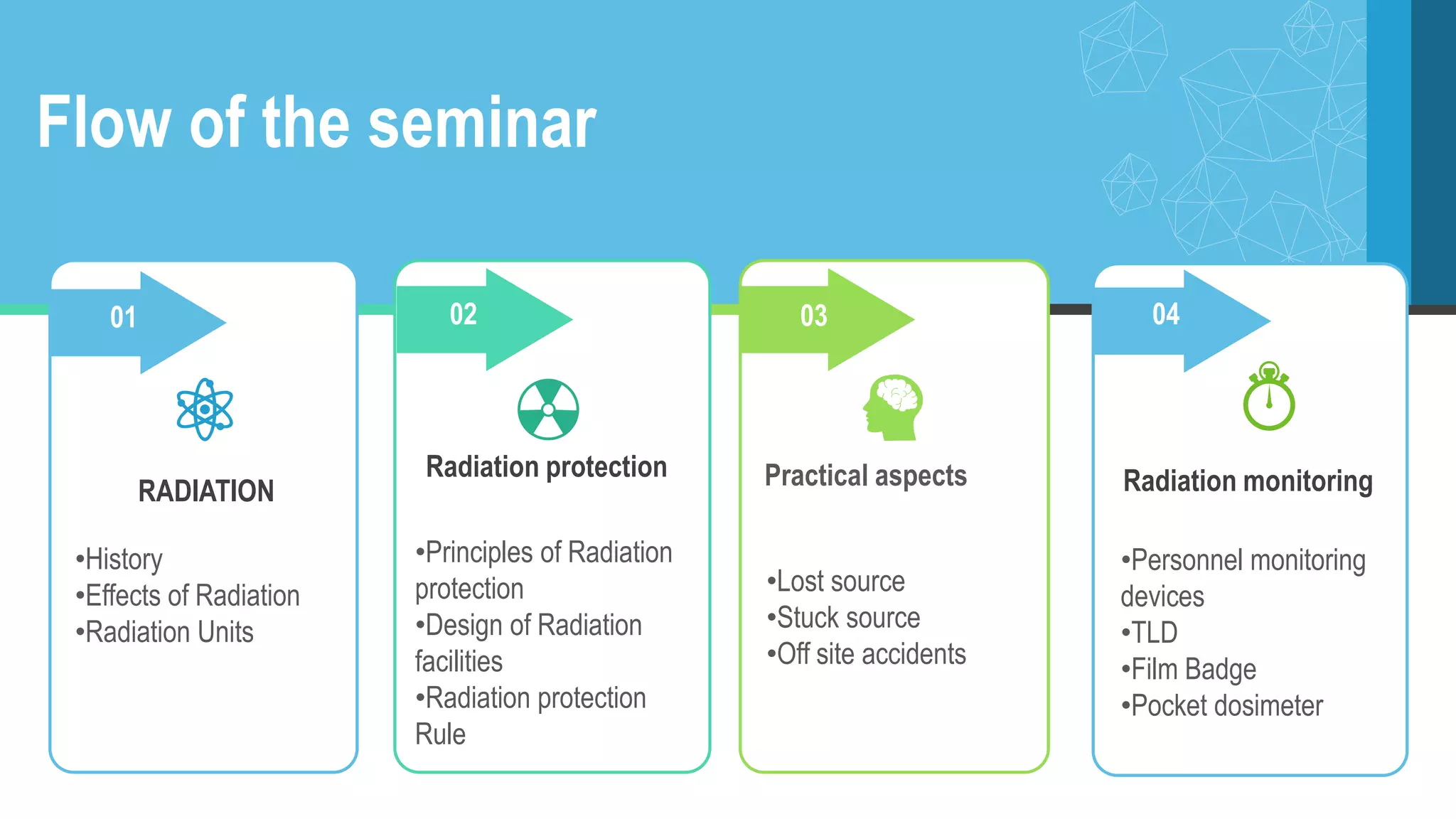
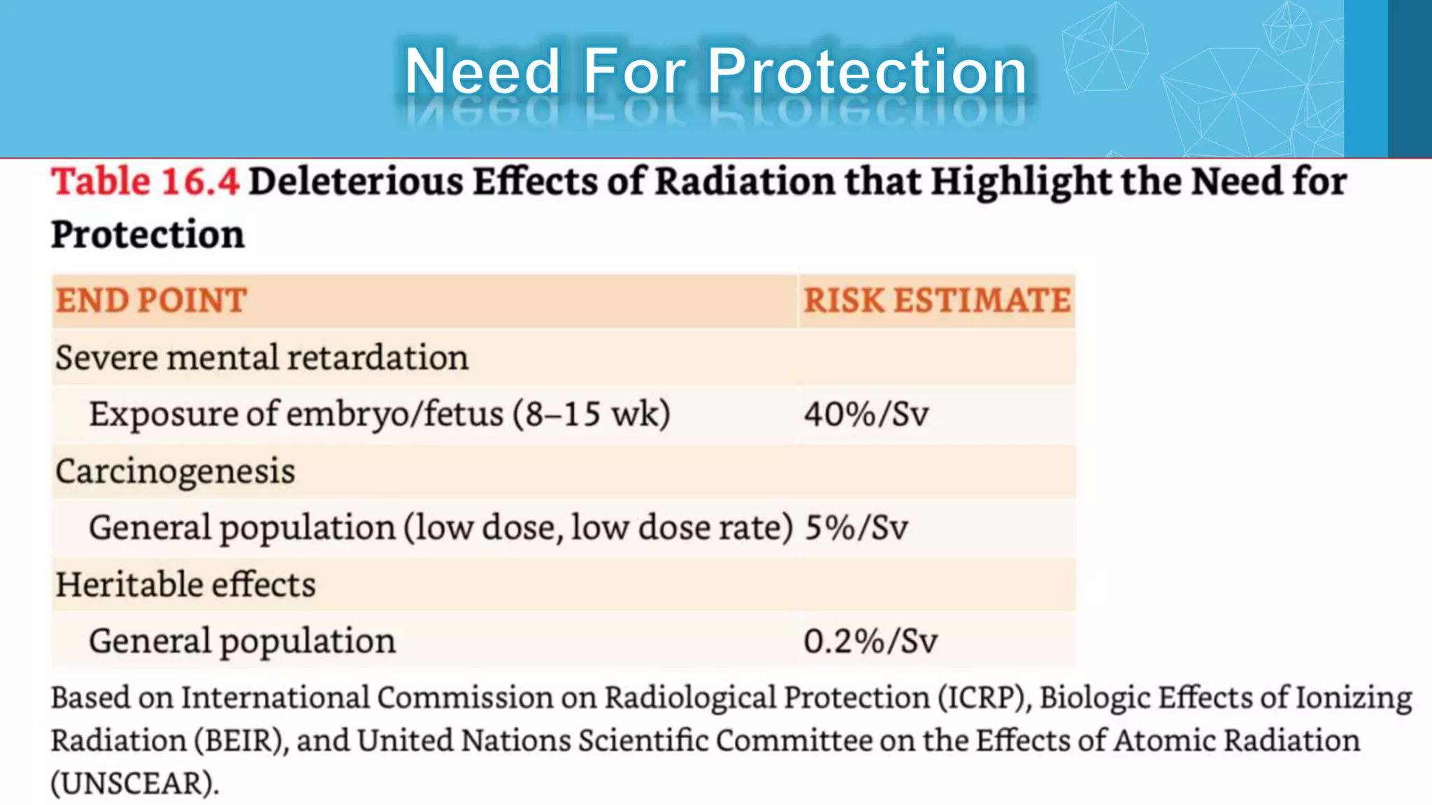
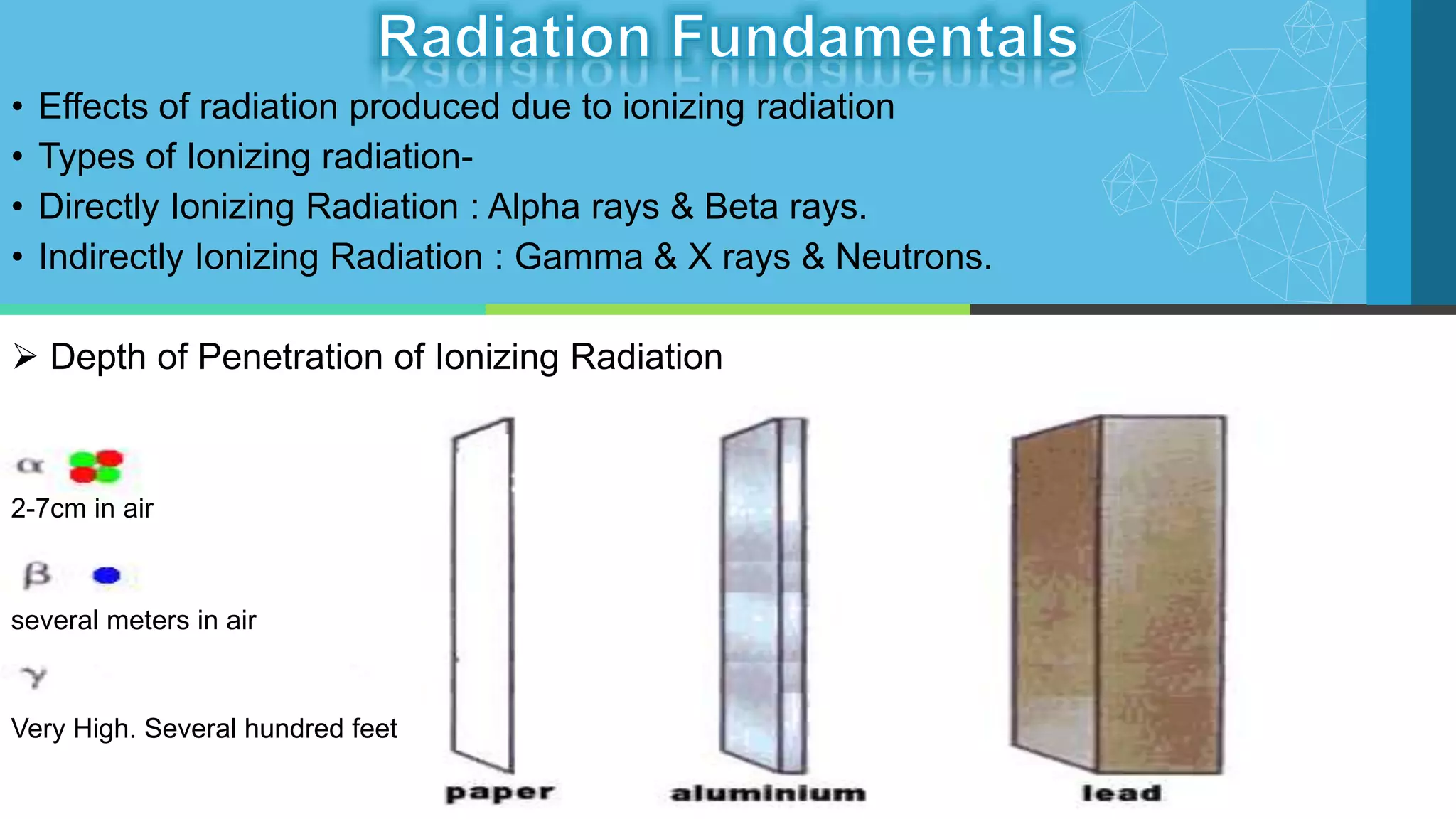
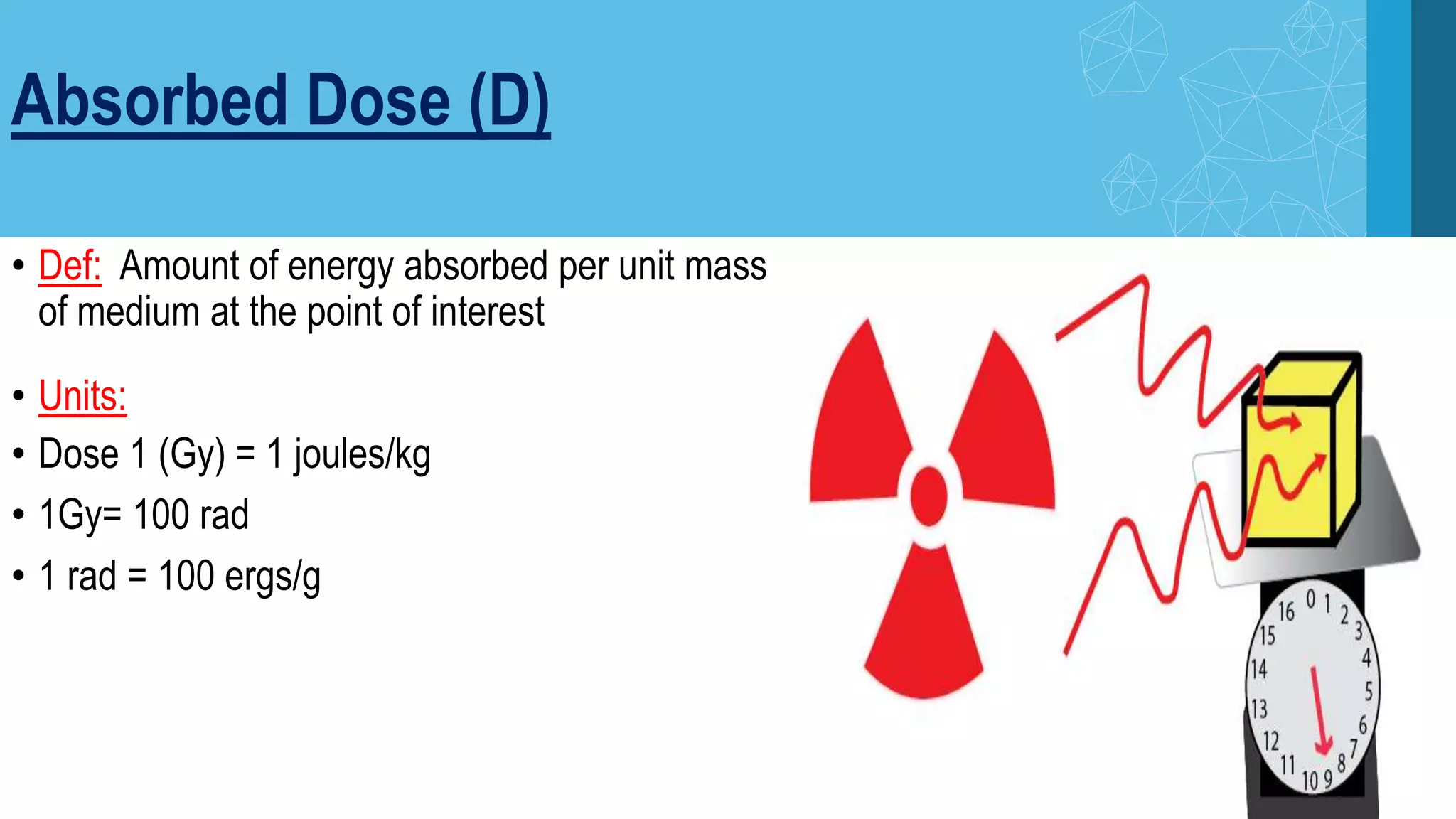
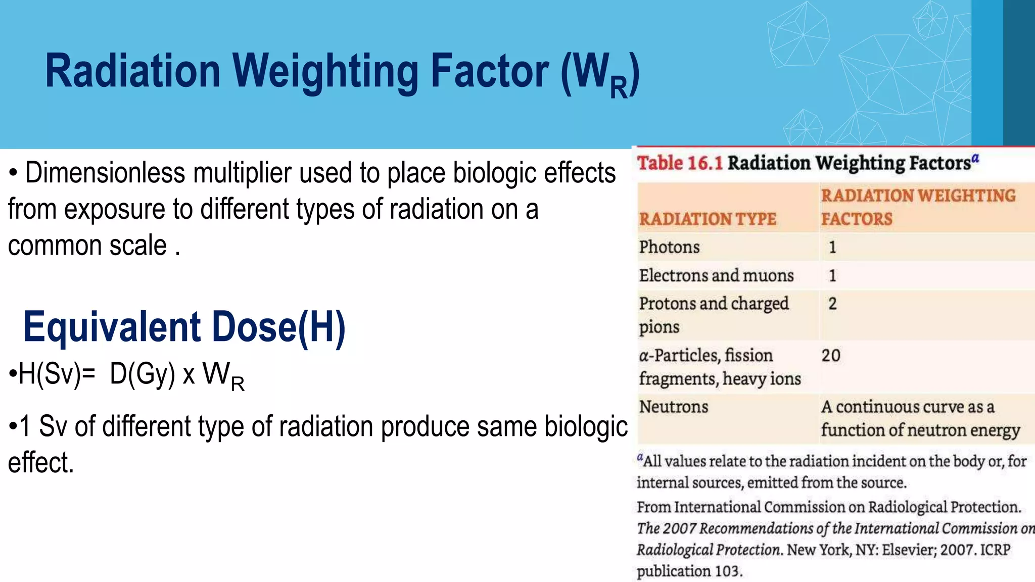
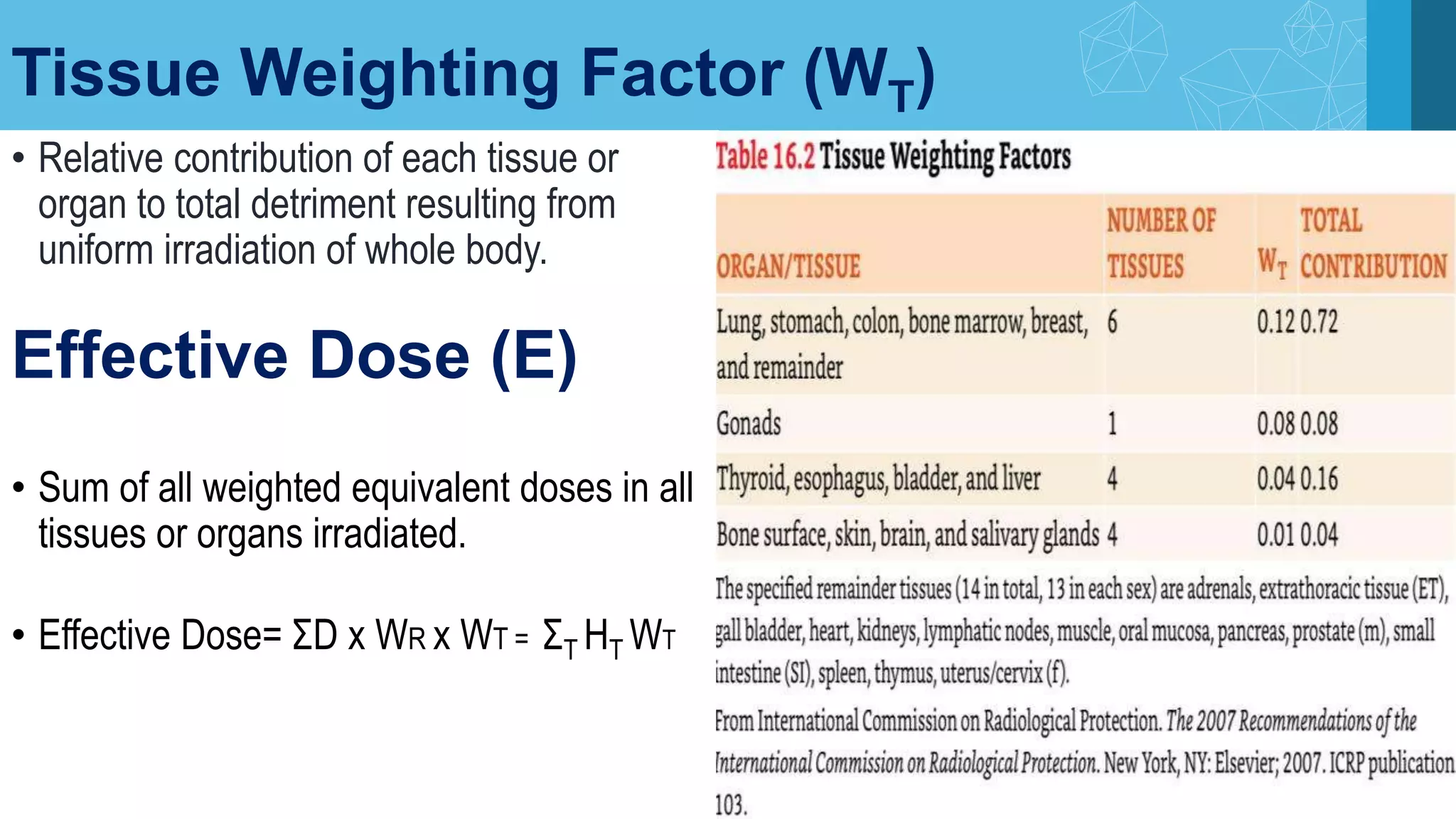
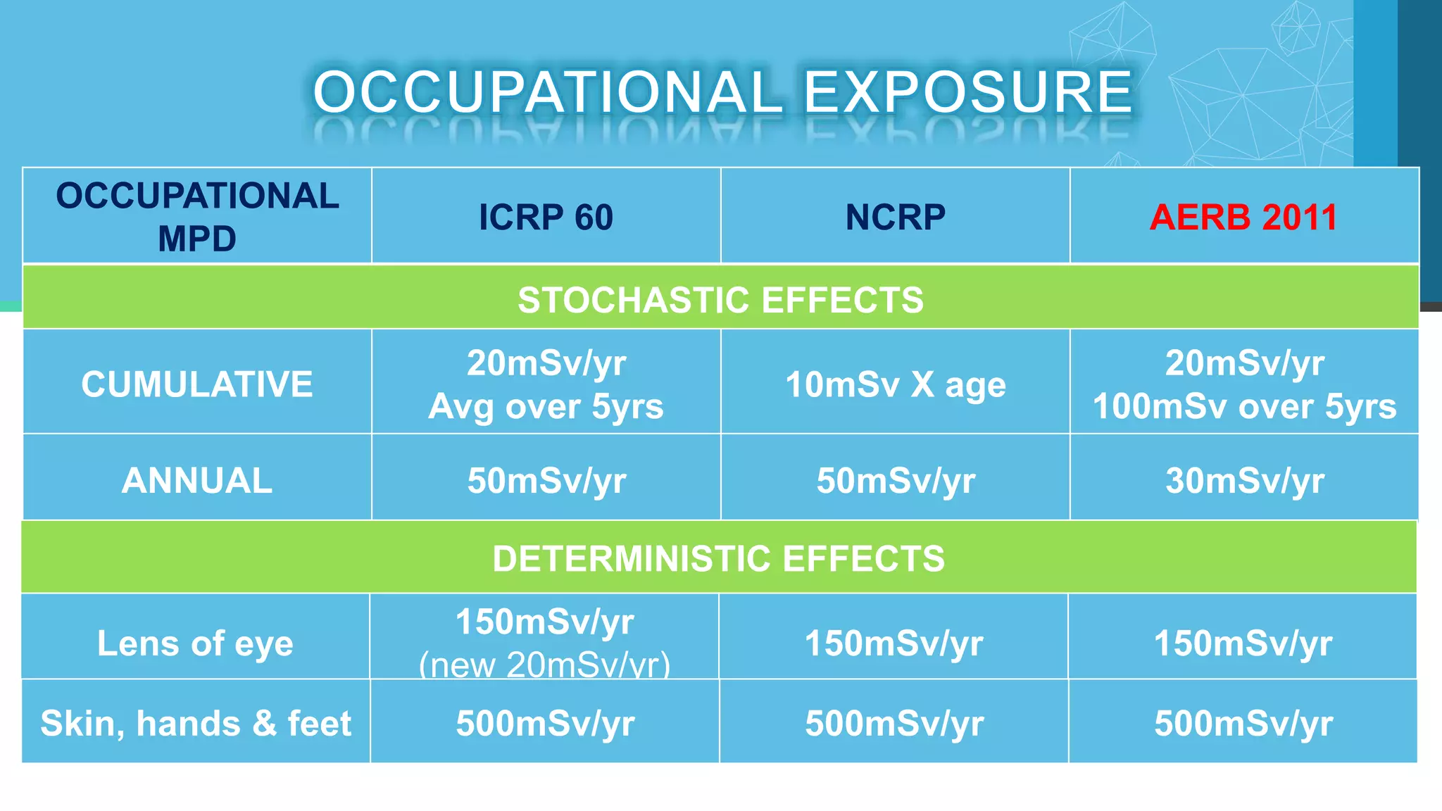
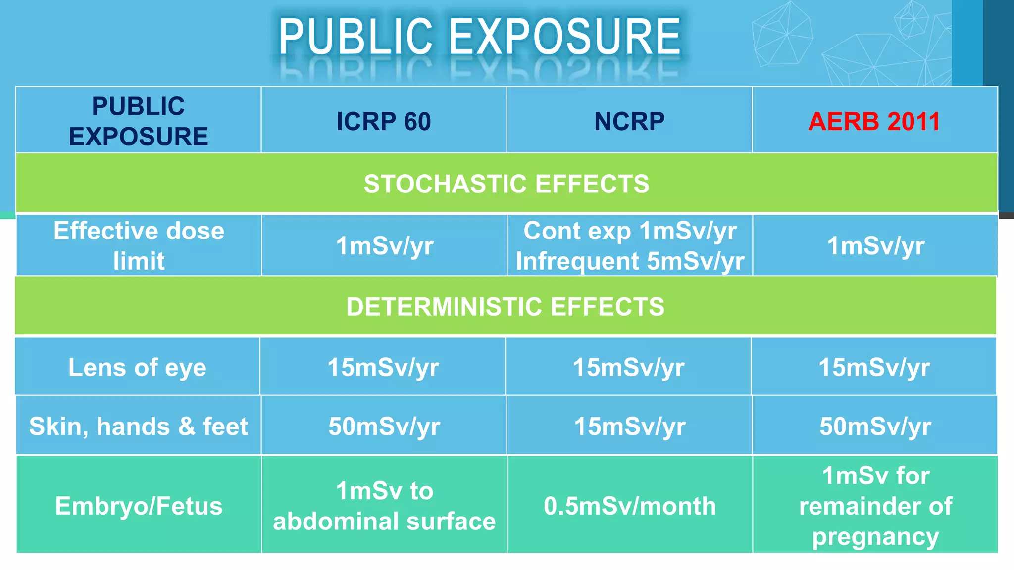
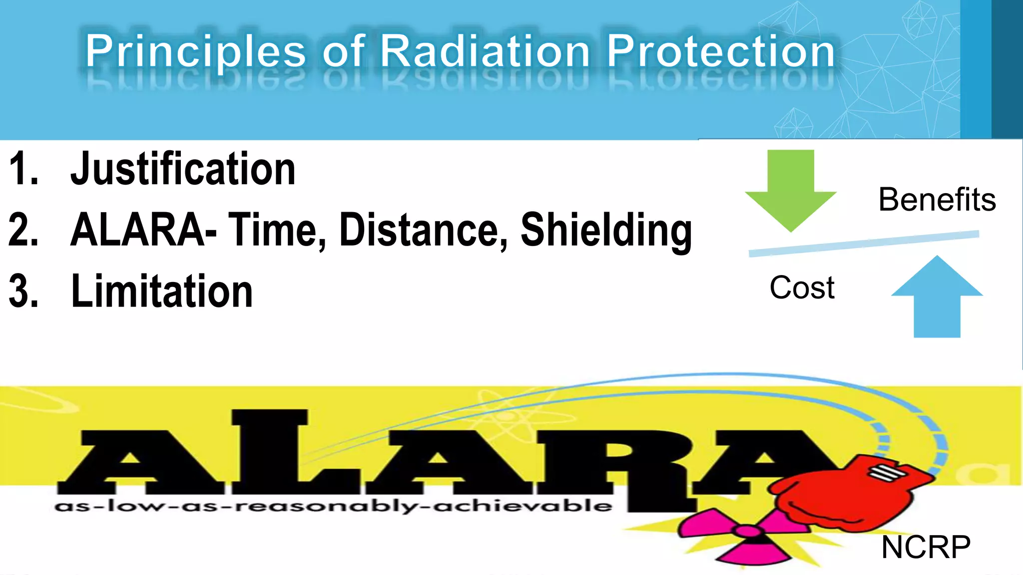
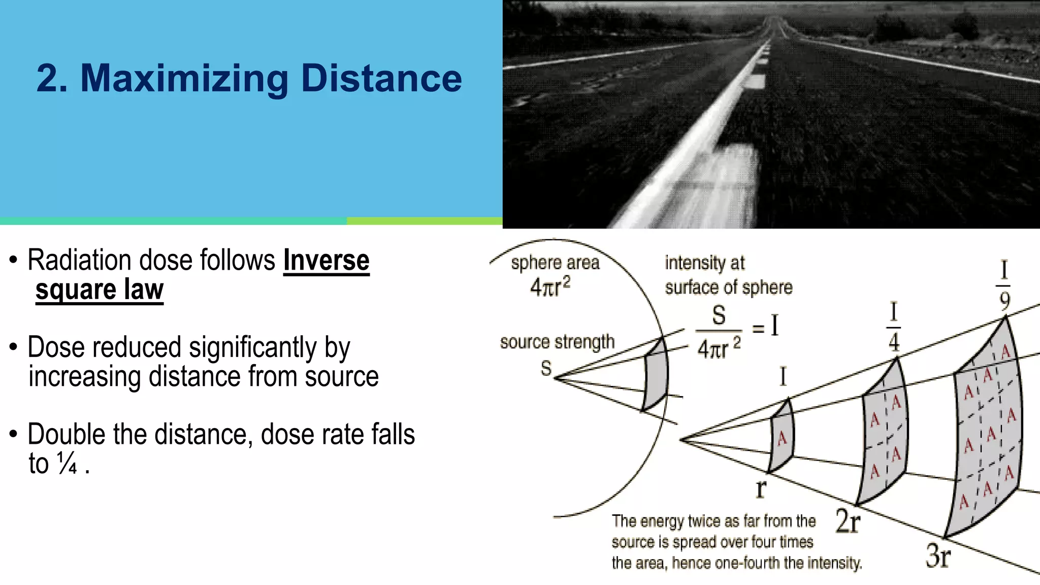
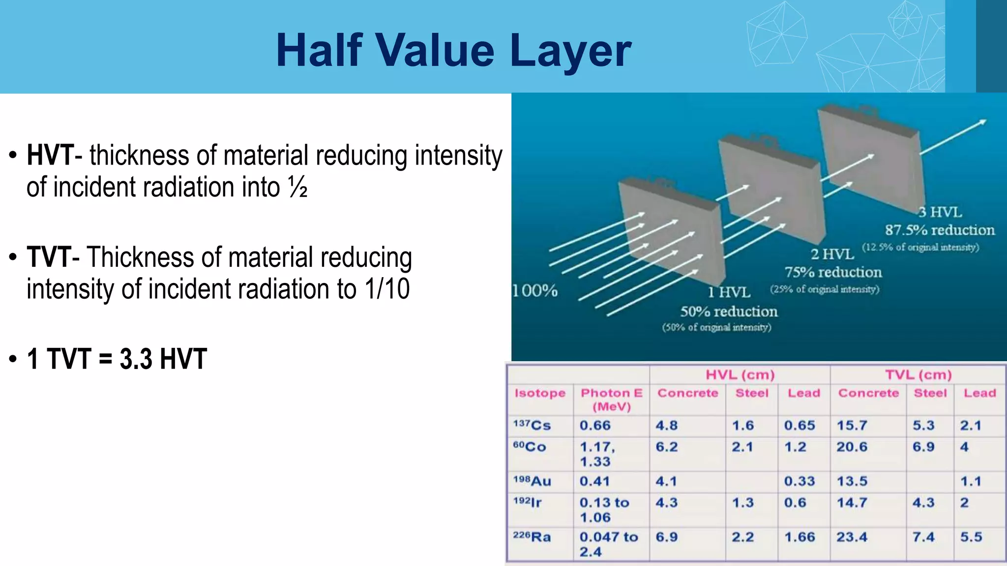
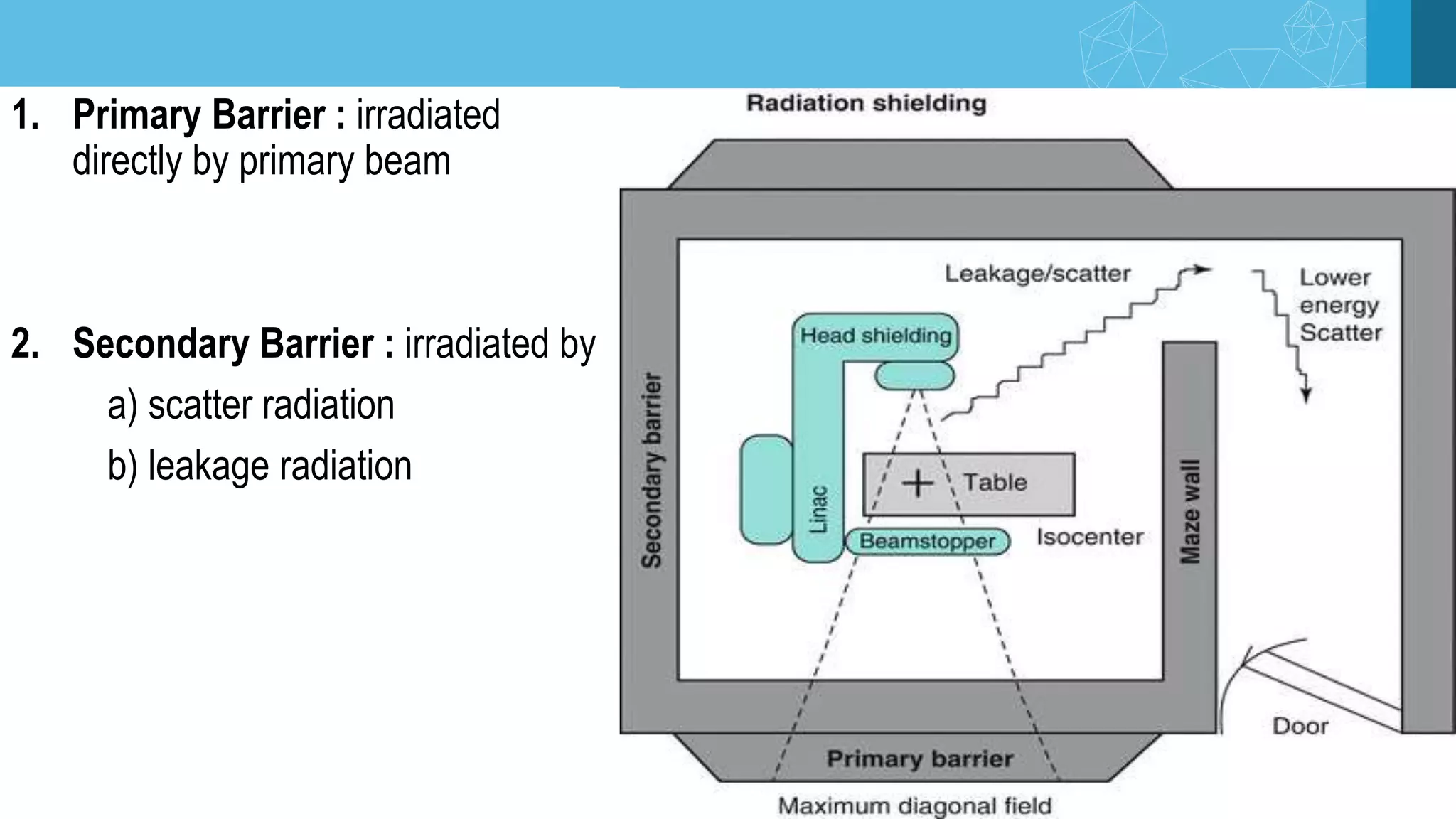
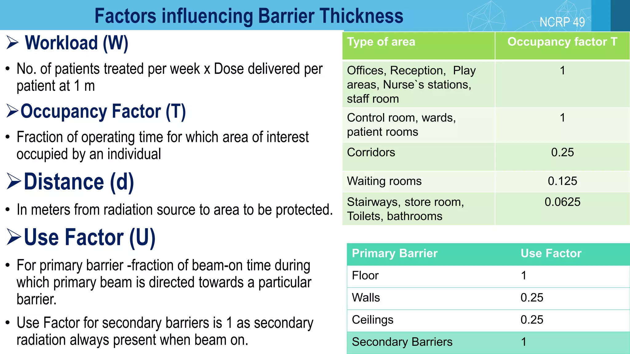
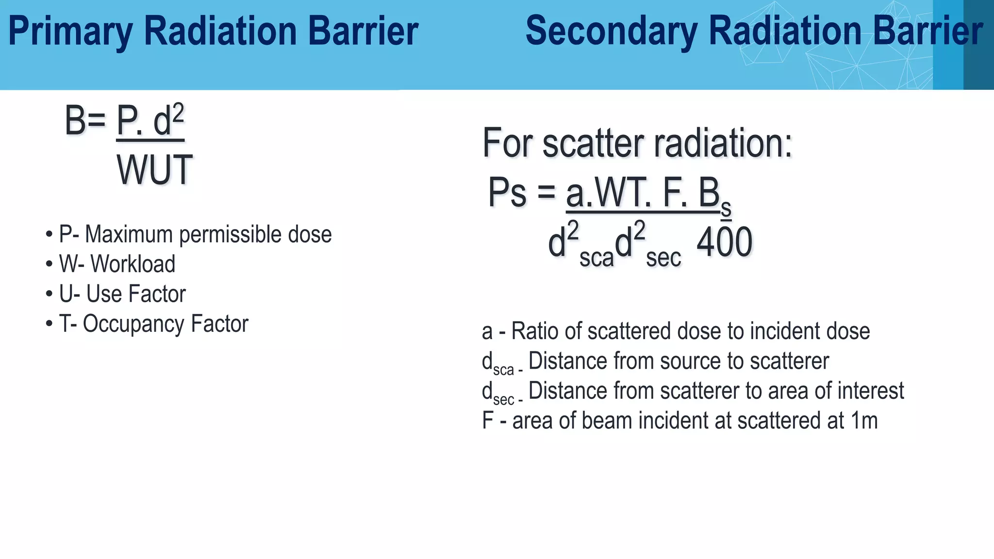
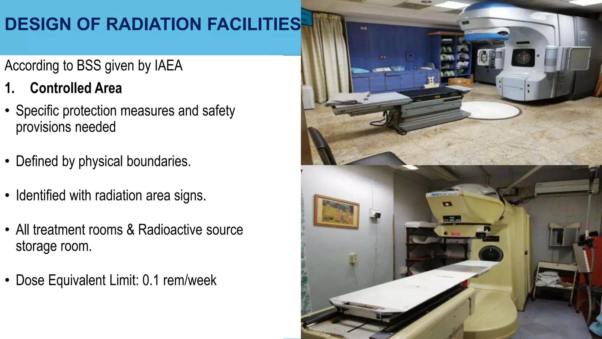
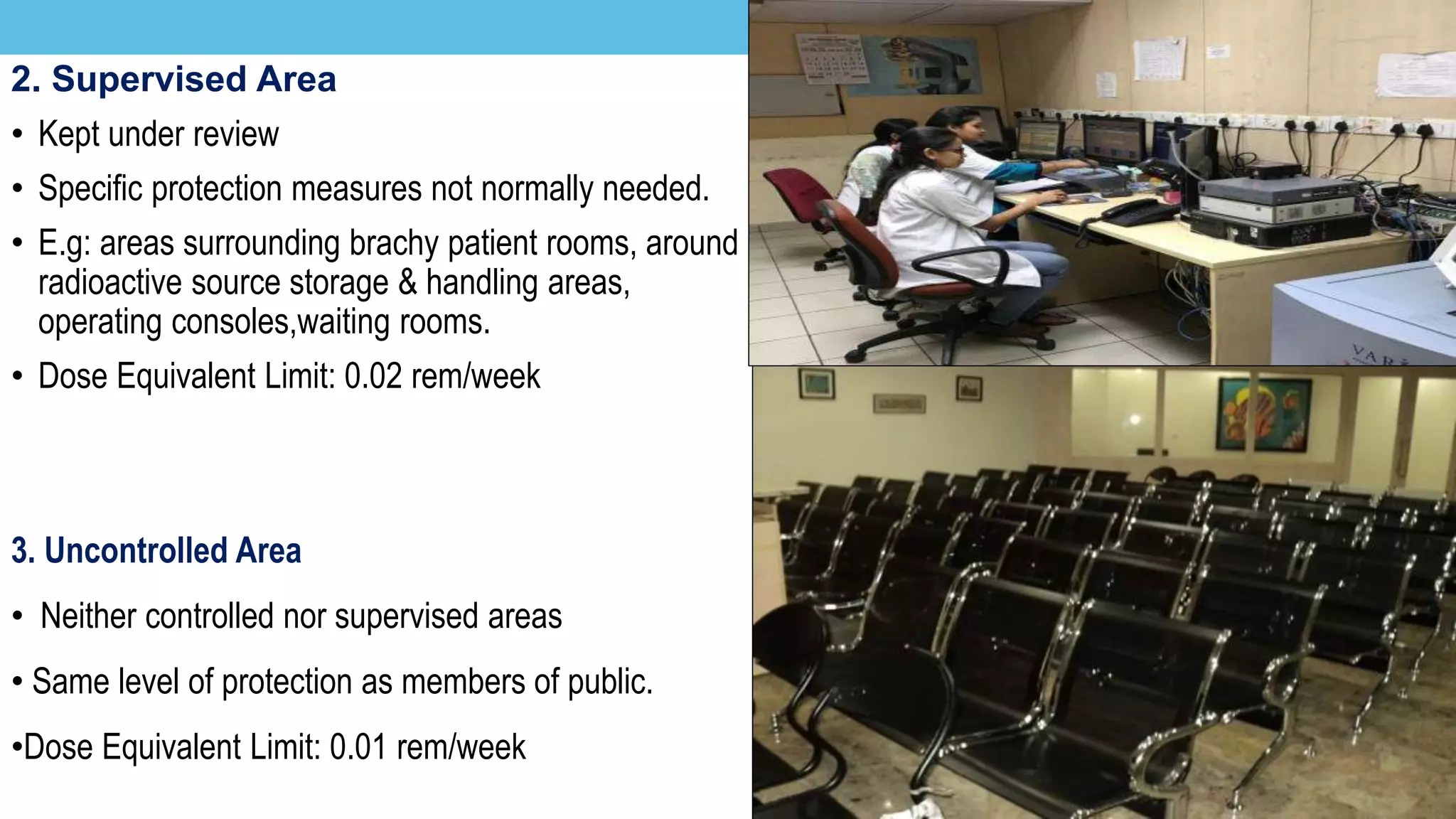
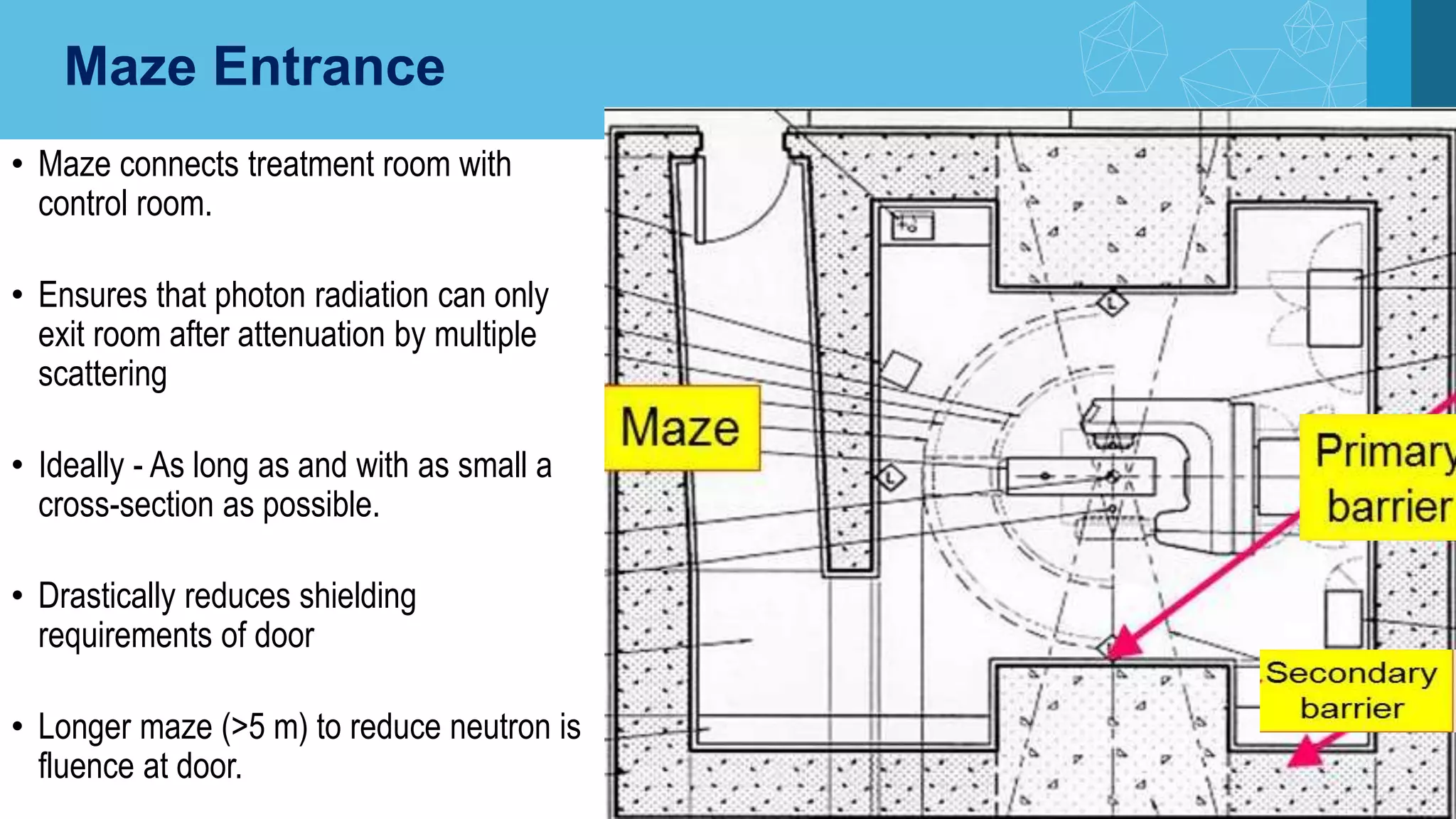
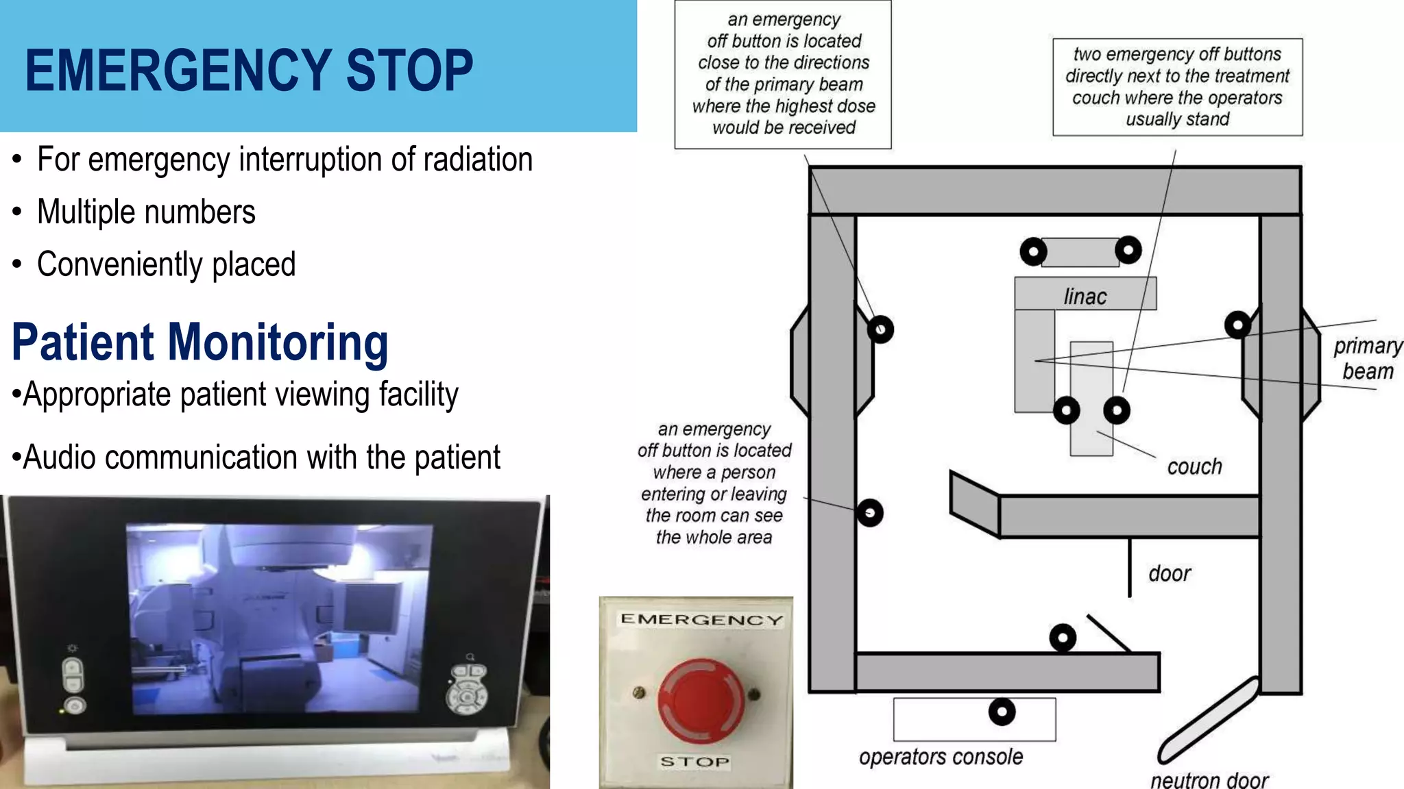
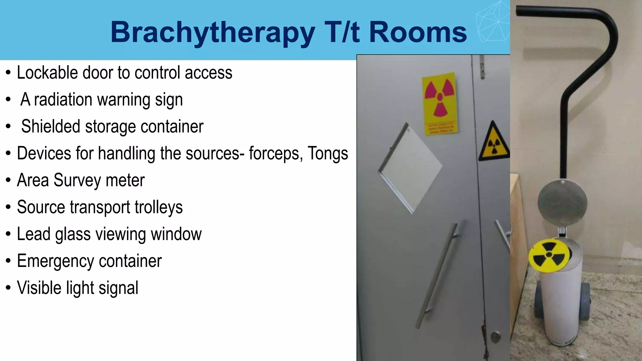
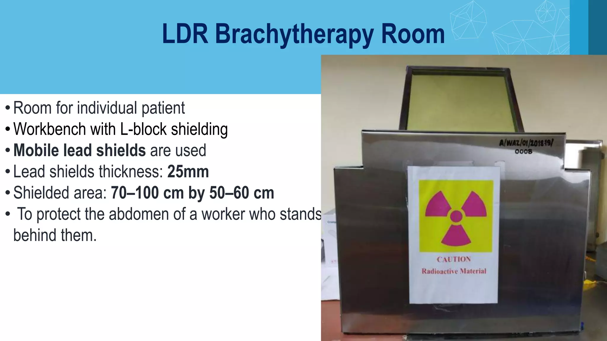
TISSUE PHANTOM RATIO - THE PHOTON BEAM QUALITY INDEXVictor Ekpo
TPR(20,10) is the recommended photon beam quality index by IAEA TRS-398 for megavoltage clinical photons generated by linear accelerators. This presentation goes through the basics of Tissue Phantom Ratio (TPR).
This power-point presentation is very important for radiology resident radiologist and radiographers and technician. this includes principles, technique , biological effects of radiation and how to protect, whats should normal radiation dose with latest update. This slide also includes ALARA PRINCIPLE thanks.
Radiation is energy that is given off by particular materials and devices.
Radiation protection, also known as radiological protection, is defined by the International Atomic Energy Agency (IAEA) as "The protection of people from harmful effects of exposure to ionizing radiation, and the means for achieving this". Exposure can be from a source of radiation external to the human body or due to internal irradiation caused by the ingestion of radioactive contamination
Radioisotopes and dose rates used for brachytherapySubhash Thakur
Radioisotopes and dose rates used for brachytherapy
This is the seminar about different radioisotopes used in brachytherapy beginning from radium to iradium and different dose rates, low dose rate, high dose rate used in brachytherapy. The significance of different dose rates and its radiobiology along with the clinical results.
Radiation protection, also known as radiological protection, is defined by the International Atomic Energy Agency (IAEA) as "The protection of people from harmful effects of exposure to ionizing radiation, and the means for achieving this". Exposure can be from a source of radiation external to the human body or due to internal irradiation caused by the ingestion of radioactive contamination.
Ionizing radiation is widely used in industry and medicine, and can present a significant health hazard by causing microscopic damage to living tissue. There are two main categories of ionizing radiation health effects. At high exposures, it can cause "tissue" effects, also called "deterministic" effects due to the certainty of them happening, conventionally indicated by the unit gray and resulting in acute radiation syndrome. For low level exposures there can be statistically elevated risks of radiation-induced cancer, called "stochastic effects" due to the uncertainty of them happening, conventionally indicated by the unit sievert.
Fundamental to radiation protection is the avoidance or reduction of dose using the simple protective measures of time, distance and shielding. The duration of exposure should be limited to that necessary, the distance from the source of radiation should be maxi mised, and the source shielded wherever possible. To measure personal dose uptake in occupational or emergency exposure, for external radiation personal dosimeters are used, and for internal dose to due to ingestion of radioactive contamination, bioassay techniques are applied.
Radioation protection.. radiology information by r midha.Rahul Midha
this ppt contains radition safety data and radition protection.
doses of radition and monitoring tools for radition data , personal protection with patient protection . radiology information by r midha.
Basic Radiation Safety Awareness Training
History of Radiation
Natural and Man-Made Background Sources of Radiation
Fundamentals
Exposure Limits & Regulations
Detection of Radiation
Safe Practices with Radiation
Biological Effects of Radiation
Where to Find Further Information
radiation control safety, role of Organization in radiation protection and environmental radiological surveillance.
Factors that affect radiation dose:
Regulations and procedures have been developed and implemented to limit radiation dose by regulating the use, storage, transport, and disposal of radioactive material by controlling time, distance and shielding
Time
The short the time spent near the source, the smaller the dose
Distance
The greater the distance the smaller the dose
Shielding
Use of materials to absorb the radiation dose
Recomendações da OMS sobre cuidados maternos e neonatais para uma experiência pós-natal positiva.
Em consonância com os ODS – Objetivos do Desenvolvimento Sustentável e a Estratégia Global para a Saúde das Mulheres, Crianças e Adolescentes, e aplicando uma abordagem baseada nos direitos humanos, os esforços de cuidados pós-natais devem expandir-se para além da cobertura e da simples sobrevivência, de modo a incluir cuidados de qualidade.
Estas diretrizes visam melhorar a qualidade dos cuidados pós-natais essenciais e de rotina prestados às mulheres e aos recém-nascidos, com o objetivo final de melhorar a saúde e o bem-estar materno e neonatal.
Uma “experiência pós-natal positiva” é um resultado importante para todas as mulheres que dão à luz e para os seus recém-nascidos, estabelecendo as bases para a melhoria da saúde e do bem-estar a curto e longo prazo. Uma experiência pós-natal positiva é definida como aquela em que as mulheres, pessoas que gestam, os recém-nascidos, os casais, os pais, os cuidadores e as famílias recebem informação consistente, garantia e apoio de profissionais de saúde motivados; e onde um sistema de saúde flexível e com recursos reconheça as necessidades das mulheres e dos bebês e respeite o seu contexto cultural.
Estas diretrizes consolidadas apresentam algumas recomendações novas e já bem fundamentadas sobre cuidados pós-natais de rotina para mulheres e neonatos que recebem cuidados no pós-parto em unidades de saúde ou na comunidade, independentemente dos recursos disponíveis.
É fornecido um conjunto abrangente de recomendações para cuidados durante o período puerperal, com ênfase nos cuidados essenciais que todas as mulheres e recém-nascidos devem receber, e com a devida atenção à qualidade dos cuidados; isto é, a entrega e a experiência do cuidado recebido. Estas diretrizes atualizam e ampliam as recomendações da OMS de 2014 sobre cuidados pós-natais da mãe e do recém-nascido e complementam as atuais diretrizes da OMS sobre a gestão de complicações pós-natais.
O estabelecimento da amamentação e o manejo das principais intercorrências é contemplada.
Recomendamos muito.
Vamos discutir essas recomendações no nosso curso de pós-graduação em Aleitamento no Instituto Ciclos.
Esta publicação só está disponível em inglês até o momento.
Prof. Marcus Renato de Carvalho
www.agostodourado.com
Prix Galien International 2024 Forum ProgramLevi Shapiro
June 20, 2024, Prix Galien International and Jerusalem Ethics Forum in ROME. Detailed agenda including panels:
- ADVANCES IN CARDIOLOGY: A NEW PARADIGM IS COMING
- WOMEN’S HEALTH: FERTILITY PRESERVATION
- WHAT’S NEW IN THE TREATMENT OF INFECTIOUS,
ONCOLOGICAL AND INFLAMMATORY SKIN DISEASES?
- ARTIFICIAL INTELLIGENCE AND ETHICS
- GENE THERAPY
- BEYOND BORDERS: GLOBAL INITIATIVES FOR DEMOCRATIZING LIFE SCIENCE TECHNOLOGIES AND PROMOTING ACCESS TO HEALTHCARE
- ETHICAL CHALLENGES IN LIFE SCIENCES
- Prix Galien International Awards Ceremony
Couples presenting to the infertility clinic- Do they really have infertility...Sujoy Dasgupta
Dr Sujoy Dasgupta presented the study on "Couples presenting to the infertility clinic- Do they really have infertility? – The unexplored stories of non-consummation" in the 13th Congress of the Asia Pacific Initiative on Reproduction (ASPIRE 2024) at Manila on 24 May, 2024.
These simplified slides by Dr. Sidra Arshad present an overview of the non-respiratory functions of the respiratory tract.
Learning objectives:
1. Enlist the non-respiratory functions of the respiratory tract
2. Briefly explain how these functions are carried out
3. Discuss the significance of dead space
4. Differentiate between minute ventilation and alveolar ventilation
5. Describe the cough and sneeze reflexes
Study Resources:
1. Chapter 39, Guyton and Hall Textbook of Medical Physiology, 14th edition
2. Chapter 34, Ganong’s Review of Medical Physiology, 26th edition
3. Chapter 17, Human Physiology by Lauralee Sherwood, 9th edition
4. Non-respiratory functions of the lungs https://academic.oup.com/bjaed/article/13/3/98/278874
Pulmonary Thromboembolism - etilogy, types, medical- Surgical and nursing man...VarunMahajani
Disruption of blood supply to lung alveoli due to blockage of one or more pulmonary blood vessels is called as Pulmonary thromboembolism. In this presentation we will discuss its causes, types and its management in depth.
HOT NEW PRODUCT! BIG SALES FAST SHIPPING NOW FROM CHINA!! EU KU DB BK substit...GL Anaacs
Contact us if you are interested:
Email / Skype : kefaya1771@gmail.com
Threema: PXHY5PDH
New BATCH Ku !!! MUCH IN DEMAND FAST SALE EVERY BATCH HAPPY GOOD EFFECT BIG BATCH !
Contact me on Threema or skype to start big business!!
Hot-sale products:
NEW HOT EUTYLONE WHITE CRYSTAL!!
5cl-adba precursor (semi finished )
5cl-adba raw materials
ADBB precursor (semi finished )
ADBB raw materials
APVP powder
5fadb/4f-adb
Jwh018 / Jwh210
Eutylone crystal
Protonitazene (hydrochloride) CAS: 119276-01-6
Flubrotizolam CAS: 57801-95-3
Metonitazene CAS: 14680-51-4
Payment terms: Western Union,MoneyGram,Bitcoin or USDT.
Deliver Time: Usually 7-15days
Shipping method: FedEx, TNT, DHL,UPS etc.Our deliveries are 100% safe, fast, reliable and discreet.
Samples will be sent for your evaluation!If you are interested in, please contact me, let's talk details.
We specializes in exporting high quality Research chemical, medical intermediate, Pharmaceutical chemicals and so on. Products are exported to USA, Canada, France, Korea, Japan,Russia, Southeast Asia and other countries.
micro teaching on communication m.sc nursing.pdfAnurag Sharma
Microteaching is a unique model of practice teaching. It is a viable instrument for the. desired change in the teaching behavior or the behavior potential which, in specified types of real. classroom situations, tends to facilitate the achievement of specified types of objectives.
Title: Sense of Taste
Presenter: Dr. Faiza, Assistant Professor of Physiology
Qualifications:
MBBS (Best Graduate, AIMC Lahore)
FCPS Physiology
ICMT, CHPE, DHPE (STMU)
MPH (GC University, Faisalabad)
MBA (Virtual University of Pakistan)
Learning Objectives:
Describe the structure and function of taste buds.
Describe the relationship between the taste threshold and taste index of common substances.
Explain the chemical basis and signal transduction of taste perception for each type of primary taste sensation.
Recognize different abnormalities of taste perception and their causes.
Key Topics:
Significance of Taste Sensation:
Differentiation between pleasant and harmful food
Influence on behavior
Selection of food based on metabolic needs
Receptors of Taste:
Taste buds on the tongue
Influence of sense of smell, texture of food, and pain stimulation (e.g., by pepper)
Primary and Secondary Taste Sensations:
Primary taste sensations: Sweet, Sour, Salty, Bitter, Umami
Chemical basis and signal transduction mechanisms for each taste
Taste Threshold and Index:
Taste threshold values for Sweet (sucrose), Salty (NaCl), Sour (HCl), and Bitter (Quinine)
Taste index relationship: Inversely proportional to taste threshold
Taste Blindness:
Inability to taste certain substances, particularly thiourea compounds
Example: Phenylthiocarbamide
Structure and Function of Taste Buds:
Composition: Epithelial cells, Sustentacular/Supporting cells, Taste cells, Basal cells
Features: Taste pores, Taste hairs/microvilli, and Taste nerve fibers
Location of Taste Buds:
Found in papillae of the tongue (Fungiform, Circumvallate, Foliate)
Also present on the palate, tonsillar pillars, epiglottis, and proximal esophagus
Mechanism of Taste Stimulation:
Interaction of taste substances with receptors on microvilli
Signal transduction pathways for Umami, Sweet, Bitter, Sour, and Salty tastes
Taste Sensitivity and Adaptation:
Decrease in sensitivity with age
Rapid adaptation of taste sensation
Role of Saliva in Taste:
Dissolution of tastants to reach receptors
Washing away the stimulus
Taste Preferences and Aversions:
Mechanisms behind taste preference and aversion
Influence of receptors and neural pathways
Impact of Sensory Nerve Damage:
Degeneration of taste buds if the sensory nerve fiber is cut
Abnormalities of Taste Detection:
Conditions: Ageusia, Hypogeusia, Dysgeusia (parageusia)
Causes: Nerve damage, neurological disorders, infections, poor oral hygiene, adverse drug effects, deficiencies, aging, tobacco use, altered neurotransmitter levels
Neurotransmitters and Taste Threshold:
Effects of serotonin (5-HT) and norepinephrine (NE) on taste sensitivity
Supertasters:
25% of the population with heightened sensitivity to taste, especially bitterness
Increased number of fungiform papillae
New Directions in Targeted Therapeutic Approaches for Older Adults With Mantl...i3 Health
i3 Health is pleased to make the speaker slides from this activity available for use as a non-accredited self-study or teaching resource.
This slide deck presented by Dr. Kami Maddocks, Professor-Clinical in the Division of Hematology and
Associate Division Director for Ambulatory Operations
The Ohio State University Comprehensive Cancer Center, will provide insight into new directions in targeted therapeutic approaches for older adults with mantle cell lymphoma.
STATEMENT OF NEED
Mantle cell lymphoma (MCL) is a rare, aggressive B-cell non-Hodgkin lymphoma (NHL) accounting for 5% to 7% of all lymphomas. Its prognosis ranges from indolent disease that does not require treatment for years to very aggressive disease, which is associated with poor survival (Silkenstedt et al, 2021). Typically, MCL is diagnosed at advanced stage and in older patients who cannot tolerate intensive therapy (NCCN, 2022). Although recent advances have slightly increased remission rates, recurrence and relapse remain very common, leading to a median overall survival between 3 and 6 years (LLS, 2021). Though there are several effective options, progress is still needed towards establishing an accepted frontline approach for MCL (Castellino et al, 2022). Treatment selection and management of MCL are complicated by the heterogeneity of prognosis, advanced age and comorbidities of patients, and lack of an established standard approach for treatment, making it vital that clinicians be familiar with the latest research and advances in this area. In this activity chaired by Michael Wang, MD, Professor in the Department of Lymphoma & Myeloma at MD Anderson Cancer Center, expert faculty will discuss prognostic factors informing treatment, the promising results of recent trials in new therapeutic approaches, and the implications of treatment resistance in therapeutic selection for MCL.
Target Audience
Hematology/oncology fellows, attending faculty, and other health care professionals involved in the treatment of patients with mantle cell lymphoma (MCL).
Learning Objectives
1.) Identify clinical and biological prognostic factors that can guide treatment decision making for older adults with MCL
2.) Evaluate emerging data on targeted therapeutic approaches for treatment-naive and relapsed/refractory MCL and their applicability to older adults
3.) Assess mechanisms of resistance to targeted therapies for MCL and their implications for treatment selection
Tom Selleck Health: A Comprehensive Look at the Iconic Actor’s Wellness Journeygreendigital
Tom Selleck, an enduring figure in Hollywood. has captivated audiences for decades with his rugged charm, iconic moustache. and memorable roles in television and film. From his breakout role as Thomas Magnum in Magnum P.I. to his current portrayal of Frank Reagan in Blue Bloods. Selleck's career has spanned over 50 years. But beyond his professional achievements. fans have often been curious about Tom Selleck Health. especially as he has aged in the public eye.
Follow us on: Pinterest
Introduction
Many have been interested in Tom Selleck health. not only because of his enduring presence on screen but also because of the challenges. and lifestyle choices he has faced and made over the years. This article delves into the various aspects of Tom Selleck health. exploring his fitness regimen, diet, mental health. and the challenges he has encountered as he ages. We'll look at how he maintains his well-being. the health issues he has faced, and his approach to ageing .
Early Life and Career
Childhood and Athletic Beginnings
Tom Selleck was born on January 29, 1945, in Detroit, Michigan, and grew up in Sherman Oaks, California. From an early age, he was involved in sports, particularly basketball. which played a significant role in his physical development. His athletic pursuits continued into college. where he attended the University of Southern California (USC) on a basketball scholarship. This early involvement in sports laid a strong foundation for his physical health and disciplined lifestyle.
Transition to Acting
Selleck's transition from an athlete to an actor came with its physical demands. His first significant role in "Magnum P.I." required him to perform various stunts and maintain a fit appearance. This role, which he played from 1980 to 1988. necessitated a rigorous fitness routine to meet the show's demands. setting the stage for his long-term commitment to health and wellness.
Fitness Regimen
Workout Routine
Tom Selleck health and fitness regimen has evolved. adapting to his changing roles and age. During his "Magnum, P.I." days. Selleck's workouts were intense and focused on building and maintaining muscle mass. His routine included weightlifting, cardiovascular exercises. and specific training for the stunts he performed on the show.
Selleck adjusted his fitness routine as he aged to suit his body's needs. Today, his workouts focus on maintaining flexibility, strength, and cardiovascular health. He incorporates low-impact exercises such as swimming, walking, and light weightlifting. This balanced approach helps him stay fit without putting undue strain on his joints and muscles.
Importance of Flexibility and Mobility
In recent years, Selleck has emphasized the importance of flexibility and mobility in his fitness regimen. Understanding the natural decline in muscle mass and joint flexibility with age. he includes stretching and yoga in his routine. These practices help prevent injuries, improve posture, and maintain mobilit
Ozempic: Preoperative Management of Patients on GLP-1 Receptor Agonists Saeid Safari
Preoperative Management of Patients on GLP-1 Receptor Agonists like Ozempic and Semiglutide
ASA GUIDELINE
NYSORA Guideline
2 Case Reports of Gastric Ultrasound
2. Flow of the seminar
•History
•Effects of Radiation
•Radiation Units
01
Radiation protection
•Principles of Radiation
protection
•Design of Radiation
facilities
•Radiation protection
Rule
02
Practical aspects
•Lost source
•Stuck source
•Off site accidents
03
Radiation monitoring
•Personnel monitoring
devices
•TLD
•Film Badge
•Pocket dosimeter
04
RADIATION
3. Thompson reported
deliberate exposure of
finger to the x-ray and
then cautioned against
overexposure.
Thomas Edison gave upon X-
rays, fearing they were too
dangerous after death of his
assistant.
Mihran Kassabian is an american
radiologist who documented his radiation
burns & published a paper on irritant
effect of X-rays.
History
5. • The ICRP Non governmental organization that sets
acceptable limits of exposure
• Members - scientists, sociologists and experts in
various fields
• The IAEA is an autonomous inter-governmental
organization under the aegis of the United Nations.
• The NCRP provides advice & recommendations on
matters pertaining to radiation protection in US.
• The AERB constituted on November 15, 1983 by the
President of India.
• Ensure use of ionizing radiation & nuclear energy in
India does not cause any risk to health of people &
environment
Regulatory bodies
6. Stochastic EffectsDeterministic Effects
1. All or nothing effect
2. Probabilistic or random in
nature
3. No threshold level
4. Probability of effects is
proportional to the dose
5. Caused by modification of
cell
6. E.g. Carcinogenesis and
hereditary effects
1. Renamed as Tissue reaction.
2. Deterministic in nature
3. Have a threshold level of dose
4. Severity of effect increases with
dose above threshold
5. Caused by cell death or delayed
cell division
6. E.g. Organ failure, Radiation
induced dermatitis, cataract
Effects of Radiation Exposure
7.
8.
9. • Effects of radiation produced due to ionizing radiation
• Types of Ionizing radiation-
• Directly Ionizing Radiation : Alpha rays & Beta rays.
• Indirectly Ionizing Radiation : Gamma & X rays & Neutrons.
Depth of Penetration of Ionizing Radiation
2-7cm in air
several meters in air
Very High. Several hundred feet
11. Absorbed Dose (D)
• Def: Amount of energy absorbed per unit mass
of medium at the point of interest
• Units:
• Dose 1 (Gy) = 1 joules/kg
• 1Gy= 100 rad
• 1 rad = 100 ergs/g
12. Radiation Weighting Factor (WR)
• Dimensionless multiplier used to place biologic effects
from exposure to different types of radiation on a
common scale .
Equivalent Dose(H)
•H(Sv)= D(Gy) x WR
•1 Sv of different type of radiation produce same biologic
effect.
13. • Relative contribution of each tissue or
organ to total detriment resulting from
uniform irradiation of whole body.
Tissue Weighting Factor (WT)
Effective Dose (E)
• Sum of all weighted equivalent doses in all
tissues or organs irradiated.
• Effective Dose= ΣD x WR x WT = ΣT HT WT
14. Committed Dose
• Committed dose: Total dose delivered throughout period of time for which radionuclide
inside body.
• Committed equivalent dose: Equivalent dose integrated over 50 years.
• Committed effective dose : Effective dose integrated over 50 years.
• Unit: Sievert
15. Collective Dose
• Collective equivalent dose : Avg equivalent dose to a population X n Persons exposed
• Collective effective dose: Avg effective dose to a population X n Persons exposed
• Unit - person- sievert .
• For population ingesting or inhaling radionuclides depositing dose over prolonged period,
integral of effective dose over entire population to a period of 50 years = Collective
committed effective dose.
16. OCCUPATIONAL
MPD
ICRP 60 NCRP AERB 2011
STOCHASTIC EFFECTS
CUMULATIVE
20mSv/yr
Avg over 5yrs
10mSv X age
20mSv/yr
100mSv over 5yrs
ANNUAL 50mSv/yr 50mSv/yr 30mSv/yr
DETERMINISTIC EFFECTS
Lens of eye
150mSv/yr
(new 20mSv/yr)
150mSv/yr 150mSv/yr
Skin, hands & feet 500mSv/yr 500mSv/yr 500mSv/yr
17. PUBLIC
EXPOSURE
ICRP 60 NCRP AERB 2011
Effective dose
limit
1mSv/yr
Cont exp 1mSv/yr
Infrequent 5mSv/yr
1mSv/yr
Lens of eye 15mSv/yr 15mSv/yr 15mSv/yr
Skin, hands & feet 50mSv/yr 15mSv/yr 50mSv/yr
STOCHASTIC EFFECTS
DETERMINISTIC EFFECTS
Embryo/Fetus
1mSv to
abdominal surface
0.5mSv/month
1mSv for
remainder of
pregnancy
18. • DE MINIMIS DOSE
• Dose below which further efforts to reduce radiation exposure to person unwarranted.
• Annual effective dose of 0.01mSv (NCRP)
20. • Radiation Dose received ∝ Time
• To ensure thorough planning before entering radiation
area
• Have all necessary tools present
• Work efficiently and swiftly.
1. Minimizing Time
21. 2. Maximizing Distance
• Radiation dose follows Inverse
square law
• Dose reduced significantly by
increasing distance from source
• Double the distance, dose rate falls
to ¼ .
22. Half Value Layer
• HVT- thickness of material reducing intensity
of incident radiation into ½
• TVT- Thickness of material reducing
intensity of incident radiation to 1/10
• 1 TVT = 3.3 HVT
24. • Parameters involved in shielding design (NCRP 151)
1. Establishing dose value (P) in occupied area
2. Estimating dose (D) in occupied area in absence of any shielding
3. Obtaining attenuation factors necessary to reduce dose value from D to P
3. SHIELDING
25. 1. Primary Barrier : irradiated
directly by primary beam
2. Secondary Barrier : irradiated by
a) scatter radiation
b) leakage radiation
26. Workload (W)
• No. of patients treated per week x Dose delivered per
patient at 1 m
Occupancy Factor (T)
• Fraction of operating time for which area of interest
occupied by an individual
Distance (d)
• In meters from radiation source to area to be protected.
Use Factor (U)
• For primary barrier -fraction of beam-on time during
which primary beam is directed towards a particular
barrier.
• Use Factor for secondary barriers is 1 as secondary
radiation always present when beam on.
Type of area Occupancy factor T
Offices, Reception, Play
areas, Nurse`s stations,
staff room
1
Control room, wards,
patient rooms
1
Corridors 0.25
Waiting rooms 0.125
Stairways, store room,
Toilets, bathrooms
0.0625
NCRP 49Factors influencing Barrier Thickness
Primary Barrier Use Factor
Floor 1
Walls 0.25
Ceilings 0.25
Secondary Barriers 1
27. Primary Radiation Barrier
B= P. d2
WUT
• P- Maximum permissible dose
• W- Workload
• U- Use Factor
• T- Occupancy Factor
For scatter radiation:
Ps = a.WT. F. Bs
d2
scad2
sec 400
a - Ratio of scattered dose to incident dose
dsca - Distance from source to scatterer
dsec - Distance from scatterer to area of interest
F - area of beam incident at scattered at 1m
Secondary Radiation Barrier
28. 6 TVL for Primary barrier
3 TVL for Secondary barrier
Thickness of Secondary Barrier:
• Beam intensity incident on scatterer
• Quality of radiation
• Area of beam on scatterer
• Scattering angle
Neutron shielding for >10MV
Shielding thickness of ordinary concrete (NCRP 151)
Rad Quality Primary Barrier (cm) Secondary Barrier (cm)
Co-60 130 65
10-25 MV 240 120
Materials used for shielding in construction of Radiation
facilities (NCRP 151)
Material Density (g/cm3
)
Ordinary concrete (RCC) 2.35
Heavy concrete (Ledite) Up to 5
Lead 11.3
steel 7.9
Polyethylene 0.95
Earth 1.5
Barite Concrete 3.2
RULE OF THUMB
29. According to BSS given by IAEA
1. Controlled Area
• Specific protection measures and safety
provisions needed
• Defined by physical boundaries.
• Identified with radiation area signs.
• All treatment rooms & Radioactive source
storage room.
• Dose Equivalent Limit: 0.1 rem/week
DESIGN OF RADIATION FACILITIES
30. 2. Supervised Area
• Kept under review
• Specific protection measures not normally needed.
• E.g: areas surrounding brachy patient rooms, around
radioactive source storage & handling areas,
operating consoles,waiting rooms.
• Dose Equivalent Limit: 0.02 rem/week
3. Uncontrolled Area
• Neither controlled nor supervised areas
• Same level of protection as members of public.
•Dose Equivalent Limit: 0.01 rem/week
31.
32. Features of Good Design in EBRT installation:
• Maze Entrance
• Door Shielding
• Beam Stopper
• LMO
• Warning sign
• Emergency stop
• Patient Monitoring
33. Maze Entrance
• Maze connects treatment room with
control room.
• Ensures that photon radiation can only
exit room after attenuation by multiple
scattering
• Ideally - As long as and with as small a
cross-section as possible.
• Drastically reduces shielding
requirements of door
• Longer maze (>5 m) to reduce neutron is
fluence at door.
34. Door Shielding
• If maze absent - door must provide shielding equivalent to wall
surrounding door.
• Low energy accelerators (≤10Mv): Heavy motorized door not
necessary for shielding against scattered photons.
• High energy accelerators (>10 MV): special doors that shield
against neutrons required
• Fast neutrons attenuated efficiently by materials with high
hydrogen content - borated polyethylene.
• Door interlock: to ensure irradiation terminated when door
open
35. Beam Stopper
• Part of primary shielding incorporated into some
machines
• retractable or permanent
• adequate to attenuate the primary radiation beam
to 0.1% of its original.
• It reduces the need for primary barrier thickness.
• Useful in installations with space constraints
• Made of lead (10-15cm thick)
36. LAST MAN OUT SWITCH
• Supposed to be pressed by last person exiting treatment room.
• Interlock system remains deactivated unless done so.
• Treatment will commence thereafter.
WARNING SIGNS
• Trefoil sign
• Warning lights
• Audible alarms
37.
38. EMERGENCY STOP
• For emergency interruption of radiation
• Multiple numbers
• Conveniently placed
Patient Monitoring
•Appropriate patient viewing facility
•Audio communication with the patient
41. Tomotherapy
Console
Corridor
Corridor
Patientwaitingarea
Hyperthermia Room
Conduit
• None of the walls have benefit of natural
earth.
• All walls surrounded by Occupied Area.
• 6MV low energy slit beam, scatter
contribution is low.
• Maze of is comparatively small.
• Beam stopper that prevents need of
adequate primary barrier thickness as
there is space constraint.
• Sky Shine Reading is measured during
Survey
43. Brachytherapy T/t Rooms
• Lockable door to control access
• A radiation warning sign
• Shielded storage container
• Devices for handling the sources- forceps, Tongs
• Area Survey meter
• Source transport trolleys
• Lead glass viewing window
• Emergency container
• Visible light signal
44. Staircase
Entrance
Conduit
Corridor
Console RT OT
Door
HBB Brachytherapy Room Design
Maze
• All walls - primary barriers.
• Concrete barriers 400–800
mm thick.
• Dose rate within room will
be > 7.5 mSv/hr
• Dose rate outside the T/t
room <2.5 mSv/hr
45. DOOR
C HDR CONTROL CONSOLE CONDUIT
TRUE BEAM CONTROL CONSOLE
HDR UNIT
EARTH
CTSIMULATOR
TRUEBEAM
Fig. Layout of a Remote After Loading Brachytherapy Facility
Annexe Brachytherapy Room Design
•All walls, floor, ceiling are primary
barriers.
•Advantage of natural earth
•Location
•No Maze
•Lead door
46. LDR Brachytherapy Room
•Room for individual patient
•Workbench with L-block shielding
•Mobile lead shields are used
•Lead shields thickness: 25mm
•Shielded area: 70–100 cm by 50–60 cm
• To protect the abdomen of a worker who stands
behind them.
48. LOST SOURCE
• It is critical for this type of event to happen
• An up to date inventory exists so that following can be determined immediately:
• Which sources are missing?
• What is their type and activity?
• Where they were last known to be, and when?
• Who last took possession of the sources?
• Restrict entry of the area where the sources were last known.
• Survey with radiation detection survey meter.
49. • Source driving mechanism to return source to shielded position
• Tell patient to get up & come out - if he is mobile.
• Restrict area from further entry.
• Obtain emergency T-bar.
• Enter the room, avoiding direct exposure to treatment beam.
• Insert end of T-bar over red indicator rod through head cover.
• Apply firm pressure to T-bar & push source back into fully shielded position.
• If Yellow colored position is entirely inside head cover fully shielded position.
• If yellow colored position is visible & Red colored position is entirely inside cover Relatively
safe position.
• Inform radiation protection officer.
STUCK SOURCE- TELECOBALT
50. Stuck source in remote control
brachytherapy units
•Observation of error message at console.
• Recovery from console with emergency stop
• Entry into room with a portable radiation survey meter.
• Opening the door acivates interlock that retracts source.
• Recovery from afterloading unit emergency stop.
• Manual retraction of source using a hand crank.
•Applicator removal and placement into emergency container.
• Patient and HDR unit , emergency container survey to confirm
source is in the safe.
•Removal of patient & subsequent survey to monitor radiation
levels in the room.
51. OFF-SITE ACCIDENT
• Rare but can happen through loss of
security of teletherapy sources not in
use.
• Can cause large scale contamination
or external irradiation
• Require action by national and
international intervening organizations.
• E.g. Mayapuri Orphaned source 2010
52. Radiation Protection Rules 2004
1. Employer
• Ensure that provisions of these rules are implemented by licensee, RSO & worker.
• Custodian of radiation sources in his possession
• Ensure physical security of sources at all times.
• Inform competent authority, within 24Hrs of any accident involving a source or loss of source.
2. Licensee
• Responsible for controlling public exposure resulting from radiotherapy practice.
• Should notify regulatory authority & submit a plan for transfer & disposal of sources not in use.
• Inform the employer & competent authority of any loss of source.
53. 3. Radiation Safety Officer
• Establish & maintain radiation protection program.
• Ensure that staff observe safe work practices.
• Planning, Radiation Protection Survey of teletherapy, brachytherapy, diagnostic and radioisotope
labs installations.
• Periodic Survey & Quality Assurance.
• Personnel monitoring & radiological protection.
• Inventory of Radioactive Sources.
• Packing, Safe transport & Disposal of Radioactive Isotopes.
• Preparation of periodic Safety Report.
• Establish procedures for management of emergency situations & conduct periodic drills to ensure
their effectiveness
55. Results of external exposure monitoring are used
• To assess workplace conditions & individual exposures.
• To ensure acceptably safe and satisfactory radiological conditions in workplace
• To keep records of monitoring over a long period of time
• For the purposes of regulation or as good practice
56. • Developed in 1962 by Cameron in University of Wisconsin
• Thermo luminescence - property exhibited by a large no. of crystalline materials which emit light from
an irradiated phosphor on heating.
• Intensity of the emitted light is proportional to radiation dose absorbed by material.
• Materials - LiB4O7:Mn, LiF, CaSO4:Dy, CaF2
• Available as - powder, rods, chips, discs
• Measurement of x-rays, gamma & beta rays
• Range-100microSv-10Sv.
• Types:
TLD
Wrist Badge Chest Badge Ring Dosimeter
57. Parts of TLD
1. TLD card
2. Ni coated Al plate
3. Cassette
4. Thin paper wrapper
5. Polythene pouch
58. Parts of TLD
1. TLD card consists of:
1. 3 CaSO4:Dy-Teflon TLD discs. Size: 13.2mm diameter
& 0.8mm thick
2. Ni coated Al plate: 52.5mm X 29.9mm X 1mm
3. 3 symmetrical circular holes 12mm in diameter
4. An asymmetric V cut provided at one end of the card to
ensure a fixed orientation of card in the TLD casstte.
59. Parts of TLD
3. Cassette
• TLD card is loaded in a cassette having suitable
metallic filters.
• Consists of 3 filters:
1. D1 between 1mm Al & 0.9mm Cu X & γ
2. D2 between pair of 1.5 mm thick plastic filters β
3. D3 under a circular open window control
• A clip attachment affixes the badge to the user`s
clothing.
60. Parts of TLD
4. Thin paper wrapper provides personal data & duration
of use
5. To protect the TLD discs from dust and mishandling, the
card along with its wrapper is sealed in a thin plastic
(polythene) pouch.
Pouch also protects the card from radioactive
contamination while working with open sources.
61. CONDUCTION BAND
VALENCE BAND
On Irradiation During reading (after heating)
Ionizing radiation
Electron traps
Energy
Mechanism of TLD
62. Advantages of TLD
• Availability of tissue equivalent Thermo
luminescent materials.
• High sensitivity & accuracy in desired dose
range.
• Can store doses for long periods.
• Reusability and therefore economy.
• Excellent resistance to environment.
• Sensitive than film.
• Worn for intervals up to 3 months at a time.
• Ease of handling
Disadvantages of TLD
• No permanent record of exposure
• Dose reading is retrospective.
• Reading & calibration is time consuming.
• Sensitivity varies with time of storage & with
sensitivity of reader
• Loss of data after heating and annealing. Only one
time reading.
63. APPLICATIONS
• Personnel Monitoring.
• Measurement of output from Co-60 units and accelerators used in medicine and industry.
• Area survey of medical and industrial radiographic installations.
• Measurement of stray and leakage radiation around X-ray tubes and source containers.
• Medical radiographic exposure measurement and population exposure survey studies.
• Estimation of activities of various radionuclides used in brachytherapy and nuclear medicine.
• To measure dose rates in rectum and bladder of patient undergoing treatment with Cobalt
on Cesium implants for carcinoma of uterine cervix.
64. GUIDELINES FOR USING TLD BADGE
1. Used only by person directly working in radiation.
2. Name, personal number, period of use, type of badge should be written in capital
letters on the front of the badge.
3. Badge issued to a person should not be used by any other person.
4. Should be compulsorily worn at chest level.
5. If lead apron is used,it should be worn underneath
6. Each Institute should keep 1 TLD badge as control to monitor background radiation
level.
7. All used/unused badges should be returned after 3 months.
66. Mechanism
Radiation Exposure
4 weeks
•Optical density measured by densitometer
•Dose under each filter is evaluated & expressed in mSv
• ADVANTAGES:
1. Discriminate between X-ray, Gamma, Beta rays and
thermal neutrons.
2. Permanent Exposure Record.
3. Good Accuracy at higher exposures.
AgBr
Developer- Quinol
Fixer- NaThiosulphate
Metallic silver remains produce blackening
• LIMITATIONS:
1. Can’t give instantaneous reading.
2. Film fades at high temperature & humidity.
3. Less accuracy to lower exposure
4. Cannot be reused.
5. Processing takes more time.
67. Pocket dosimeter
Microscope
Eyepiece• The dosimeter records total exposure from the initial
charging to the time of reading.
•Ranges: 200mR, 500mR, 1R, 5R, 10R, 100R.
• Parts:
1. Ionization chamber .
2. Quartz electrometer - To measure the charge.
3. A capacitor- highly insulated to share the charge with
the electrometer.
4. The electrometer embodies two electrodes, one of
which a fixed support & the other movable quartz fiber.
5. Microscope- to read the fiber image off a reticle.
Reticle
Microscope
Objective
Lens
Ionisation
chamber
Protective
Barrel
Insulation
Quartz Fiber
Electrometer
Capacitor
Insulated
Charging pin
Glass
bottom seal
Bellows
68. Radiation Exposure
Ionization of gases
Discharges the capacitor
Current produced is directly proportional to exposure
Read as deflection of wire from original position
Quartz Fiber casts shadow on the scale provided
Advantages:
• Active device – Immediate reading
• Handy and portable
• Helpful in fluoroscopy, radioactive source
installations etc.
Disadvantages:
• Poor useful range & Poor sensitivity
• Charge leakage problems
Mechanism