Al-Quds University
School of Medicine
Vascular disorders
1st laboratory
Prepared by : LAYTH HUSSEIN
3rd year-spring semester
2.
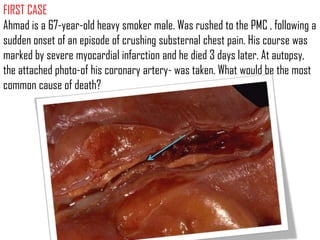
FIRST CASE
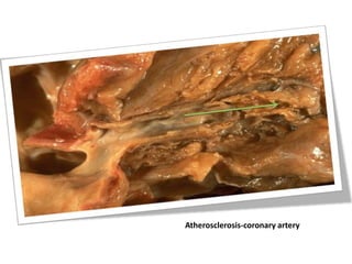
Ahmad isa 67-year-old heavy smoker male. Was rushed to the PMC , following a
sudden onset of an episode of crushing substernal chest pain. His course was
marked by severe myocardial infarction and he died 3 days later. At autopsy,
the attached photo-of his coronary artery- was taken. What would be the most
common cause of death?
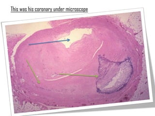
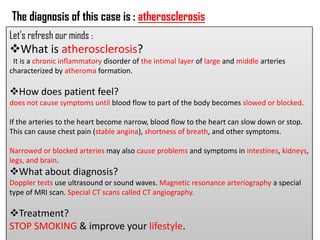
The diagnosis ofthis case is : atherosclerosis
Let’s refresh our minds :
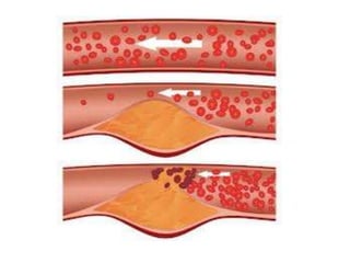
What is atherosclerosis?
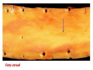
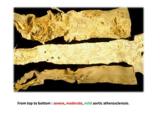
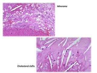
It is a chronic inflammatory disorder of the intimal layer of large and middle arteries
characterized by atheroma formation.
How does patient feel?
does not cause symptoms until blood flow to part of the body becomes slowed or blocked.
If the arteries to the heart become narrow, blood flow to the heart can slow down or stop.
This can cause chest pain (stable angina), shortness of breath, and other symptoms.
Narrowed or blocked arteries may also cause problems and symptoms in intestines, kidneys,
legs, and brain.
What about diagnosis?
Doppler tests use ultrasound or sound waves. Magnetic resonance arteriography a special
type of MRI scan. Special CT scans called CT angiography.
Treatment?
STOP SMOKING & improve your lifestyle.
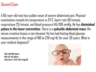
Second Case
A 65-year-oldman has sudden onset of severe abdominal pain. Physical
examination reveals his temperature is 37 C, heart rate 110/minute,
respirations 25/minute, and blood pressure 145/100 mmHg. He has diminished
pulses in the lower extremities. There is a pulsatile abdominal mass. His
serum creatine kinase is not elevated. He has had fasting blood glucose
measurements in the range of 180 to 220 mg/dL for over 20 years. What is
your medical diagnosis?
HR: 60-80 bpm.
RR:12-18 pbm.
Glucose: 110-125 mg/dl
13.
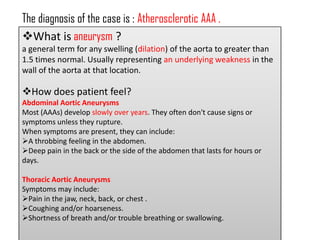
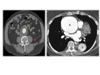
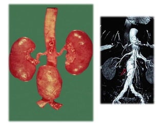
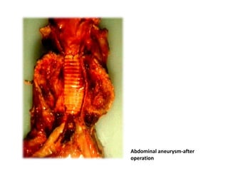
The diagnosis ofthe case is : Atherosclerotic AAA .
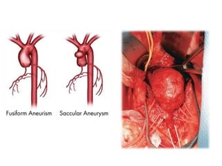
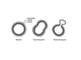
What is aneurysm ?
a general term for any swelling (dilation) of the aorta to greater than
1.5 times normal. Usually representing an underlying weakness in the
wall of the aorta at that location.
How does patient feel?
Abdominal Aortic Aneurysms
Most (AAAs) develop slowly over years. They often don't cause signs or
symptoms unless they rupture.
When symptoms are present, they can include:
A throbbing feeling in the abdomen.
Deep pain in the back or the side of the abdomen that lasts for hours or
days.
Thoracic Aortic Aneurysms
Symptoms may include:
Pain in the jaw, neck, back, or chest .
Coughing and/or hoarseness.
Shortness of breath and/or trouble breathing or swallowing.
14.
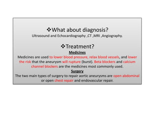
What about diagnosis?
Ultrasound and Echocardiography ,CT ,MRI ,Angiography.
Treatment?
Medicines
Medicines are used to lower blood pressure, relax blood vessels, and lower
the risk that the aneurysm will rupture (burst). Beta blockers and calcium
channel blockers are the medicines most commonly used.
Surgery
The two main types of surgery to repair aortic aneurysms are open abdominal
or open chest repair and endovascular repair.
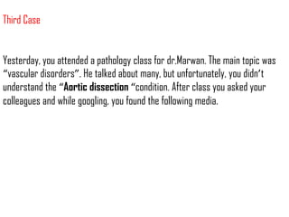
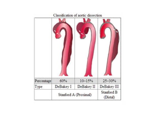
Third Case
Yesterday, youattended a pathology class for dr.Marwan. The main topic was
“vascular disorders”. He talked about many, but unfortunately, you didn’t
understand the “Aortic dissection “condition. After class you asked your
colleagues and while googling, you found the following media.
25.
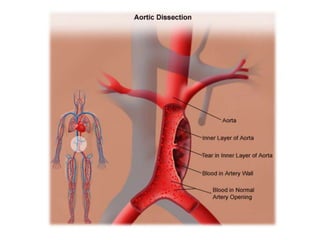
Hypertension.
Marfan syndrome.
Trauma.
Pregnancy.
Anterograde or retrograde
80% and 50%
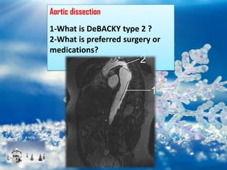
Surgery and medications.
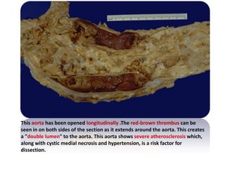
Aortic dissection-grossly
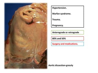
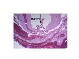
28.
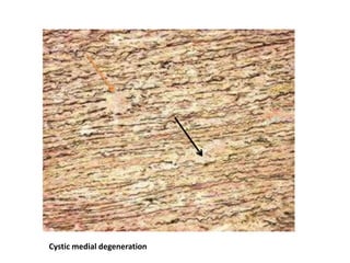
This aorta hasbeen opened longitudinally .The red-brown thrombus can be
seen in on both sides of the section as it extends around the aorta. This creates
a "double lumen" to the aorta. This aorta shows severe atherosclerosis which,
along with cystic medial necrosis and hypertension, is a risk factor for
dissection.
30.
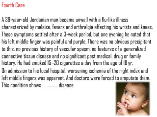
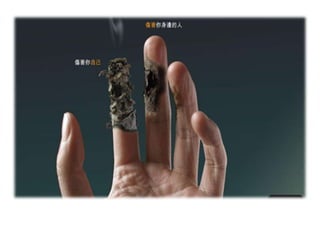
Fourth Case
A 39‐year‐oldJordanian man became unwell with a flu‐like illness
characterized by malaise, fevers and arthralgia affecting his wrists and knees.
These symptoms settled after a 3‐week period, but one evening he noted that
his left middle finger was painful and purple. There was no obvious precipitant
to this, no previous history of vascular spasm, no features of a generalized
connective tissue disease and no significant past medical, drug or family
history. He had smoked 15–20 cigarettes a day from the age of 18 yr.
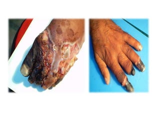
On admission to his local hospital, worsening ischemia of the right index and
left middle fingers was apparent. And doctors were forced to amputate them.
This condition shows …………… disease.
31.
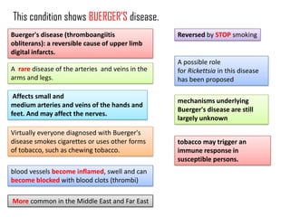
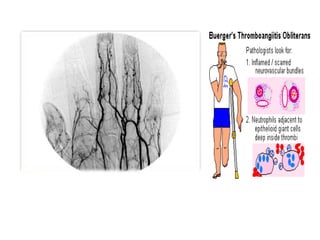
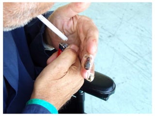
This condition showsBUERGER’S disease.
Buerger's disease (thromboangiitis Reversed by STOP smoking
obliterans): a reversible cause of upper limb
digital infarcts.
A possible role
A rare disease of the arteries and veins in the for Rickettsia in this disease
arms and legs. has been proposed
Affects small and
mechanisms underlying
medium arteries and veins of the hands and
Buerger's disease are still
feet. And may affect the nerves.
largely unknown
Virtually everyone diagnosed with Buerger's
disease smokes cigarettes or uses other forms tobacco may trigger an
of tobacco, such as chewing tobacco. immune response in
susceptible persons.
blood vessels become inflamed, swell and can
become blocked with blood clots (thrombi)
More common in the Middle East and Far East
36.
Fifth case
While doingyour elective stage in Hamburg university hospital, a 12-month-
Japanese male, was admitted with a high fever greater than 38 °C which
lasted for 5 days. He didn’t not respond to normal doses of acetaminophen
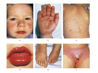
(Tylenol) or ibuprofen. On physical examination you founded the following:
Extremely red eyes (without pus or drainage). Bright red, chapped, and cracked
lips. Red mucous membranes in the mouth. Strawberry tongue, with negative
culture. Red palms of the hands and the soles of the feet. Skin rashes on the
middle of the body, NOT blister-like. According to your pathology knowledge,
detect the proper diagnosis.
37.
The diagnosis ofthe case is : Kawasaki disease /川崎病.
It is called also Mucocutaneous lymph node syndrome; Infantile
polyarteritis.
Is a rare condition in children that involves inflammation of the
blood vessels.
Kawasaki disease occurs most frequently in Japan, after
congenital heart defects, Kawasaki disease is the leading cause of
heart disease in children. Most of patients are younger than age 5.
The disease occurs more often in boys than in girls.
Kawasaki disease is a poorly understood illness. The cause has
not been determined. It may be an autoimmune disorder.
S.aureus may also have a role.
The disorder affects the mucus membranes, lymph nodes, walls
of the blood vessels, especially the coronary arteries. And the
heart .
No tests specifically diagnose Kawasaki disease. The diagnosis is
usually made based on the patient having most of the classic
symptoms.
38.
Coronary artery aneurysms,or ectasia,
develop in 15%–25% of untreated children.
Treatment
I.V gamma globulin is the standard
treatment. It is given in high doses.
The child's condition usually
greatly improves within 24 hours of
treatment with IV gamma globulin.
And aspirin…..as soon as possible.
Dose: 2 gm/kg . within 10 days of onset of
symptoms.
40.
Sixth Case
Hassan isyour 12th –year- favorite neighbor, complains from severe
pain in the tip of his fingers and toes especially when dealing with
cold materials. He stated that after playing in snow, the painful
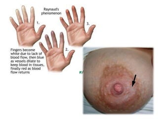
episode attacked him and a discoloration of his fingers progressed
from white (pallor) to blue (cyanosis) then the normal red color was
restored. What is your medical diagnosis?
41.
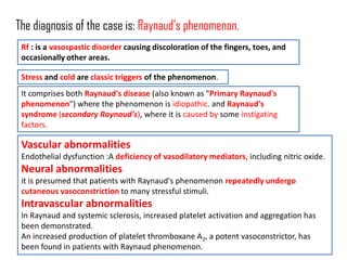
The diagnosis ofthe case is: Raynaud’s phenomenon.
Rf : is a vasospastic disorder causing discoloration of the fingers, toes, and
occasionally other areas.
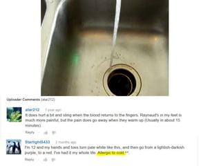
Stress and cold are classic triggers of the phenomenon.
It comprises both Raynaud's disease (also known as "Primary Raynaud's
phenomenon") where the phenomenon is idiopathic. and Raynaud's
syndrome (secondary Raynaud's), where it is caused by some instigating
factors.
Vascular abnormalities
Endothelial dysfunction :A deficiency of vasodilatory mediators, including nitric oxide.
Neural abnormalities
it is presumed that patients with Raynaud's phenomenon repeatedly undergo
cutaneous vasoconstriction to many stressful stimuli.
Intravascular abnormalities
In Raynaud and systemic sclerosis, increased platelet activation and aggregation has
been demonstrated.
An increased production of platelet thromboxane A2, a potent vasoconstrictor, has
been found in patients with Raynaud phenomenon.
44.
TEST TIME
A T H E R O S C L E R O S I S
N K W E R T Y H U I O P K R J
E A E R T B U E R G E R Y A M
U W A S D F G H J K L I U Y N
R A A S D F V G J G R T Y N M
Y S A W D R G T A R E T Y U N
S A A X C F G H A E W R U D D
M K Q W E F G R Z W Q E Y S S
D I S S E C T I O N M Y E W W
M A S D F G H J K L B V C S S