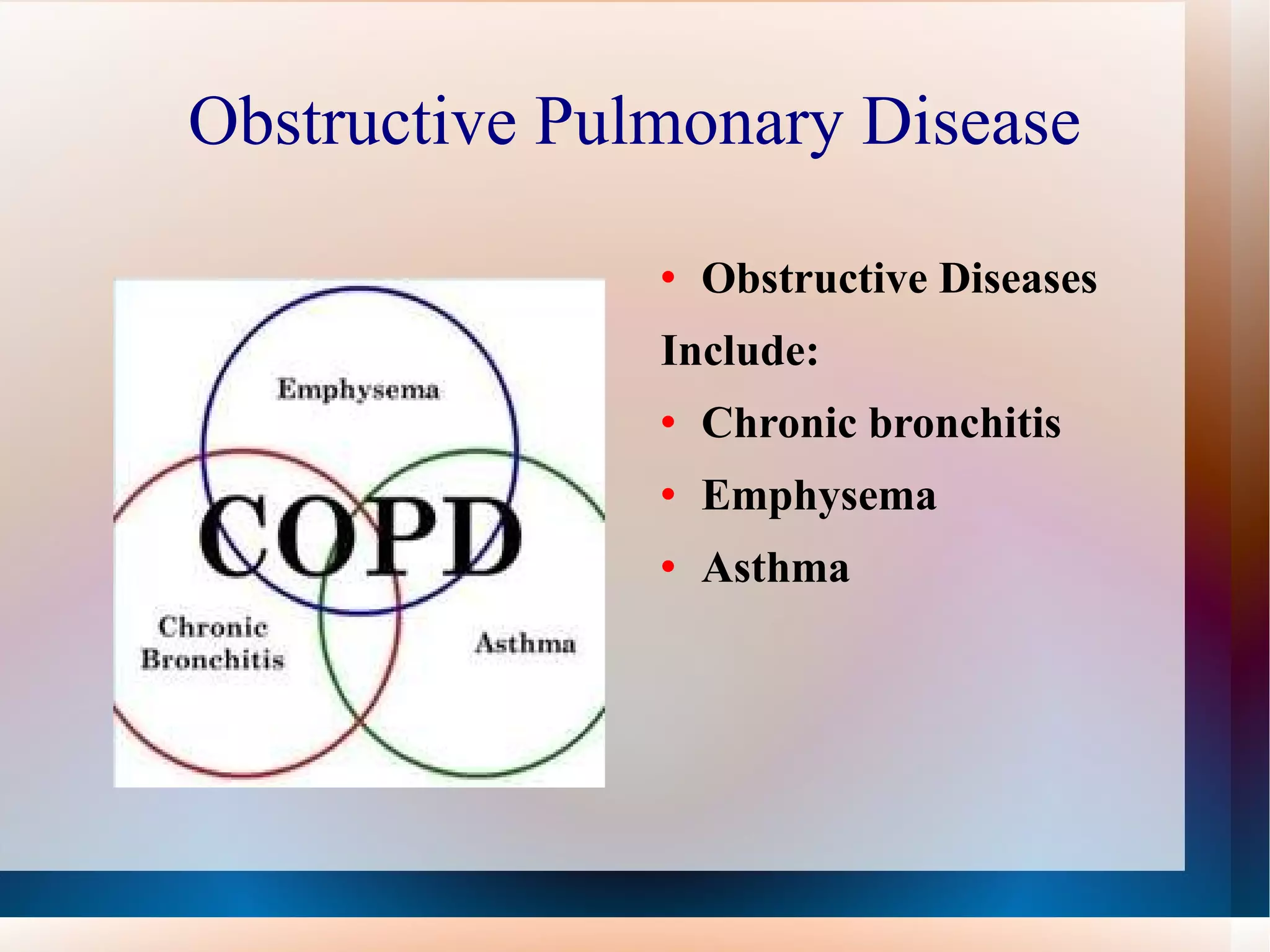
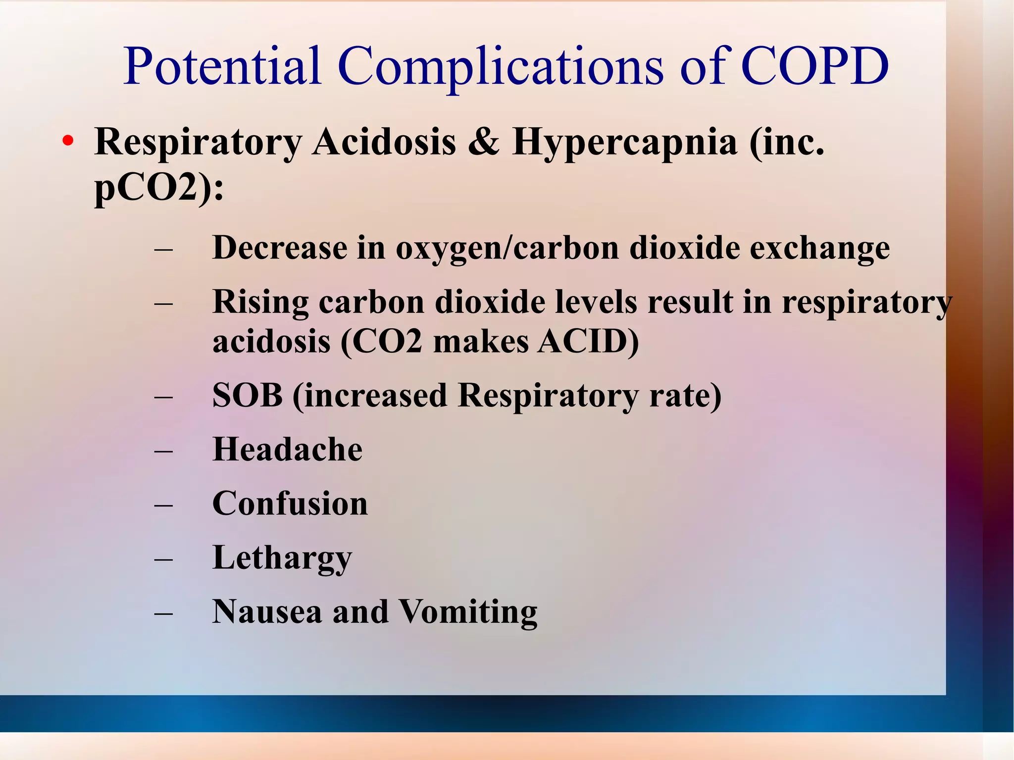
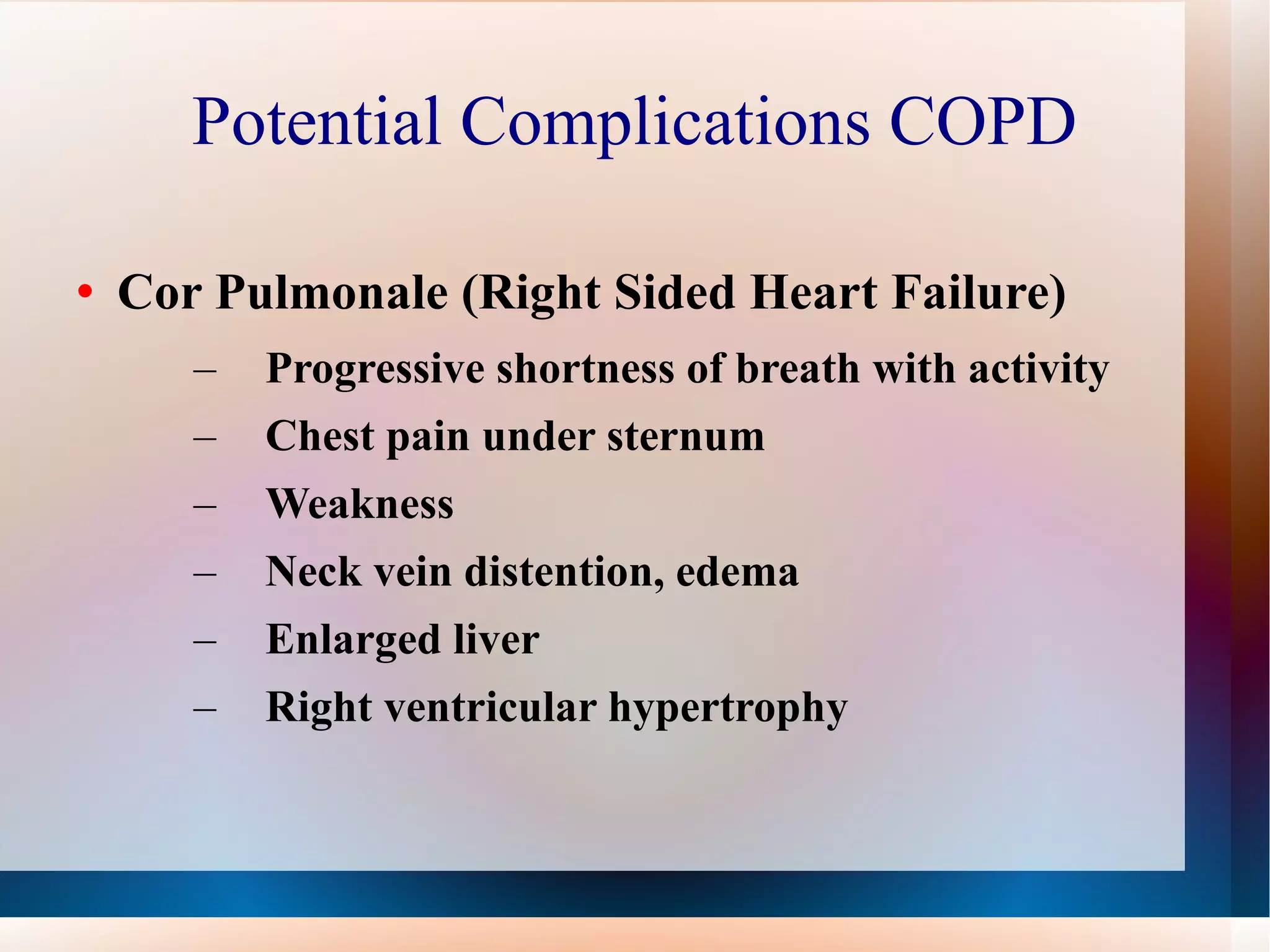
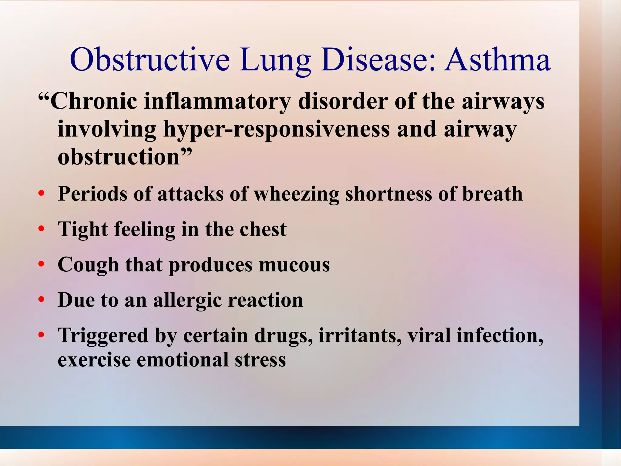
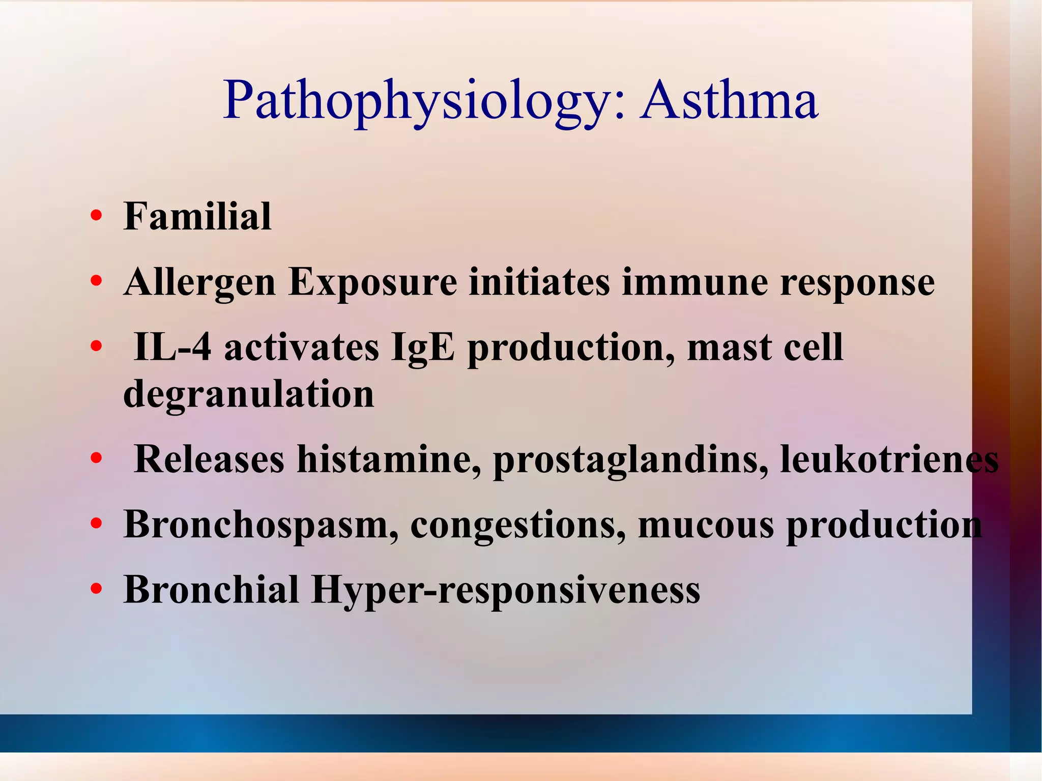
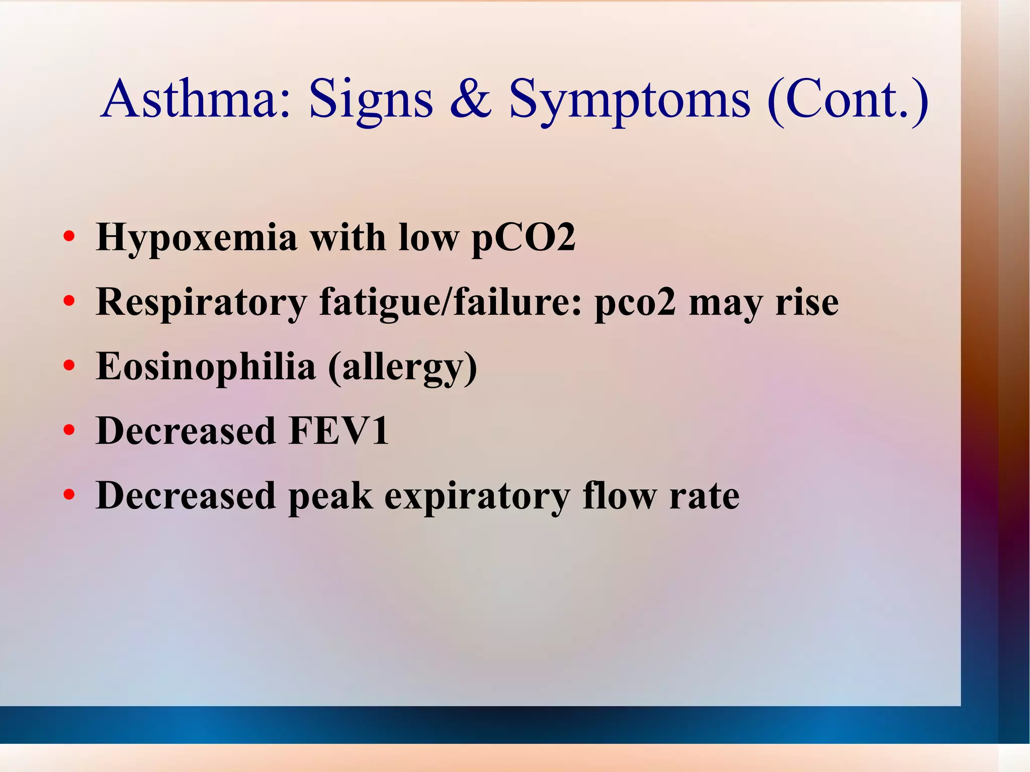
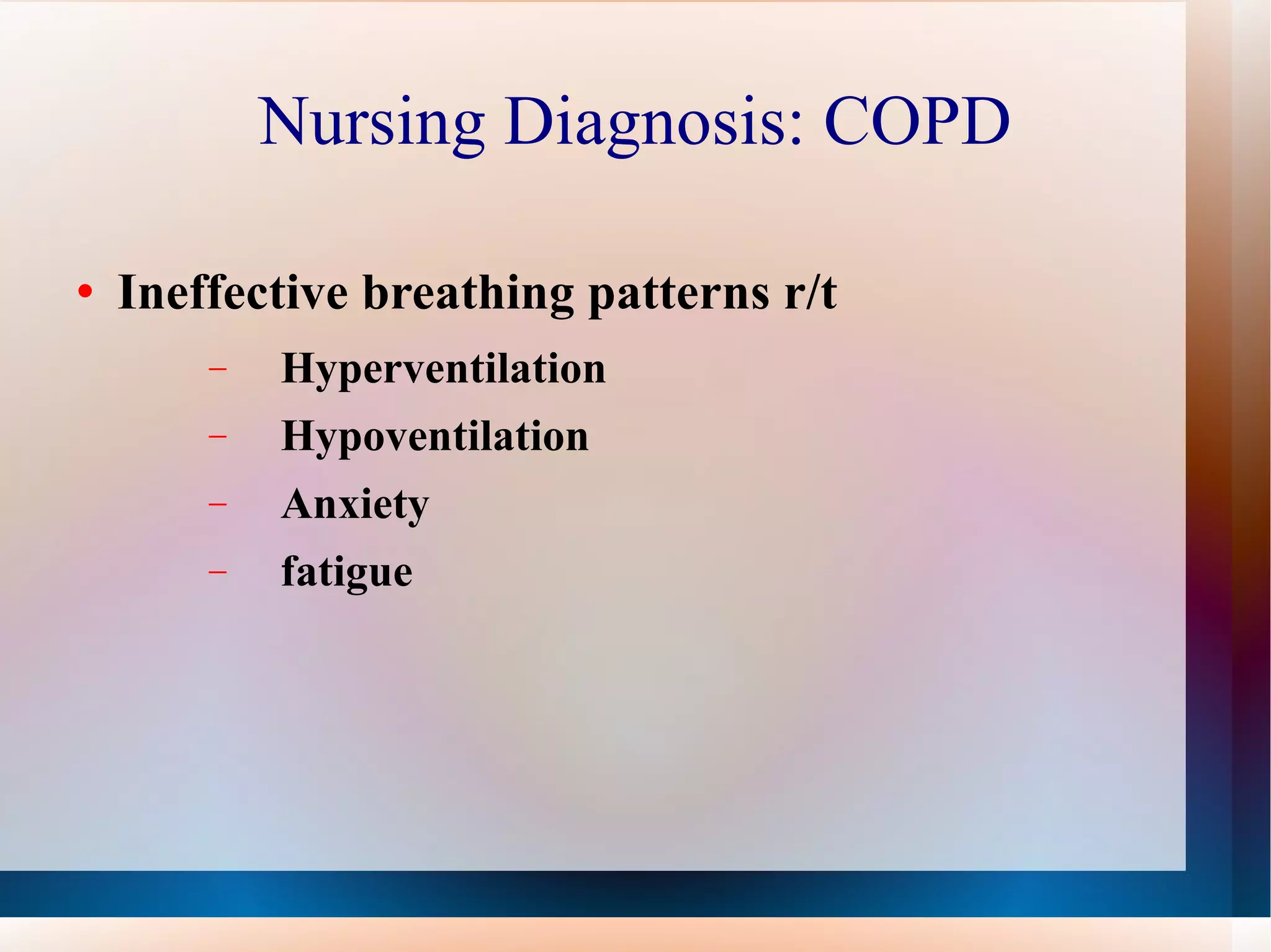
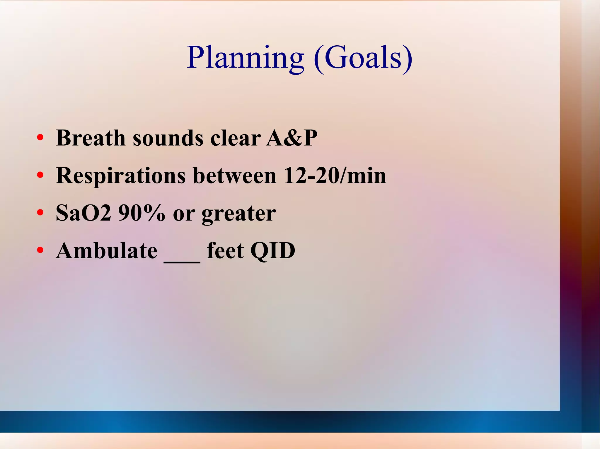
Obstructive Lung Disease KimberlyAugustine BSRN
Objectives Define Obstructive Lung Disease Epidemiology  Pathophysiology  Identify Clinical manifestations Identify Risk factors  Discuss Evaluation & Treatment
Definition Several different definitions have existed for COPD. “ A disease state characterized by airflow limitation that is not fully reversible. The airflow limitation is usually both progressive and associated with an abnormal inflammatory response of the lung to noxious particles or gases.”-GOLD A group of diseases that cause airflow blockage and breathing-related problems. It includes emphysema, chronic bronchitis, and in some cases asthma.-CDC “ Airway obstruction:most common obstructive diseases  are asthma, chronic bronchitis, emphysema-McCane & Huether
Obstructive Pulmonary Disease Obstructive Diseases  Include: Chronic bronchitis Emphysema Asthma
Epidemiology 4th leading cause of death in the U. S.  12.1 million U.S. adults were estimated to have COPD  Women have exceeded men in the number of deaths attributable to COPD 2010, $49.9 billion COPD health care costs Worldwide leading cause of death & disability 2020, predicted 3 rd  leading cause of death
Pathophysiology: Video http://www.youtube.com/watch?v=lYW_2Rfuii8&feature=related
Obstructive Lung Disease: Emphysema  “  A condition which the lungs lose elasticity and alveoli enlarge that disrupts function” Destruction of lung parenchyma Loss of elastic recoil Alveolar gas is trapped in expiration Gas exchange is compromised
Pathophysiology: Emphysema Begins with the destruction of air sacs (alveoli) in the lungs where oxygen from the air is exchanged for carbon dioxide in the blood.  The walls of the air sacs are thin and fragile. Damage to the air sacs is irreversible and results in permanent "holes" in the tissues of the lower lungs.  As air sacs are destroyed, the lungs are able to transfer less and less oxygen to the bloodstream, causing shortness of breath.  The lungs also lose their elasticity, which is important to keep airways open. The patient experiences great difficulty exhaling.
COPD: Emphysema
COPD: Emphysema Signs & Symptoms Dyspnea  Little sputum production or cough Tachypnea with prolonged expiration Use of accessory muscles for ventilation Increased anteroposterior diameter of thorax (Barrel Chest)  Pierced Lips to prevent expiratory airway collapse Cardiac enlargement Hyperresonant (loud, low) sound with chest percussion d/t hyperinflation
Obstructive Lung Disease:  Chronic Bronchitis “ The presence of a mucus-producing cough three months of a year for two consecutive years without other underlying disease to explain the cough.” Inflammation and eventual scarring of the lining of the bronchial tubes  Inflamed, infected bronchi allow for less air to flow to and from the lungs and a heavy mucus or phlegm is coughed up
Pathophysiology: Chronic Bronchitis  Increased mucous production Increase in size, number goblet cells  Impaired Ciliary function Bronchospasm, permanent narrowing of airways Decreased ventilation Tidal Volume Decreased Hypoventilation Hypercapnia
COPD: Chronic Bronchitis
COPD: Chronic Bronchitis Signs & Symptoms Wheezing and shortness of breath Productive cough “smoker's cough” Decreased tolerance, hypoxic with exercise Frequent pulmonary infections Decreased FEV1, FEC FRC & RV increased Increased Paco2, Hypoxemia, Polycythemia  Cyanosis “Blue Bloater”
Risk Factors: COPD  (Emphysema & Chronic Bronchitis) Smoking predominant Cause  Alpha-1antitrypsin deficiency Occupational exposure, pollution Diet deficient in vitamin C Low Birth weight Childhood respiratory infections Pre-existing bronchial hyper-responsiveness  Low social class
Global Initiative: COPD
Potential Complications: COPD Hypoxemia (paO2 of 55mmHg or less with an oxygen saturation of 85% or less) Cor Pulmonale (Right Sided Heart Failure) Respiratory Acidosis & Hypercapnia (increased paCO2):
Oxyhemoglobin Dissociation Curve The oxyhemoglobin dissociation curve is an important tool for understanding how our blood carries and releases oxygen. Specifically, the oxyhemoglobin dissociation curve relates oxygen saturation (SO 2 ) and partial pressure of oxygen in the blood (PO 2 ), and is determined by what is called "hemoglobin's affinity for oxygen," that is, how readily hemoglobin acquires and releases oxygen molecules from its surrounding tissue.
Potential Complications: COPD Hypoxemia (paO2 of 55mmHg or less with an oxygen saturation of 85% or less) Mood changes Forgetfulness Inability to concentrate Cyanosis a late sign of hypoxia
Potential Complications of COPD Respiratory Acidosis & Hypercapnia (inc. pCO2): Decrease in oxygen/carbon dioxide exchange Rising carbon dioxide levels result in respiratory acidosis (CO2 makes ACID) SOB (increased Respiratory rate) Headache Confusion Lethargy Nausea and Vomiting
Potential Complications COPD Cor Pulmonale (Right Sided Heart Failure) Progressive shortness of breath with activity Chest pain under sternum Weakness Neck vein distention, edema Enlarged liver Right ventricular hypertrophy
Obstructive Lung Disease: Asthma “ Chronic inflammatory disorder of the airways involving hyper-responsiveness and airway obstruction” Periods of attacks of wheezing shortness of breath Tight feeling in the chest Cough that produces mucous Due to an allergic reaction Triggered by certain drugs, irritants, viral infection, exercise emotional stress
Pathophysiology: Asthma Familial  Allergen Exposure initiates immune response IL-4 activates IgE production, mast cell degranulation Releases histamine, prostaglandins, leukotrienes  Bronchospasm, congestions, mucous production Bronchial Hyper-responsiveness
Asthma: Signs & Symptoms Asymptomatic between attacks Chest constriction Expiratory Wheezing  Dyspnea Non productive cough Tachycardia, tachypnea Pulsus Paradoxus
Asthma: Signs & Symptoms (Cont.) Hypoxemia with low pCO2 Respiratory fatigue/failure: pco2 may rise Eosinophilia (allergy) Decreased FEV1 Decreased peak expiratory flow rate
Risk Factors: Asthma
Asthma: EvaluationTreatment Treat precipitating event Oxygen therapy Hydration Antibotics (with infection) Meds: bronchodilators, steroids, mast cell stabilizers, methylxanthines
Nursing Diagnosis: COPD Ineffective airway clearance r/t Airway spasm Retained secretions Excessive mucous Fatigue Impaired gas exchange r/t  Descreased lung expansion Decreased LOC Presence of pulmonary secretions
Nursing Diagnosis: COPD Ineffective breathing patterns r/t  Hyperventilation Hypoventilation Anxiety fatigue
Planning (Goals) Breath sounds clear A&P Respirations between 12-20/min SaO2 90% or greater Ambulate ___ feet QID
Implementation: Promoting Lung expansion Positioning Breathing exercises Chest Physiotherapy Oxygen Therapy
Implementation: Promoting Lung expansion Positioning: change at least Q2 hrs
Implementation: Promoting Lung expansion Breathing exercises: to expel secretions from lungs CDB Q2 hrs Pursed lip breathing Helps COPD patients to evacuate more air by breathing out against pressure Abdominal Breathing (diaphragmatic) Promotes alveoli expansion and emptying
Implementation: Mobilizing Pulmonary Secretions Hydration Keeps pulmonary secretions moist, easy to expectorate Fluid intake 1500-2000 cc/day Humidification Air or oxygen with increased humidity will help to keep airways moist to loosen and mobilize pulmonary secretions Nebulization Adding fine drops of moisture to the respiratory tract
Implementation: Mobilizing Pulmonary Secretions Chest physiotherapy Chest percussion (cupping) Vibration: fine shaking pressure applied to chest wall only during exhalation (helps get rid of trapped air) vest
Implementation: Mobilizing Pulmonary Secretions Chest physiotherapy Postural Drainage: positioning  (not good for emphysema/bronchitis don’t tolerate asthma not needed. Just for CF-bronchitis w/o emphesema
Case Study
Journal Article: COPD the role of the nurse by Barnett Nurses have a key role in the prevention and treatment of COPD in advising and supporting patients living with this condition.
Nurses Role Prevention & Treatment Recognize clinical symptoms Recognize Associated Risk Factors Medications Available Effectiveness(Questions) Patient Education:  Smoking Nutrition Activity Vaccination
Discussion Patient Factors for COPD/Single Most Factor Clinical Manifestations of Bronchitis/Emphysema COPD Staging Arterial Blood Gas indicative of which Serious Condition Pulmonary HTN/Cor Pulmonale Clinical Manifestations
Restrictive vs Obstructive Disease http:// www.youtube.com/watch?v = wbcjFpyxkpc&feature =related  http://www.youtube.com/watch?v=wbcjFpyxkpc&feature=related
References Barnett, Margaret.  (2006, February). COPD: the role of the nurse. Journal of  Community Nursing, 20(2), 18-20,22.  Retrieved October 26, 2010, from  Research Library. (Document ID: 989426231). Bauldoff, G. (2009). When breathing is a burden: how to help patients with  COPD. American Nurse Today, 4(9), 17-22. Retrieved from CINAHL  database. National Heart Lung and Blood Institute  http://www.nhlbi.nih.gov/health/public/lung/copd/ . American Lung Association  http://www.lungusa.org/lung-disease/copd/resources/facts-figures/COPD-Fact-Sheet.html

Advanced Practice Powerpoint

  • 1.
    Obstructive Lung DiseaseKimberlyAugustine BSRN
  • 2.
    Objectives Define ObstructiveLung Disease Epidemiology Pathophysiology Identify Clinical manifestations Identify Risk factors Discuss Evaluation & Treatment
  • 3.
    Definition Several differentdefinitions have existed for COPD. “ A disease state characterized by airflow limitation that is not fully reversible. The airflow limitation is usually both progressive and associated with an abnormal inflammatory response of the lung to noxious particles or gases.”-GOLD A group of diseases that cause airflow blockage and breathing-related problems. It includes emphysema, chronic bronchitis, and in some cases asthma.-CDC “ Airway obstruction:most common obstructive diseases are asthma, chronic bronchitis, emphysema-McCane & Huether
  • 4.
    Obstructive Pulmonary DiseaseObstructive Diseases Include: Chronic bronchitis Emphysema Asthma
  • 5.
    Epidemiology 4th leadingcause of death in the U. S. 12.1 million U.S. adults were estimated to have COPD Women have exceeded men in the number of deaths attributable to COPD 2010, $49.9 billion COPD health care costs Worldwide leading cause of death & disability 2020, predicted 3 rd leading cause of death
  • 6.
  • 7.
    Obstructive Lung Disease:Emphysema “ A condition which the lungs lose elasticity and alveoli enlarge that disrupts function” Destruction of lung parenchyma Loss of elastic recoil Alveolar gas is trapped in expiration Gas exchange is compromised
  • 8.
    Pathophysiology: Emphysema Beginswith the destruction of air sacs (alveoli) in the lungs where oxygen from the air is exchanged for carbon dioxide in the blood. The walls of the air sacs are thin and fragile. Damage to the air sacs is irreversible and results in permanent "holes" in the tissues of the lower lungs. As air sacs are destroyed, the lungs are able to transfer less and less oxygen to the bloodstream, causing shortness of breath. The lungs also lose their elasticity, which is important to keep airways open. The patient experiences great difficulty exhaling.
  • 9.
  • 10.
    COPD: Emphysema Signs& Symptoms Dyspnea Little sputum production or cough Tachypnea with prolonged expiration Use of accessory muscles for ventilation Increased anteroposterior diameter of thorax (Barrel Chest) Pierced Lips to prevent expiratory airway collapse Cardiac enlargement Hyperresonant (loud, low) sound with chest percussion d/t hyperinflation
  • 11.
    Obstructive Lung Disease: Chronic Bronchitis “ The presence of a mucus-producing cough three months of a year for two consecutive years without other underlying disease to explain the cough.” Inflammation and eventual scarring of the lining of the bronchial tubes Inflamed, infected bronchi allow for less air to flow to and from the lungs and a heavy mucus or phlegm is coughed up
  • 12.
    Pathophysiology: Chronic Bronchitis Increased mucous production Increase in size, number goblet cells Impaired Ciliary function Bronchospasm, permanent narrowing of airways Decreased ventilation Tidal Volume Decreased Hypoventilation Hypercapnia
  • 13.
  • 14.
    COPD: Chronic BronchitisSigns & Symptoms Wheezing and shortness of breath Productive cough “smoker's cough” Decreased tolerance, hypoxic with exercise Frequent pulmonary infections Decreased FEV1, FEC FRC & RV increased Increased Paco2, Hypoxemia, Polycythemia Cyanosis “Blue Bloater”
  • 15.
    Risk Factors: COPD (Emphysema & Chronic Bronchitis) Smoking predominant Cause Alpha-1antitrypsin deficiency Occupational exposure, pollution Diet deficient in vitamin C Low Birth weight Childhood respiratory infections Pre-existing bronchial hyper-responsiveness Low social class
  • 16.
  • 17.
    Potential Complications: COPDHypoxemia (paO2 of 55mmHg or less with an oxygen saturation of 85% or less) Cor Pulmonale (Right Sided Heart Failure) Respiratory Acidosis & Hypercapnia (increased paCO2):
  • 18.
    Oxyhemoglobin Dissociation CurveThe oxyhemoglobin dissociation curve is an important tool for understanding how our blood carries and releases oxygen. Specifically, the oxyhemoglobin dissociation curve relates oxygen saturation (SO 2 ) and partial pressure of oxygen in the blood (PO 2 ), and is determined by what is called "hemoglobin's affinity for oxygen," that is, how readily hemoglobin acquires and releases oxygen molecules from its surrounding tissue.
  • 19.
    Potential Complications: COPDHypoxemia (paO2 of 55mmHg or less with an oxygen saturation of 85% or less) Mood changes Forgetfulness Inability to concentrate Cyanosis a late sign of hypoxia
  • 20.
    Potential Complications ofCOPD Respiratory Acidosis & Hypercapnia (inc. pCO2): Decrease in oxygen/carbon dioxide exchange Rising carbon dioxide levels result in respiratory acidosis (CO2 makes ACID) SOB (increased Respiratory rate) Headache Confusion Lethargy Nausea and Vomiting
  • 21.
    Potential Complications COPDCor Pulmonale (Right Sided Heart Failure) Progressive shortness of breath with activity Chest pain under sternum Weakness Neck vein distention, edema Enlarged liver Right ventricular hypertrophy
  • 22.
    Obstructive Lung Disease:Asthma “ Chronic inflammatory disorder of the airways involving hyper-responsiveness and airway obstruction” Periods of attacks of wheezing shortness of breath Tight feeling in the chest Cough that produces mucous Due to an allergic reaction Triggered by certain drugs, irritants, viral infection, exercise emotional stress
  • 23.
    Pathophysiology: Asthma Familial Allergen Exposure initiates immune response IL-4 activates IgE production, mast cell degranulation Releases histamine, prostaglandins, leukotrienes Bronchospasm, congestions, mucous production Bronchial Hyper-responsiveness
  • 24.
    Asthma: Signs &Symptoms Asymptomatic between attacks Chest constriction Expiratory Wheezing Dyspnea Non productive cough Tachycardia, tachypnea Pulsus Paradoxus
  • 25.
    Asthma: Signs &Symptoms (Cont.) Hypoxemia with low pCO2 Respiratory fatigue/failure: pco2 may rise Eosinophilia (allergy) Decreased FEV1 Decreased peak expiratory flow rate
  • 26.
  • 27.
    Asthma: EvaluationTreatment Treatprecipitating event Oxygen therapy Hydration Antibotics (with infection) Meds: bronchodilators, steroids, mast cell stabilizers, methylxanthines
  • 28.
    Nursing Diagnosis: COPDIneffective airway clearance r/t Airway spasm Retained secretions Excessive mucous Fatigue Impaired gas exchange r/t Descreased lung expansion Decreased LOC Presence of pulmonary secretions
  • 29.
    Nursing Diagnosis: COPDIneffective breathing patterns r/t Hyperventilation Hypoventilation Anxiety fatigue
  • 30.
    Planning (Goals) Breathsounds clear A&P Respirations between 12-20/min SaO2 90% or greater Ambulate ___ feet QID
  • 31.
    Implementation: Promoting Lungexpansion Positioning Breathing exercises Chest Physiotherapy Oxygen Therapy
  • 32.
    Implementation: Promoting Lungexpansion Positioning: change at least Q2 hrs
  • 33.
    Implementation: Promoting Lungexpansion Breathing exercises: to expel secretions from lungs CDB Q2 hrs Pursed lip breathing Helps COPD patients to evacuate more air by breathing out against pressure Abdominal Breathing (diaphragmatic) Promotes alveoli expansion and emptying
  • 34.
    Implementation: Mobilizing PulmonarySecretions Hydration Keeps pulmonary secretions moist, easy to expectorate Fluid intake 1500-2000 cc/day Humidification Air or oxygen with increased humidity will help to keep airways moist to loosen and mobilize pulmonary secretions Nebulization Adding fine drops of moisture to the respiratory tract
  • 35.
    Implementation: Mobilizing PulmonarySecretions Chest physiotherapy Chest percussion (cupping) Vibration: fine shaking pressure applied to chest wall only during exhalation (helps get rid of trapped air) vest
  • 36.
    Implementation: Mobilizing PulmonarySecretions Chest physiotherapy Postural Drainage: positioning (not good for emphysema/bronchitis don’t tolerate asthma not needed. Just for CF-bronchitis w/o emphesema
  • 37.
  • 38.
    Journal Article: COPDthe role of the nurse by Barnett Nurses have a key role in the prevention and treatment of COPD in advising and supporting patients living with this condition.
  • 39.
    Nurses Role Prevention& Treatment Recognize clinical symptoms Recognize Associated Risk Factors Medications Available Effectiveness(Questions) Patient Education: Smoking Nutrition Activity Vaccination
  • 40.
    Discussion Patient Factorsfor COPD/Single Most Factor Clinical Manifestations of Bronchitis/Emphysema COPD Staging Arterial Blood Gas indicative of which Serious Condition Pulmonary HTN/Cor Pulmonale Clinical Manifestations
  • 41.
    Restrictive vs ObstructiveDisease http:// www.youtube.com/watch?v = wbcjFpyxkpc&feature =related http://www.youtube.com/watch?v=wbcjFpyxkpc&feature=related
  • 42.
    References Barnett, Margaret. (2006, February). COPD: the role of the nurse. Journal of Community Nursing, 20(2), 18-20,22.  Retrieved October 26, 2010, from Research Library. (Document ID: 989426231). Bauldoff, G. (2009). When breathing is a burden: how to help patients with COPD. American Nurse Today, 4(9), 17-22. Retrieved from CINAHL database. National Heart Lung and Blood Institute http://www.nhlbi.nih.gov/health/public/lung/copd/ . American Lung Association http://www.lungusa.org/lung-disease/copd/resources/facts-figures/COPD-Fact-Sheet.html