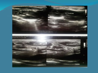
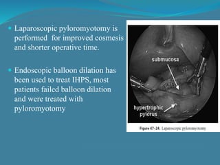
Dr raghavendra.
Fellow in neonatology
Pramod
Pt name: Promod, age : 1month 18 days
s/o nagraj
kumbarpet
DOB:17/12/2014
DOA :05/02/2015.
Single/live/term /AGA term male baby born via naturalis at WCH
hospital, davangere on 17/12/2014 at 08.45 AM.
Baby cried immediately after birth.
Birth wt – 2.3 kg
 C/C : Vomiting after each feed since 7days projectile,
non- bilious in nature.
Distension of abdomen since 3 days .
 Obstetric history: Regular ANCs taken, three scan
done USGs normal,. taken iron and folicacid
supplements, taken 2 dose of TT. Pregnancy
uneventful..
 Family history: ML: 5 years, 3rd NCM
G2P2L1, No h/o of similar complains in parents
 GPE: 48 day old male baby with vomiting & excessive
crying with visible lump in epigastrium well hydrated
and alert.
Vitals: HR-128 bpm
RR- 38 cpm
CFT < 3secs
Head to toe examination:
AF- at level
Visible lump over epigastrium
Visible gastric peristalsis from lt to rt hypochondrium
 Anthropometry :
Wt-2.3 kg
HC-34cm
Length-47cm
 O/E :
Colour - pink
Cry - Good
Activity - good
 S/E:
o CVS: S1 & S2 heard, no murmur
o R/S: B/L air entry equal, no added sounds.
o CNS: Normal
o P/A: Inspection- visible lump over epigastrium,
visible gastric peristalsis from lt to rt hypochondrium
Palpation – palpable mobile mass, firm in cosistency
in epigastrium measuring about 3 cm below liver edge
Auscultation- bowel sounds present
INVESTIGATIONS
 Complete hemogram
TLC- 10740 cells/cumm N- 24%
Hb- 11.3 gm/dl L- 58%
PCV- 37.9%
Platelet count- 3.5 lakhs/cumm
 CRP - Non Reactive
 PT, APTT – Normal.
 BBG: AB positive.
 HBsAg:negative.
 HIV:negative
Discussion
 48 days old male baby presenting with projectile non
bilious vomiting immediately after feed with visible lump
over epigastrium, visible gastric peristalsis from lt to rt
hypochondrium with ABG showing metabolic
alkalosis,hypokalemia & USG abdomen showing
hypertrophic elongated pylorus with dilated stomach
favours the diagnosis of IHPS
Treatment
 IV antibiotics and IV fluids Isolyte-P maintainence for
20hrs.
Surgery – laparoscopic pyloromyotomy done on
06/02/2015.
Post operative 5ml feeds BM started after 5 hrs of surgery,
increasing slowly every 6 hrs.
Introduction
 Infantile hypertrophic pyloric stenosis (IHPS) is the most
common cause of gastric outlet obstruction in children &
is one of the most frequent conditions requiring surgery in
infants.
 IHPS is an acquired condition in which the
circumferential muscle layer of the pyloric sphinter
becomes thickened, resulting in narrowing & elongation
of the pyloric channel.
 This produces a high-grade gastric outlet obstruction with
compensatory dilation, hypertrophy & hyperperistalsis of
stomach
Epidemology
 The incidence of IHPS is approximately 2 to 5 per 1,000
births per year in most white populations.
 Less common in India, and among black and Asian
populations, with a frequency that is one third to one-fifth
that in the white population.
 The male-to-female ratio is approximately 4:1.
 There is a familial link, but a hereditary propensity to the
development of IHPS is likely polygenic with no single
locus accounting for the fivefold increase in the risk of
first-degree relatives.
 Male and female children of affected fathers carry a risk
of 5% and 2.5% respectively, of developing IHPS
 Male and female children of affected mothers carry a risk
of 19% and 7%, respectively of developing IHPS
 Concordance in monozygotic twins is 0.25-0.44 and that
in dizygotic twins is 0.05-0.10
Normal pylorus
 In IHPS the pyloric ring is no longer a clearly defined
separation between the pyloric canal and duodenum
 Instead the muscle of the pyloric antrum is hypertrophic (3
or more mm), which separates the normal antrum (1mm
thickness) from the duodenum
 The lumen is filled with compressed and redundant
mucosa, which obstructs the passage of gastric contents
IHPS anatomy
Etiology
 It has been found that, when compared to controls, in
IHPS specimens, the muscle layer is deficient in:
– the quantity of nerve terminals
–markers for nerve-supporting cells (Neurotrophins-
peptides that govern survival of ENS neurons)
– nitric oxide synthase activity
–mRNA production for nitric oxide synthase.
 Axonal degeneration in both myentric plexus &
intramuscular nerves
 One more hypothesis is that there is dyscoordination b/w
gastric peristalsis & pyloric relaxation resulting in,
simultaneous gastric & pyloric contraction, & work
hypertrophy of the pyloric muscle.
 More recently an association b/w maternal & infant
exposure to erythromycin that stimulates phase 3 MMC
(migrating myoelectric complex) activity exposing
immature pylorus to high gastric pressures was found
leading to development of IHPS .
 Gastric outlet obstruction due to pylorospasm has been
reported in neonates receiving PGE infusions
Clinical presentation
 The typical clinical presentation in a term male infant
between 3-6 wks of age having progressive, nonbilious,
projectile vomiting and demands to be re-fed soon
afterwards ("hungry vomiter")
 Vomiting consistently follows every feeding & is forceful
leading to significant dehydration.
 Starvation can exacerbate diminished hepatic glucoronyl
transferase activity, and indirect hyperbilirubinemia may
be seen in 1-2% of affected infants.
 Prolonged vomiting leads to the loss of large quantities of
gastric secretions rich in H+ and Cl- .
 As a result of dehydration, the kidney attempts to
conserve Na+ to maintain volume, by exchanging them
for K+ and H+ (paradoxical aciduria).
 The net result is a loss of H+ and K+, which results in
hypokalemic, hypochloremic metabolic alkalosis.
 Significant hypoglycemia can occur which may
precipitate seizures
Diagnosis
 Initially suggested by the typical clinical presentation.
 The hallmark of diagnosis is the finding of a small, mobile,
ovoid mass referred to as an olive in the epigastrium in a calm
infant with relaxed abdominal muscles best palpated from the
left, located in the midepigastrium beneath the liver edge..
 The olive can be felt to roll under the fingertips during
sweeping motion- no other structure gives this sensation.
 If the olive is not palpable in an infant who has a clinical picture
suggestive of IHPS, further studies are warranted.
 Ultrasonography is used to measure the thickness of the
pyloric wall and the length of the pyloric canal
– normal wall thickness <2mm, IHPS >4 mm
– normal length of the pyloric canal <10 mm,
IHPS >17 mm
- diameter in IHPS > 17mm.
 If USG is not diagnostic and IHPS remains a concern, the
next test of choice is an upper GI contrast studies.
 The canal is outlined by a string of contrast material
coursing through spaces between redundant mucosa
(“string sign”)
Double track signShoulder signString sign
Differential diagnosis
 Overfeeding
 GER
 Hiatal hernia
 Pylorospasm
 Preampulary duodenal obstruction
 Salt-wasting adrenogenital syndrome
Treatment
 The preoperative treatment is directed towards correcting the
fluid, acid-base, and electrolyte losses.
 Intravenous fluid therapy is begun with 0.45–0.9% saline, in 5–
10% dextrose, with the addition of KCl in concentrations of
30–50mEq/L.
 the infant is rehydrated till normal urine output and the serum
bicarbonate concentration is less than 30mEq/dL, which
implies that the alkalosis has been corrected.
 Correction of the alkalosis is essential to prevent postoperative
apnea, which may be associated with anaesthesia
 Infant can undergo the Fredet-Ramstedt pylormyotomy,
which is the procedure of choice.
 If the mucosa is entered (usually on the duodenal side), it
can be primarily repaired and reinforced with an omental
patch.
 Large perforations are managed by closing the
pyloromyotomy, rotating the pylorus 90°, and repeating
the myotomy.
 Mortality and morbidity of less that 0.5%
Fredet-Ramstedt pylormyotomy
 Highly effective simple, elegant, and inexpensive
operation.
 Described as 'one of the most easy and gratifying
procedures performed by pediatric surgeons’, the most
consistently successful operation ever described.
 Post-operative complications:
–Wound infection
– Incomplete myotomy, treated by repeat myotomy or
endoscopic balloon dilation
 Laparoscopic pyloromyotomy is
performed for improved cosmesis
and shorter operative time.
 Endoscopic balloon dilation has
been used to treat IHPS, most
patients failed balloon dilation
and were treated with
pyloromyotomy
 Feeding can be resumed within 6 to 12 hours
postoperatively with small volume of sugar water
advancing volume & osmolarity every 2-3 hrs until infant
is taking breast milk
 Post-operative vomiting may occur in up to 50% of infants,
thought to be secondary to edema of the pylorus at the
incision site
 Most infants will tolerate full feeds within 24 to 48 hrs
Non-surgical treatment
 Injection of botulinum toxin, Antispasmodics to relieve
pylorospasm
 Transpyloric nasoduodenal feeding
 Atropine: Oral or IV atropine has been used in a dose of
0.06 mg/kg/day in eight divided doses, increased by 0.15
mg/kg/day till vomiting ceased and remained so for a
period of 24 hours at a stretch, then substituted by oral
atropine at double the effective IV dose for 3 weeks
 Atropine temporarily suppresses spastic contractions of
pyloric muscle in pyloric stenosis, and it resulted in
cessation of vomiting and eventual regression of pyloric
hypertrophy.
 Medical management may be successful in less severe
cases, but treatment takes longer and the complication rate
from medical management was higher.
 Surgical therapy is considered superior to conservative
management, since complications are seldom, long-term
results are equal, and patients get better sooner.
Prognosis
 Most children treated for HPS have excellent short &
long-term outcomes
 Postoperative USG studies have documented a return to
normal muscle thickness within 4 wks a/w healing of
pyloric muscle & return of function
References
 Christine A Gleason, Sherin U Devaskar. Avery’s diseases
of newborn: 9th edition. 2012; 984-85
 Richard J Martin, Avroy A Fanaroff, Michel C Walsh.
Fanaroff & Martin’s Neonatal and Perinatal medicine: 9th
edition 2011; 1415-18
 Ashcraft, Keith W; Murphy.J Patrick. Pediatric Surgery:
6th edition.2000; 391-94
 Keith T.Oldham, Paul M.Colombani, Robert P.Foglia,
Michael A.Skinner. Principles & Practice of Pediatric
Surgery: 2005; 1168-72
 Google images
References
 Christine A Gleason, Sherin U Devaskar. Avery’s
diseases of newborn: 9th edition. 2012; 984-85
 Richard J Martin, Avroy A Fanaroff, Michel C Walsh.
Fanaroff & Martin’s Neonatal and Perinatal medicine:
9th edition 2011; 1415-18
 Ashcraft, Keith W; Murphy.J Patrick. Pediatric Surgery:
6th edition.2000; 391-94
 Keith T.Oldham, Paul M.Colombani, Robert P.Foglia,
Michael A.Skinner. Principles & Practice of Pediatric
Surgery: 2005; 1168-72
 Google images

Case ihps

  • 1.
  • 2.
  • 3.
    Pt name: Promod,age : 1month 18 days s/o nagraj kumbarpet DOB:17/12/2014 DOA :05/02/2015. Single/live/term /AGA term male baby born via naturalis at WCH hospital, davangere on 17/12/2014 at 08.45 AM. Baby cried immediately after birth. Birth wt – 2.3 kg
  • 4.
     C/C :Vomiting after each feed since 7days projectile, non- bilious in nature. Distension of abdomen since 3 days .  Obstetric history: Regular ANCs taken, three scan done USGs normal,. taken iron and folicacid supplements, taken 2 dose of TT. Pregnancy uneventful..  Family history: ML: 5 years, 3rd NCM G2P2L1, No h/o of similar complains in parents
  • 5.
     GPE: 48day old male baby with vomiting & excessive crying with visible lump in epigastrium well hydrated and alert. Vitals: HR-128 bpm RR- 38 cpm CFT < 3secs Head to toe examination: AF- at level Visible lump over epigastrium Visible gastric peristalsis from lt to rt hypochondrium
  • 6.
     Anthropometry : Wt-2.3kg HC-34cm Length-47cm  O/E : Colour - pink Cry - Good Activity - good
  • 7.
     S/E: o CVS:S1 & S2 heard, no murmur o R/S: B/L air entry equal, no added sounds. o CNS: Normal o P/A: Inspection- visible lump over epigastrium, visible gastric peristalsis from lt to rt hypochondrium Palpation – palpable mobile mass, firm in cosistency in epigastrium measuring about 3 cm below liver edge Auscultation- bowel sounds present
  • 8.
    INVESTIGATIONS  Complete hemogram TLC-10740 cells/cumm N- 24% Hb- 11.3 gm/dl L- 58% PCV- 37.9% Platelet count- 3.5 lakhs/cumm  CRP - Non Reactive  PT, APTT – Normal.  BBG: AB positive.  HBsAg:negative.  HIV:negative
  • 10.
    Discussion  48 daysold male baby presenting with projectile non bilious vomiting immediately after feed with visible lump over epigastrium, visible gastric peristalsis from lt to rt hypochondrium with ABG showing metabolic alkalosis,hypokalemia & USG abdomen showing hypertrophic elongated pylorus with dilated stomach favours the diagnosis of IHPS
  • 11.
    Treatment  IV antibioticsand IV fluids Isolyte-P maintainence for 20hrs. Surgery – laparoscopic pyloromyotomy done on 06/02/2015. Post operative 5ml feeds BM started after 5 hrs of surgery, increasing slowly every 6 hrs.
  • 12.
    Introduction  Infantile hypertrophicpyloric stenosis (IHPS) is the most common cause of gastric outlet obstruction in children & is one of the most frequent conditions requiring surgery in infants.  IHPS is an acquired condition in which the circumferential muscle layer of the pyloric sphinter becomes thickened, resulting in narrowing & elongation of the pyloric channel.  This produces a high-grade gastric outlet obstruction with compensatory dilation, hypertrophy & hyperperistalsis of stomach
  • 13.
    Epidemology  The incidenceof IHPS is approximately 2 to 5 per 1,000 births per year in most white populations.  Less common in India, and among black and Asian populations, with a frequency that is one third to one-fifth that in the white population.  The male-to-female ratio is approximately 4:1.  There is a familial link, but a hereditary propensity to the development of IHPS is likely polygenic with no single locus accounting for the fivefold increase in the risk of first-degree relatives.
  • 14.
     Male andfemale children of affected fathers carry a risk of 5% and 2.5% respectively, of developing IHPS  Male and female children of affected mothers carry a risk of 19% and 7%, respectively of developing IHPS  Concordance in monozygotic twins is 0.25-0.44 and that in dizygotic twins is 0.05-0.10
  • 15.
  • 16.
     In IHPSthe pyloric ring is no longer a clearly defined separation between the pyloric canal and duodenum  Instead the muscle of the pyloric antrum is hypertrophic (3 or more mm), which separates the normal antrum (1mm thickness) from the duodenum  The lumen is filled with compressed and redundant mucosa, which obstructs the passage of gastric contents
  • 17.
  • 18.
    Etiology  It hasbeen found that, when compared to controls, in IHPS specimens, the muscle layer is deficient in: – the quantity of nerve terminals –markers for nerve-supporting cells (Neurotrophins- peptides that govern survival of ENS neurons) – nitric oxide synthase activity –mRNA production for nitric oxide synthase.  Axonal degeneration in both myentric plexus & intramuscular nerves
  • 19.
     One morehypothesis is that there is dyscoordination b/w gastric peristalsis & pyloric relaxation resulting in, simultaneous gastric & pyloric contraction, & work hypertrophy of the pyloric muscle.  More recently an association b/w maternal & infant exposure to erythromycin that stimulates phase 3 MMC (migrating myoelectric complex) activity exposing immature pylorus to high gastric pressures was found leading to development of IHPS .  Gastric outlet obstruction due to pylorospasm has been reported in neonates receiving PGE infusions
  • 20.
    Clinical presentation  Thetypical clinical presentation in a term male infant between 3-6 wks of age having progressive, nonbilious, projectile vomiting and demands to be re-fed soon afterwards ("hungry vomiter")  Vomiting consistently follows every feeding & is forceful leading to significant dehydration.  Starvation can exacerbate diminished hepatic glucoronyl transferase activity, and indirect hyperbilirubinemia may be seen in 1-2% of affected infants.
  • 21.
     Prolonged vomitingleads to the loss of large quantities of gastric secretions rich in H+ and Cl- .  As a result of dehydration, the kidney attempts to conserve Na+ to maintain volume, by exchanging them for K+ and H+ (paradoxical aciduria).  The net result is a loss of H+ and K+, which results in hypokalemic, hypochloremic metabolic alkalosis.  Significant hypoglycemia can occur which may precipitate seizures
  • 22.
    Diagnosis  Initially suggestedby the typical clinical presentation.  The hallmark of diagnosis is the finding of a small, mobile, ovoid mass referred to as an olive in the epigastrium in a calm infant with relaxed abdominal muscles best palpated from the left, located in the midepigastrium beneath the liver edge..  The olive can be felt to roll under the fingertips during sweeping motion- no other structure gives this sensation.  If the olive is not palpable in an infant who has a clinical picture suggestive of IHPS, further studies are warranted.
  • 23.
     Ultrasonography isused to measure the thickness of the pyloric wall and the length of the pyloric canal – normal wall thickness <2mm, IHPS >4 mm – normal length of the pyloric canal <10 mm, IHPS >17 mm - diameter in IHPS > 17mm.  If USG is not diagnostic and IHPS remains a concern, the next test of choice is an upper GI contrast studies.  The canal is outlined by a string of contrast material coursing through spaces between redundant mucosa (“string sign”)
  • 24.
  • 25.
    Differential diagnosis  Overfeeding GER  Hiatal hernia  Pylorospasm  Preampulary duodenal obstruction  Salt-wasting adrenogenital syndrome
  • 26.
    Treatment  The preoperativetreatment is directed towards correcting the fluid, acid-base, and electrolyte losses.  Intravenous fluid therapy is begun with 0.45–0.9% saline, in 5– 10% dextrose, with the addition of KCl in concentrations of 30–50mEq/L.  the infant is rehydrated till normal urine output and the serum bicarbonate concentration is less than 30mEq/dL, which implies that the alkalosis has been corrected.  Correction of the alkalosis is essential to prevent postoperative apnea, which may be associated with anaesthesia
  • 27.
     Infant canundergo the Fredet-Ramstedt pylormyotomy, which is the procedure of choice.  If the mucosa is entered (usually on the duodenal side), it can be primarily repaired and reinforced with an omental patch.  Large perforations are managed by closing the pyloromyotomy, rotating the pylorus 90°, and repeating the myotomy.  Mortality and morbidity of less that 0.5%
  • 28.
  • 29.
     Highly effectivesimple, elegant, and inexpensive operation.  Described as 'one of the most easy and gratifying procedures performed by pediatric surgeons’, the most consistently successful operation ever described.  Post-operative complications: –Wound infection – Incomplete myotomy, treated by repeat myotomy or endoscopic balloon dilation
  • 30.
     Laparoscopic pyloromyotomyis performed for improved cosmesis and shorter operative time.  Endoscopic balloon dilation has been used to treat IHPS, most patients failed balloon dilation and were treated with pyloromyotomy
  • 31.
     Feeding canbe resumed within 6 to 12 hours postoperatively with small volume of sugar water advancing volume & osmolarity every 2-3 hrs until infant is taking breast milk  Post-operative vomiting may occur in up to 50% of infants, thought to be secondary to edema of the pylorus at the incision site  Most infants will tolerate full feeds within 24 to 48 hrs
  • 32.
    Non-surgical treatment  Injectionof botulinum toxin, Antispasmodics to relieve pylorospasm  Transpyloric nasoduodenal feeding  Atropine: Oral or IV atropine has been used in a dose of 0.06 mg/kg/day in eight divided doses, increased by 0.15 mg/kg/day till vomiting ceased and remained so for a period of 24 hours at a stretch, then substituted by oral atropine at double the effective IV dose for 3 weeks
  • 33.
     Atropine temporarilysuppresses spastic contractions of pyloric muscle in pyloric stenosis, and it resulted in cessation of vomiting and eventual regression of pyloric hypertrophy.  Medical management may be successful in less severe cases, but treatment takes longer and the complication rate from medical management was higher.  Surgical therapy is considered superior to conservative management, since complications are seldom, long-term results are equal, and patients get better sooner.
  • 34.
    Prognosis  Most childrentreated for HPS have excellent short & long-term outcomes  Postoperative USG studies have documented a return to normal muscle thickness within 4 wks a/w healing of pyloric muscle & return of function
  • 35.
    References  Christine AGleason, Sherin U Devaskar. Avery’s diseases of newborn: 9th edition. 2012; 984-85  Richard J Martin, Avroy A Fanaroff, Michel C Walsh. Fanaroff & Martin’s Neonatal and Perinatal medicine: 9th edition 2011; 1415-18  Ashcraft, Keith W; Murphy.J Patrick. Pediatric Surgery: 6th edition.2000; 391-94  Keith T.Oldham, Paul M.Colombani, Robert P.Foglia, Michael A.Skinner. Principles & Practice of Pediatric Surgery: 2005; 1168-72  Google images
  • 36.
    References  Christine AGleason, Sherin U Devaskar. Avery’s diseases of newborn: 9th edition. 2012; 984-85  Richard J Martin, Avroy A Fanaroff, Michel C Walsh. Fanaroff & Martin’s Neonatal and Perinatal medicine: 9th edition 2011; 1415-18  Ashcraft, Keith W; Murphy.J Patrick. Pediatric Surgery: 6th edition.2000; 391-94  Keith T.Oldham, Paul M.Colombani, Robert P.Foglia, Michael A.Skinner. Principles & Practice of Pediatric Surgery: 2005; 1168-72  Google images