1. Volvulus is a condition where a loop of intestine twists around its mesentery, obstructing the bowel. This can occur in the sigmoid colon, cecum, stomach, or small intestine.
2. Patients present with symptoms of intestinal obstruction like abdominal pain, distension, and constipation. Risk factors include old age, constipation, Hirschsprung's disease, pregnancy, and abdominal adhesions.
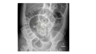
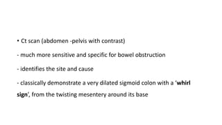
3. Diagnosis involves abdominal x-rays, CT scans, and barium enemas to identify the site of obstruction. Treatment depends on the severity but may include decompression via sigmoidoscopy or surgery to correct ischemia, perforation, or recurrent cases. Comp