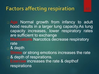
This document provides information on assessing and interpreting vital signs, including temperature, pulse, respiration, blood pressure, and pain. It describes the normal ranges for each vital sign and factors that can influence them. The procedures for measuring each vital sign are outlined, including the appropriate equipment and sites on the body. Reasons for routinely measuring vital signs and guidelines for documentation are also discussed.
Learning Objectives
Identify normal vital sign ranges in the adults. Determine appropriate response to unstable or abnormal vital signs. Identify early trends in vital signs indicative of sepsis, hypoxia, and myocardial infarction. Explain the importance of accurate I&Os and daily weights in the patient's plan of care.
vitals sign is the basic parameter used for all the patients to know the vital and general parameter for the patients and any changes in this parameter can cause the life threatening condition for the patients or clients life the proper technique and its alternatives assessment knowledge can help the nurses to improve academic performance and can be apply this knowledge in their clinical practices
Temperature is the balance between the heat production and heat loss.
A brief outline of diffrent aspects regarding body temperature is discussed here under following headings
*Normal body temperature regulation
*Fever of unknown origin
*Hyperthermia
*Hypothermia
*Frost bite
Fever is an elevation of body temperature that exceeds
normally daily variation and occurs in conjunction with an
increase in the hypothalamic set point for e.g. 37⁰C-
39⁰C.
SIGNAGE for HospitalSIGNAGE for HospitalSIGNAGE for HospitalSIGNAGE for HospitalSIGNAGE for HospitalSIGNAGE for HospitalSIGNAGE for HospitalSIGNAGE for HospitalSIGNAGE for HospitalSIGNAGE for HospitalSIGNAGE for HospitalSIGNAGE for HospitalSIGNAGE for HospitalSIGNAGE for HospitalSIGNAGE for HospitalSIGNAGE for HospitalSIGNAGE for HospitalSIGNAGE for HospitalSIGNAGE for HospitalSIGNAGE for HospitalSIGNAGE for HospitalSIGNAGE for HospitalSIGNAGE for HospitalSIGNAGE for HospitalSIGNAGE for HospitalSIGNAGE for HospitalSIGNAGE for HospitalSIGNAGE for HospitalSIGNAGE for HospitalSIGNAGE for HospitalSIGNAGE for HospitalSIGNAGE for HospitalSIGNAGE for HospitalSIGNAGE for HospitalSIGNAGE for HospitalSIGNAGE for HospitalSIGNAGE for HospitalSIGNAGE for HospitalSIGNAGE for HospitalSIGNAGE for HospitalSIGNAGE for HospitalSIGNAGE for HospitalSIGNAGE for HospitalSIGNAGE for HospitalSIGNAGE for HospitalSIGNAGE for HospitalSIGNAGE for HospitalSIGNAGE for HospitalSIGNAGE for HospitalSIGNAGE for HospitalSIGNAGE for HospitalSIGNAGE for HospitalSIGNAGE for HospitalSIGNAGE for HospitalSIGNAGE for HospitalSIGNAGE for HospitalSIGNAGE for HospitalSIGNAGE for HospitalSIGNAGE for HospitalSIGNAGE for HospitalSIGNAGE for HospitalSIGNAGE for HospitalSIGNAGE for HospitalSIGNAGE for HospitalSIGNAGE for HospitalSIGNAGE for HospitalSIGNAGE for HospitalSIGNAGE for HospitalSIGNAGE for HospitalSIGNAGE for HospitalSIGNAGE for HospitalSIGNAGE for HospitalSIGNAGE for HospitalSIGNAGE for HospitalSIGNAGE for HospitalSIGNAGE for HospitalSIGNAGE for HospitalSIGNAGE for HospitalSIGNAGE for HospitalSIGNAGE for HospitalSIGNAGE for HospitalSIGNAGE for HospitalSIGNAGE for HospitalSIGNAGE for HospitalSIGNAGE for HospitalSIGNAGE for HospitalSIGNAGE for HospitalSIGNAGE for HospitalSIGNAGE for HospitalSIGNAGE for HospitalSIGNAGE for HospitalSIGNAGE for HospitalSIGNAGE for HospitalSIGNAGE for HospitalSIGNAGE for HospitalSIGNAGE for HospitalSIGNAGE for HospitalSIGNAGE for HospitalSIGNAGE for HospitalSIGNAGE for HospitalSIGNAGE for Hospital
AN APPROACH TO SEDATION IN ICU
Sedative medications should be titrated to maintain a light rather than a deep level of sedation in adult ICU patients, unless contraindicated.
keep patients comfortable and safe using the minimum possible amount of sedation.
use protocolised care with sedation score monitoring.
Adverse Drug Recation-Adverse Drug Reaction (ADR): Any noxious change which is suspected to be due to a drug, occurs at doses normally used in man, requires treatment or decrease in dose or indicates caution in future use of the same drug.
Medication Administration
Policy & Process
Medication Administration
Policy & Process
Medication Administration
Policy & Process
Medication Administration
Policy & Process
Glasgow Coma Scale (GCS) assessment is an important aspect in neurological assessment and its management. It helps in the objective assessment of the patients and facilitates accurate interpersonal communication.
Objectives of learning pressure ulcer
evaluate the strengths and limitations of pressure ulcer guidelines; discuss the challenges related to clinical trials in the domain of pressure ulcers; discuss methods and educational strategies for implementing pressure ulcer prevention and treatment protocols in practice.
R3 Stem Cells and Kidney Repair A New Horizon in Nephrology.pptxR3 Stem Cell
R3 Stem Cells and Kidney Repair: A New Horizon in Nephrology" explores groundbreaking advancements in the use of R3 stem cells for kidney disease treatment. This insightful piece delves into the potential of these cells to regenerate damaged kidney tissue, offering new hope for patients and reshaping the future of nephrology.
Deep Leg Vein Thrombosis (DVT): Meaning, Causes, Symptoms, Treatment, and Mor...The Lifesciences Magazine
Deep Leg Vein Thrombosis occurs when a blood clot forms in one or more of the deep veins in the legs. These clots can impede blood flow, leading to severe complications.
Navigating Challenges: Mental Health, Legislation, and the Prison System in B...Guillermo Rivera
This conference will delve into the intricate intersections between mental health, legal frameworks, and the prison system in Bolivia. It aims to provide a comprehensive overview of the current challenges faced by mental health professionals working within the legislative and correctional landscapes. Topics of discussion will include the prevalence and impact of mental health issues among the incarcerated population, the effectiveness of existing mental health policies and legislation, and potential reforms to enhance the mental health support system within prisons.
Global launch of the Healthy Ageing and Prevention Index 2nd wave – alongside...ILC- UK
The Healthy Ageing and Prevention Index is an online tool created by ILC that ranks countries on six metrics including, life span, health span, work span, income, environmental performance, and happiness. The Index helps us understand how well countries have adapted to longevity and inform decision makers on what must be done to maximise the economic benefits that comes with living well for longer.
Alongside the 77th World Health Assembly in Geneva on 28 May 2024, we launched the second version of our Index, allowing us to track progress and give new insights into what needs to be done to keep populations healthier for longer.
The speakers included:
Professor Orazio Schillaci, Minister of Health, Italy
Dr Hans Groth, Chairman of the Board, World Demographic & Ageing Forum
Professor Ilona Kickbusch, Founder and Chair, Global Health Centre, Geneva Graduate Institute and co-chair, World Health Summit Council
Dr Natasha Azzopardi Muscat, Director, Country Health Policies and Systems Division, World Health Organisation EURO
Dr Marta Lomazzi, Executive Manager, World Federation of Public Health Associations
Dr Shyam Bishen, Head, Centre for Health and Healthcare and Member of the Executive Committee, World Economic Forum
Dr Karin Tegmark Wisell, Director General, Public Health Agency of Sweden
How many patients does case series should have In comparison to case reports.pdfpubrica101
Pubrica’s team of researchers and writers create scientific and medical research articles, which may be important resources for authors and practitioners. Pubrica medical writers assist you in creating and revising the introduction by alerting the reader to gaps in the chosen study subject. Our professionals understand the order in which the hypothesis topic is followed by the broad subject, the issue, and the backdrop.
https://pubrica.com/academy/case-study-or-series/how-many-patients-does-case-series-should-have-in-comparison-to-case-reports/
Defecation
Normal defecation begins with movement in the left colon, moving stool toward the anus. When stool reaches the rectum, the distention causes relaxation of the internal sphincter and an awareness of the need to defecate. At the time of defecation, the external sphincter relaxes, and abdominal muscles contract, increasing intrarectal pressure and forcing the stool out
The Valsalva maneuver exerts pressure to expel faeces through a voluntary contraction of the abdominal muscles while maintaining forced expiration against a closed airway. Patients with cardiovascular disease, glaucoma, increased intracranial pressure, or a new surgical wound are at greater risk for cardiac dysrhythmias and elevated blood pressure with the Valsalva maneuver and need to avoid straining to pass the stool.
Normal defecation is painless, resulting in passage of soft, formed stool
CONSTIPATION
Constipation is a symptom, not a disease. Improper diet, reduced fluid intake, lack of exercise, and certain medications can cause constipation. For example, patients receiving opiates for pain after surgery often require a stool softener or laxative to prevent constipation. The signs of constipation include infrequent bowel movements (less than every 3 days), difficulty passing stools, excessive straining, inability to defecate at will, and hard feaces
IMPACTION
Fecal impaction results from unrelieved constipation. It is a collection of hardened feces wedged in the rectum that a person cannot expel. In cases of severe impaction the mass extends up into the sigmoid colon.
DIARRHEA
Diarrhea is an increase in the number of stools and the passage of liquid, unformed feces. It is associated with disorders affecting digestion, absorption, and secretion in the GI tract. Intestinal contents pass through the small and large intestine too quickly to allow for the usual absorption of fluid and nutrients. Irritation within the colon results in increased mucus secretion. As a result, feces become watery, and the patient is unable to control the urge to defecate. Normally an anal bag is safe and effective in long-term treatment of patients with fecal incontinence at home, in hospice, or in the hospital. Fecal incontinence is expensive and a potentially dangerous condition in terms of contamination and risk of skin ulceration
HEMORRHOIDS
Hemorrhoids are dilated, engorged veins in the lining of the rectum. They are either external or internal.
FLATULENCE
As gas accumulates in the lumen of the intestines, the bowel wall stretches and distends (flatulence). It is a common cause of abdominal fullness, pain, and cramping. Normally intestinal gas escapes through the mouth (belching) or the anus (passing of flatus)
FECAL INCONTINENCE
Fecal incontinence is the inability to control passage of feces and gas from the anus. Incontinence harms a patient’s body image
PREPARATION AND GIVING OF LAXATIVESACCORDING TO POTTER AND PERRY,
An enema is the instillation of a solution into the rectum and sig
India Clinical Trials Market: Industry Size and Growth Trends [2030] Analyzed...Kumar Satyam
According to TechSci Research report, "India Clinical Trials Market- By Region, Competition, Forecast & Opportunities, 2030F," the India Clinical Trials Market was valued at USD 2.05 billion in 2024 and is projected to grow at a compound annual growth rate (CAGR) of 8.64% through 2030. The market is driven by a variety of factors, making India an attractive destination for pharmaceutical companies and researchers. India's vast and diverse patient population, cost-effective operational environment, and a large pool of skilled medical professionals contribute significantly to the market's growth. Additionally, increasing government support in streamlining regulations and the growing prevalence of lifestyle diseases further propel the clinical trials market.
Growing Prevalence of Lifestyle Diseases
The rising incidence of lifestyle diseases such as diabetes, cardiovascular diseases, and cancer is a major trend driving the clinical trials market in India. These conditions necessitate the development and testing of new treatment methods, creating a robust demand for clinical trials. The increasing burden of these diseases highlights the need for innovative therapies and underscores the importance of India as a key player in global clinical research.
2. Describe the procedures used to assess the vital
signs: Temperature, Pulse, Respiration, And
Blood Pressure, Pain Assessment.
Identify factors that can influence each vital sign.
Identify equipment routinely used to assess vital
signs.
Identify rationales for using different routes for
assessing temperature.
Take vital signs and interpret the finding.
Document the vital signs.
2
3. Introduction
•Vital sign are a basic component of assessment of
physiological and psychological health of a client.
•body temperature, pulse, respiration, blood pressure
and Pain are the sign of life
•Assessment of vitals sign allow the nurses to identify
specific life threatening conditions and plan the
needed nursing intervention
•Detect changes in the client health status
4. Vital signs reflect the body’s physiologic
status and provide information critical to
4
evaluating homeostatic balance.
Includes:
temperature,
Pulse Rate,
Respiratory Rate),
Blood Pressure)
Pain
5. To obtain base line data about the patient
condition
For diagnostic purpose
For therapeutic purpose
5
6. Vital sign tray
Stethoscope
Sphygmomanometer
Thermometer
Hand watch (With Sec. Counter)
Red and blue pen
Pencil;
Vital sign sheet
Cotton swab in bowel
Disposable gloves if available
Dirty receiver kidney dish
6
7. On admission – to obtain baseline date
When a client has a change in health status or reports
symptoms such as chest pain or fainting
According to a nursing or medical order
Before and after the administration of certain
medications that could affect RR or BP (Respiratory and
CVS
Before and after surgery or an invasive diagnostic
procedures
Before and after any nursing intervention that could
affect the vital signs. E.g. Ambulation
According to hospital /other health institution policy.
7
8. Body temperature may be defined as the degree
of heat maintained by the body. It is the balance
between heat production & heat loss of the body
Normal body temperature using oral 370 Celsius
or 98.6 0 F.
8
9. Mechanism of temperature regulation
Thermogenesis – a chemical regulation by the
production of heat
Thermolysis – a physical regulation by loss of heat
our internal boady temperature is regulated by a part of
our brain called the hypothalamus. The hypothalamus
checks our current temperature and compares it with
the normal temperature of about 37 Degree Celsius. If
our temp. is too low the hypothalamus
Makes sure that the boady generates and maintain heat.
Temp. is too high heat is given off or sweat is produced
to cool the skin.
10. 1. Core Temperature
Is the temperature of internal organs
and it remains constant most of the
time (37oc); with range of 36.5-37.5oc.
Is the Temperature of the deep tissues
of the body Remains relatively constant
measure with thermometer
10
11. 2. Surface Temperature:
11
o Surface body temperature: - is the
temperature of the skin, subcutaneous tissue
& fat cells and itrises & falls in response to
the environment
o (Ranges b/n 20-40oc).
o It doesn’t indicate internal physiology.
12. Way of producing heat in the body
Oxidation of the food
Specific dynamic action of food
Exercise
Strong emotions
Hormonal effects
Change in the environment and
atmospheric
conditions
Diseased conditions
13. Way of loosing heat from the body
• Through the skin
• Through the lungs
• Through the kidneys
• Through the bowels
14. Normal variation in the body temperature
Time of the days
Time of the month
Age of the person
Part of the body where the temperature is taken
Emotions
Exercise
Fasting
Environmental factors
15. Normal body temperature is 370 C or 98.6 0F (range
is 36-38 0c (96.8 – 98.6 0F))
Body temperature may be abnormal due to fever
(high temperature) or hypothermia (low
temperature).
Pyrexia, fever: a body temperature above the
normal ranges 38 0c – 410 c (100.4 –105.8 F)
Hyper pyrexia: a very high fever, such as 410 C > 42
0c leads to death.
Hypothermia: – body temperature between 34 0c –
35 0c, < 34 0c is death
15
17. Oral
Rectal
Auxiliary
Tympanic
Thermometer: is an instrument used
to
measure body temperature
17
18.
19. Types of fever
Onset or invasion : onset or invasion of fever is the period when
the
body temperature is raising and it may be a sudden or gradual
process.
Fastigium or stadium : fastigium or stadium of fever is the
period when the body temperature has reached its maximum
and remainsfairly constant at a high level.
Defervescence or decline : defervescence or decline of the fever
is period when the elevated temperature is returning to normal.
The fever may subside suddenly (decline by crisis) or gradually
(decline by lysis)
Crisis : crisis is a sudden return to normal temperature from a
very high temperature within a few hours or days
20. •True crises : the temperature falls suddenly within few
hours and touches normal, accompanied by a marked
improvement in the clients conditions
•False crises : the sudden falls in temperature not
accompanied by a improvement in the clients
conditions is called false crises.
•Lysis : the temperature falls in zig-zag manner for 2 or
3 days or a week before reaching normal, during which
time the other symptom also gradually disappear
•Constant fever or continuous fever : constant fever
or continuous fever is one in which the temperature
varies not more than two degree between morning and
evening and it does not reach normal for a period of
days or week
21. Remittent fever : remitting fever is a fever
characterized by variation of more than two degree
between morning and evening but does not reach
normal
Intermittent or quotidian fever : the temperature
rises from normal or sub normal to high fever and back
at regular interval. Usually the temperature is higher in
the evening then in the morning
Inverse fever: the highest range of temperature is
recorded in the morning hours and the lowest in the
evening which is contrary to that found in the normal
course of fever
hectic or swinging fever : when the difference between
the high and low points is very great, the fever is called
hectic fever.
22. Relapsing fever: relapsing fever is one in which there
are brief febrile period followed by one or more days
of normal temperature
Irregular fever: when the fever is entirely irregular
in its course, it cannot be classified under any one of
the fever described above is called irregular fever
Rigor : rigor is a sudden severe attack of shivering in
which the body temperature rises rapidly to a stage of
hyperpyrexia as seen in malaria
23. Nursing care in fever
1. Regulation of body temperature
2. Meeting the nutritional need
3. Maintenance of personal hygiene
4. Providing rest and sleep
5. Maintenance of personal hygiene
6. Safety factor
7. Observation of the client
24. Nursing care in rigor
1. Stage one or cold stage
2. Stage two or hot stage
3. Stage three or stage of sweating
26. Pulse is a wave of blood created by the
contraction of left ventricle.
pulse reflects the heart beat
Stroke volume and the compliance of
arterial wall are the two important factors
influencing pulse rate.
Pulse rate is regulated by autonomic
nervous system.
26
27. Peripheral Pulse: is a pulse located in the
periphery of the body e.g. in the foot, and or neck
Apical Pulse (central pulse): it is located at the
apex of the heart
The PR is expressed in beats/ minute (BPM)
The difference between peripheral and apical pulse
is called pulse deficit, and it is usually zero.
27
28. Pulse is assessedfor
28
• rate (60-100bpm),
• rhythm (regularity orirregularity),
• Volume,
• elasticity of arterialwall.
The pulse is commonly assessed by palpation
(feeling) and auscultation (hearing using a
stethoscope).
29. Age
The average pulse rate of an infant ranges from
100 to 160BPM
The normal range of the pulse in an adult is 60
to 100 BPM
Sex: Sex: after puberty the average males
PRis slightly lower than female
30
30. Autonomic Nervous systemactivity
30
Stimulation of the parasympathetic nervous
system results in decrease in the PR
Stimulation of sympathetic nervous system
results in an increased pulse rate
Sympathetic nervous system activation occurs
on response to a variety of stimuli including
▪ Pain ,anxiety ,Exercise, Fever
▪ Ingestion of caffeinated beverages
▪ Change in intravascular volume
31. Exercise: PRincrease with exercise Fever:
increases PRin response to the lowered
B/P that results from peripheral
vasodilatation – increased metabolic rate
Heat: increase PRas acompensatory
mechanism
Stress: increases the sympathetic nerve
stimulation
31
32. * Position changes:
a sitting or standing position blood usually
pools in dependent vessels of the venous
system. B/c of decrease in the venous blood
return to heart and subsequent decrease in BP
increases heart rate.
32
33. * Medication
33
o Cardiac medication such as digoxin decrease heart
rate
o Medications that decrease intravascular volume
such as diuretics may increase pulse rate
o Atropine inhibits impulses to the heart from the
parasympathetic nervous system, causing increased
pulse rate
o Propranolol blocks sympathetic nervous system
action resulting in decreased heart rate sites used
for measuring pulse rate
34. Carotid: at the side of the neck below tube of the
ear (where the carotid artery runs between the
trachea and the sternocleidomastoid muscle)
Temporal: the pulse is taken at temporalbone
area.
Apical: at the apex of the heart: routinely used
for infant and children < 3 yrs
In adults – Left mid-clavicular line under the
4th, 5th, 6th intercostalspace
34
35. Brachial: at the inner aspect of the biceps muscle
of the arm or medially in the antecubital space
(elbow crease)
Radial: on the thumb side of the inner aspect of
the wrist – readily available and routinely used
Femoral: along the inguinal ligament. Used or
infants and children
Popiliteal: behind the knee. By flexing the knee
slightly
Posterior tibial: on the medial surface of the
ankle
Pedal (Dorsal Pedis): palpated by feeling
thedorsum
35
36.
37.
38. A wave of
blood flow
created by
a
contraction
of
theheart. .
.
A.
39
B.
D.
E.
F
.
C. G.
H.
39. Pulse: is commonly assessed by
palpation (feeling) or
auscultation(hearing)
49
The middle 3 fingertips are used with
moderate pressure for palpation of all pulses
except apical;
Assess the pulsefor
Rate
Rhythm
Volume
Elasticity of the arterial wall
41. Normal Pulse Rate is 60-100 b/min)
41
Adult PR>100BPM is calledtachycardia
Adult PR<60 BPM is calledbradycardia
42. Pulse Rhythm
The pattern and interval between the beats,
random, irregular beats– dysrythymia
PulseVolume
the force of blood with each beat
Anormal pulse can be felt with moderate
pressure of thefingers
Full or bounding pulse forceful or full blood
volume destroy with difficulty
Weak, feeble readily destroy with pressure
from the fingertips
42
43. Elasticity of arterialwall
Ahealthy, normal artery feels, straight,
smooth, soft, easilybent
Reflects the status of the clients
vascular
system
43
44. If the pulse is regular, measure (count) for
30 seconds and multiply by 2
If it is irregular count for 1 full minute.
Each heart beat consists of two sounds
s1 - is caused by closure of the mitral and
tricuspid
valves separating the atria from the ventricles
S2 – is caused by the closure of the plutonic
and aortic values
The sounds are often described as a muffled
“lub – bub”
44
46. Respiration rate (RR):-Respiration is the
act of breathing and includes the intake of
oxygen and removal of carbon-dioxide.
Ventilation is also another word, which
refers
to movement of air in and out of the lung.
Hyperventilation: - is a very deep, rapid
respiration.
Hypoventilation: -is a very shallow
respiration.
46
47. 1. Costal (thoracic)
Observed by the movement of the chest up
ward and down ward.
Commonly used for adults
2. Diaphragmatic
(abdominal)
Involves the contraction and relaxation of the
diaphragm, observed by the movement of
abdomen.
Commonly used for children.
47
48. Age Normal growth from infancy to adult
hood results in a larger lung capacity.As lung
capacity increases, lower respiratory rates
are sufficient to exchange
Medications Narcotics decrease respiratory
rate
& depth
Stress or strong emotions increases the rate
& depth of respirations.
Exercise increases the rate & depthof
respirations
48
49. Altitude The rate & depth of respirations at
higher elevations (altitude) increase to
improve the supply of oxygen available to
the body tissues
Gender Men may have a lower
respirations rate than women because
men normally have a larger rung capacity
than women
Fever increases respiratoryrate
50
50. o The client should be at
rest
50
o Assessed by watching the movement of the
chest or abdomen.
o Rate,
o rhythm,
o depth and
ospecial characteristics of respiration
are assessed
51. Rate:
Is described in rate per minute (RPM)
Healthy adult RR= 15- 20/ min. is measured
for full minute, if regular for 30 seconds.
As the age decreases the respiratory rate
increases.
Eupnea- normal breathing rate and depth
Bradypnea- slowrespiration
Tachypnea - fastbreathing
Apnea - temporary cessation of breathing
51
52. 52
Age Average Range/Min
New born 30-80
Early childhood 20-40
Late childhood 15-25
Adulthood-male 14-18
Female 16-20
53. Rhythm:
is the regularity of expiration and
inspiration Normal breathing is automatic &
effortless.
Depth:
described as normal, deep or shallow.
Deep: a large volume of air inhaled &
exhaled, inflates most of the lungs.
Shallow: exchange of a small volume of air
minimal use of lung tissue.
53
54. It is the force exerted by the blood against
the walls of the arteries in which it is flowing.
It is expressedin terms of millimeters of
mercury (mm ofHg).
54
55. Systolic pressure is the maximum of the
pressure against the wall of the vessel
following ventricularcontraction.
Diastolic pressure is the minimum
pressure of the blood against the walls of
the vessels following closure of aortic
valve (ventricular relaxation).
55
56. BP is measured by using an instrument
called Bp cuff (sphygmomanometer) &
stethoscope and
the average normal value is 120/80mmHg
for
adults.
brachial artery and popliteal artery are most
commonly used.
It is measured by securing the Bp cuff to the
upper arm & thigh placing the stethoscope
on brachial artery in the antecubital space &
popliteal artery at the back of the knee.
Pulse pressure: is the difference between
the
56
58. Upperarm (using brachial artery
(commonest)
Thigh around poplitealartery
Fore-arm using radialartery
Legusing posterior tibial or dorsal pedis
58
59. Apersistently high Bp, measured for greater
than three times is called hypertension & that
persistently less than normal range is called
hypotension.
Because of many factors influencing Bp a
single
measurement is not necessarily significant to
confirm hypertension.
When the cause of hypertension is known it is
called secondary hypertension and when the
cause is unknown is calledprimary/essential
hypertension. 60
60. Purpose
To obtain base line measure of arterial
blood pressure for subsequentevaluation
To determine the clients homodynamic
status
To identify and monitor changes in blood
pressure.
60
64. Explain the procedure to the patient &
remove
any light cloth from patient’s arm
Make sure that the client has not smoked or
ingested caffeine, within 30 minutes prior to
measurement.
Position the patient on lying, sitting or
standing position, but always ensure that
the sphygmomanometer is at the level of the
heart with the arm supported & the palm
facing upwards.
64
65. apply cuff snugly/securely around the arm ,
2.5cm above the antecubital space/fossa, at
the level of the heart (for every cm the cuff
sites above or below the level of the heart
the BP varies by 0.8mmHg)
Palpate the radial pulse and inflate the cuff
until the radial pulse can no longer be felt,
this provides an estimation of systolic
pressure.
Inflate cuff 30mmHg higher than estimated
systolic pressure.
65
66. palpate the brachial artery & place the bell of
the stethoscope over the site & the ear
pieces on ear, apply enough pressure to
keep the stethoscope in place (the bell of the
stethoscope is designed to amplify/intensify
low frequency sounds)
Deflate the cuff 2-4mmHg per second.
The first pulse heard is the systolic reading,
continue to deflate until there is a change in
tone to a muffled beat, this is the diastolic
reading.
66
67. Deflate & remove cuff roll neatly and
replace.
Record the systolic and diastolic pressure
on vital singsheet and compare the
present reading with previous reading.
report or treat any change
Clear ear pieces and bell of the
stethoscope with antiseptic swab and
return all equipments.
67