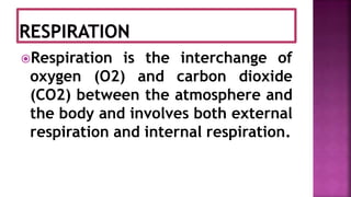
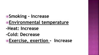
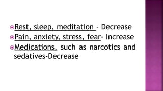
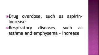
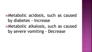
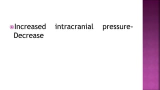
PREPARED BY:
USHA RANI KANDULA,
ASSISTANT PROFESSOR,
DEPARTMENT OF ADULT HEALTH
NURSING,
ARSI UNIVERSITY,ASELLA,ETHIOPIA,
SOUTH EAST AFRICA.
Respiration is the interchange of
oxygen (O2) and carbon dioxide
(CO2) between the atmosphere and
the body and involves both external
respiration and internal respiration.
Respiration is the exchange
of these gases between the
lungs’ alveoli and the blood
found in the capillaries that
surround the alveoli.
Internal respiration is the
process of exchanging gases
between the circulating blood
and the tissue cells that make
up the body.
The movement of air into and out
of the lungs is known as ventilation.
The mechanics of respiration involve
the act of breathing in, termed as
Inhalation or inspiration,
and breathing out, described as
Exhalation or expiration.
Although the act of breathing is a
function of the respiratory system, it is
regulated by the central nervous system,
specifically by the medulla oblongata and
the pons, two structures of the
brainstem, and by chemoreceptors in the
carotid and aortic bodies.
The carotid body is a section of the
carotid artery wall, and the aortic
body is a section of the aortic arch
wall, both of which contain chemical
receptor sites that detect decreases
in the blood level of O2, known as
Hypoxemia.
When hypoxemia is detected,
electrical impulses are generated
and sent via nerves to the medulla,
which will then increase the
respiratory rate or depth as needed
to correct the hypoxemia.
To effect inspiration, the medulla
sends an impulse via the phrenic
nerves to the diaphragm muscle
and along the intercostals nerves to
the intercostal muscles, telling the
muscles to contract.
The contraction of the diaphragm
causes it to flatten and move
downward, while the contraction of
the intercostals muscles results in
pulling the ribs upward and
outward, enlarging the chest cavity.
Once the medulla stops sending the
motor impulses to inhale, the
intercostal and diaphragm muscles
begin to relax, once again shrinking
the thoracic or chest cavity to the
smaller, pre-inhalation state and
compressing the lungs.
 The elastic connective tissue of
the alveoli recoils somewhat like a
rubber band, forcing the air that is
mostly CO2 out of the alveoli so
that it can be exhaled and returned
to the atmosphere.
The only equipment required
to assess respiration is a
watch with a second hand.
Rate per minute
Depth
Rhythm
Pattern
Respiratory effort
Because individuals can
voluntarily control their
breathing for short intervals
of time, it is best to assess
the respirations without the
patient’s awareness.
This may be accomplished by
positioning the patient’s arm
across his or her chest or
abdomen.
Feel for the radial pulse and
hold the pulse site while you
assess first the respirations
and then the radial pulse.
 It will appear that you are
simply taking the patient’s
pulse, and this will help to
distract the patient from
concentrating on his or her
breathing.
While pressing two fingers against
the pulse site, count the
respiratory rate for 30 seconds and
multiply times two for a 1-minute
rate, and then continue to palpate
the radial pulse site to assess the
pulse rate.
 If the patient is very ill or
respirations deviate in any
manner from the norm,
assess for a full minute.
Each respiration consists of one
Inspiration and one expiration.
Observe the rise and fall of the
chest or abdomen to count the
rate.
The normal rate for adults is
between 12 and 20 bpm.
When the rate, depth,
rhythm, pattern, and
respiratory effort fall within
normal parameters, the term
eupnea is used to describe
the respirations.
Bradypnea describes a
respiratory rate below 12
respirations per minute;
when the rate exceeds 20
respirations per minute, the
term is Tachypnea.
When the breathing rate
slows, it results in a
decreased intake of O2 that
can result in a deficiency of
O2 in the tissues and cells.
When respirations cease or are
absent, this is known as Apnea.
You have only a brief window of 3
to 5 minutes in which to restore
respirations before brain damage
and death occur.
Because the heart and lungs work
together to provide circulation of
nutrients and oxygen, the factors
that affect the heart rate generally
affect the respiratory rate as well.
For example, as fever raises the
pulse rate, it will also increase the
respiratory rate.
Breathing speeds up in an
attempt to meet the body’s
increased metabolic needs
and to remove excess heat.
For every 1°F rise in body
temperature, the respiratory rate
increases approximately four breaths
per minute.
The depth of respiration is observed
by the amount of chest expansion
with each breath and is related to
the volume of air that is inhaled.
The average amount of air inhaled in
one breath is between 300 and 500
mL and is known as the tidal volume,
which can be assessed with special
equipment.
Without special equipment, you can
onlyn observe the rise and fall of the
chest to provide a subjective
measurement of the depth, usually
described as shallow, normal, or
deep.
The rhythm of normal respirations is
one that is regular, or has evenly
spaced intervals between the
respirations.
When the intervals are not
consistent, you assess them as
irregular.
When the rhythm is irregular,
further assess the respirations to
determine if the respirations fall
into certain patterns.
Respiratory effort refers to the
amount of work required to breathe.
Normally, the act of breathing is
effortless and is performed
unconsciously.
An individual who is having
labored or difficult breathing
is said to be having Dyspnea.
The severely dyspneic
patient will usually appear
frightened, anxious, or
worried.
The first thing you may
notice when you look at the
patient is a “wide-eyed” or
startled appearance.
This may be indicative that the
impaired gas exchange has
progressed to the point of
hypoxemia or hypoxia.
Hypoxemia is the term used
to denote a decreased oxygen
level in the blood.
 If hypoxemia is not relieved,
hypoxia, or decreased
delivery of oxygen to the
tissues and cells, occurs.
Often the patient does not appear
to have dyspnea until he or she
begins to exert energy, which
increases the body’s metabolic
demand for oxygen.
Exertional dyspnea only occurs
during activities such as speaking,
eating, repositioning, or
ambulating.
Another sign of difficult breathing
is use of the accessory respiratory
muscles: the neck and abdominal
muscles.
It is important to assess which
activities result in exertional
dyspnea, document findings in the
patient’s record, and report such
findings to the RN supervisor.
Some patients find it too difficult to
breathe unless positioned in an
upright position, such as sitting or
standing.
This condition is known as
orthopnea.
Other respiratory assessments go
hand in hand with those previously
detailed, including the audible
sounds during ventilation and the
breath sounds that are auscultated
with a stethoscope:
Stertorous breathing refers to
noisy, snoring, labored respirations
that are audible without a
stethoscope.
Adventitious sounds are abnormal
sounds that may be heard when
auscultating the lungs with a
stethoscope and include wheezes,
rales or crackles, rhonchi, and
stridor.
Wheezes are musical, whistling
sounds that may be audible without
a stethoscope or heard only during
auscultation.
Rales, or crackles, are adventitious
sounds that may be auscultated
with a stethoscope and are the
result of air moving over secretions
in the lungs.
Rales are short, choppy, popping,
snapping, or raspy sounds that may
resemble the sound made by
rubbing strands of hair between
your thumb and index finger.
Rales may be classified as fine or
coarse depending on their
characteristics.
Rhonchi are continuous, low-
pitched, rattling or bubbling,
snoring or sonorous wheezing
sounds that can be auscultated
when there is partial obstruction of
the larger airways due to secretions
or tumor.
Stridor is an audible, high-pitched
crowing sound that results from
partial obstruction of the airways.
Newborns: 30–60
Infants: 20–40
Children: 20–30
Adolescents: 14–25
Adults: 12–20
Smoking - Increase
Environmental temperature
-Heat: Increase
-Cold: Decrease
Exercise, exertion - Increase
Rest, sleep, meditation - Decrease
Pain, anxiety, stress, fear- Increase
Medications, such as narcotics and
sedatives-Decrease
Drug overdose, such as aspirin-
Increase
Respiratory diseases, such as
asthma and emphysema - Increase
Metabolic acidosis, such as caused
by diabetes - Increase
Metabolic alkalosis, such as caused
by severe vomiting - Decrease
Increased intracranial pressure-
Decrease
Respirations begin shallow, gradually
increase in depth and frequency to
a peak,
Then begin to decrease in depth and
frequency until slow and shallow;
this is followed by a period of
apnea lasting from 10 to 60
seconds.
Pattern is repetitious.
Respirations are increased in rate
and depth, with long, strong,
blowing or grunting exhalations.
Respirations are grouped as several
shallow breaths followed by
variable-length periods of apnea.
Vital signs - Respiration

Vital signs - Respiration

  • 1.
    PREPARED BY: USHA RANIKANDULA, ASSISTANT PROFESSOR, DEPARTMENT OF ADULT HEALTH NURSING, ARSI UNIVERSITY,ASELLA,ETHIOPIA, SOUTH EAST AFRICA.
  • 2.
    Respiration is theinterchange of oxygen (O2) and carbon dioxide (CO2) between the atmosphere and the body and involves both external respiration and internal respiration.
  • 3.
    Respiration is theexchange of these gases between the lungs’ alveoli and the blood found in the capillaries that surround the alveoli.
  • 4.
    Internal respiration isthe process of exchanging gases between the circulating blood and the tissue cells that make up the body.
  • 5.
    The movement ofair into and out of the lungs is known as ventilation.
  • 6.
    The mechanics ofrespiration involve the act of breathing in, termed as Inhalation or inspiration, and breathing out, described as Exhalation or expiration.
  • 7.
    Although the actof breathing is a function of the respiratory system, it is regulated by the central nervous system, specifically by the medulla oblongata and the pons, two structures of the brainstem, and by chemoreceptors in the carotid and aortic bodies.
  • 8.
    The carotid bodyis a section of the carotid artery wall, and the aortic body is a section of the aortic arch wall, both of which contain chemical receptor sites that detect decreases in the blood level of O2, known as Hypoxemia.
  • 9.
    When hypoxemia isdetected, electrical impulses are generated and sent via nerves to the medulla, which will then increase the respiratory rate or depth as needed to correct the hypoxemia.
  • 10.
    To effect inspiration,the medulla sends an impulse via the phrenic nerves to the diaphragm muscle and along the intercostals nerves to the intercostal muscles, telling the muscles to contract.
  • 11.
    The contraction ofthe diaphragm causes it to flatten and move downward, while the contraction of the intercostals muscles results in pulling the ribs upward and outward, enlarging the chest cavity.
  • 12.
    Once the medullastops sending the motor impulses to inhale, the intercostal and diaphragm muscles begin to relax, once again shrinking the thoracic or chest cavity to the smaller, pre-inhalation state and compressing the lungs.
  • 13.
     The elasticconnective tissue of the alveoli recoils somewhat like a rubber band, forcing the air that is mostly CO2 out of the alveoli so that it can be exhaled and returned to the atmosphere.
  • 15.
    The only equipmentrequired to assess respiration is a watch with a second hand.
  • 16.
  • 17.
    Because individuals can voluntarilycontrol their breathing for short intervals of time, it is best to assess the respirations without the patient’s awareness.
  • 18.
    This may beaccomplished by positioning the patient’s arm across his or her chest or abdomen.
  • 19.
    Feel for theradial pulse and hold the pulse site while you assess first the respirations and then the radial pulse.
  • 20.
     It willappear that you are simply taking the patient’s pulse, and this will help to distract the patient from concentrating on his or her breathing.
  • 21.
    While pressing twofingers against the pulse site, count the respiratory rate for 30 seconds and multiply times two for a 1-minute rate, and then continue to palpate the radial pulse site to assess the pulse rate.
  • 22.
     If thepatient is very ill or respirations deviate in any manner from the norm, assess for a full minute.
  • 23.
    Each respiration consistsof one Inspiration and one expiration. Observe the rise and fall of the chest or abdomen to count the rate. The normal rate for adults is between 12 and 20 bpm.
  • 24.
    When the rate,depth, rhythm, pattern, and respiratory effort fall within normal parameters, the term eupnea is used to describe the respirations.
  • 25.
    Bradypnea describes a respiratoryrate below 12 respirations per minute; when the rate exceeds 20 respirations per minute, the term is Tachypnea.
  • 26.
    When the breathingrate slows, it results in a decreased intake of O2 that can result in a deficiency of O2 in the tissues and cells.
  • 27.
    When respirations ceaseor are absent, this is known as Apnea. You have only a brief window of 3 to 5 minutes in which to restore respirations before brain damage and death occur.
  • 28.
    Because the heartand lungs work together to provide circulation of nutrients and oxygen, the factors that affect the heart rate generally affect the respiratory rate as well.
  • 29.
    For example, asfever raises the pulse rate, it will also increase the respiratory rate.
  • 30.
    Breathing speeds upin an attempt to meet the body’s increased metabolic needs and to remove excess heat.
  • 31.
    For every 1°Frise in body temperature, the respiratory rate increases approximately four breaths per minute.
  • 32.
    The depth ofrespiration is observed by the amount of chest expansion with each breath and is related to the volume of air that is inhaled.
  • 33.
    The average amountof air inhaled in one breath is between 300 and 500 mL and is known as the tidal volume, which can be assessed with special equipment.
  • 34.
    Without special equipment,you can onlyn observe the rise and fall of the chest to provide a subjective measurement of the depth, usually described as shallow, normal, or deep.
  • 35.
    The rhythm ofnormal respirations is one that is regular, or has evenly spaced intervals between the respirations.
  • 36.
    When the intervalsare not consistent, you assess them as irregular.
  • 37.
    When the rhythmis irregular, further assess the respirations to determine if the respirations fall into certain patterns.
  • 38.
    Respiratory effort refersto the amount of work required to breathe. Normally, the act of breathing is effortless and is performed unconsciously.
  • 39.
    An individual whois having labored or difficult breathing is said to be having Dyspnea.
  • 40.
    The severely dyspneic patientwill usually appear frightened, anxious, or worried.
  • 41.
    The first thingyou may notice when you look at the patient is a “wide-eyed” or startled appearance.
  • 42.
    This may beindicative that the impaired gas exchange has progressed to the point of hypoxemia or hypoxia.
  • 43.
    Hypoxemia is theterm used to denote a decreased oxygen level in the blood.
  • 44.
     If hypoxemiais not relieved, hypoxia, or decreased delivery of oxygen to the tissues and cells, occurs.
  • 45.
    Often the patientdoes not appear to have dyspnea until he or she begins to exert energy, which increases the body’s metabolic demand for oxygen.
  • 46.
    Exertional dyspnea onlyoccurs during activities such as speaking, eating, repositioning, or ambulating.
  • 47.
    Another sign ofdifficult breathing is use of the accessory respiratory muscles: the neck and abdominal muscles.
  • 48.
    It is importantto assess which activities result in exertional dyspnea, document findings in the patient’s record, and report such findings to the RN supervisor.
  • 49.
    Some patients findit too difficult to breathe unless positioned in an upright position, such as sitting or standing. This condition is known as orthopnea.
  • 51.
    Other respiratory assessmentsgo hand in hand with those previously detailed, including the audible sounds during ventilation and the breath sounds that are auscultated with a stethoscope:
  • 52.
    Stertorous breathing refersto noisy, snoring, labored respirations that are audible without a stethoscope.
  • 53.
    Adventitious sounds areabnormal sounds that may be heard when auscultating the lungs with a stethoscope and include wheezes, rales or crackles, rhonchi, and stridor.
  • 54.
    Wheezes are musical,whistling sounds that may be audible without a stethoscope or heard only during auscultation.
  • 55.
    Rales, or crackles,are adventitious sounds that may be auscultated with a stethoscope and are the result of air moving over secretions in the lungs.
  • 56.
    Rales are short,choppy, popping, snapping, or raspy sounds that may resemble the sound made by rubbing strands of hair between your thumb and index finger.
  • 57.
    Rales may beclassified as fine or coarse depending on their characteristics.
  • 58.
    Rhonchi are continuous,low- pitched, rattling or bubbling, snoring or sonorous wheezing sounds that can be auscultated when there is partial obstruction of the larger airways due to secretions or tumor.
  • 59.
    Stridor is anaudible, high-pitched crowing sound that results from partial obstruction of the airways.
  • 61.
    Newborns: 30–60 Infants: 20–40 Children:20–30 Adolescents: 14–25 Adults: 12–20
  • 62.
    Smoking - Increase Environmentaltemperature -Heat: Increase -Cold: Decrease Exercise, exertion - Increase
  • 63.
    Rest, sleep, meditation- Decrease Pain, anxiety, stress, fear- Increase Medications, such as narcotics and sedatives-Decrease
  • 64.
    Drug overdose, suchas aspirin- Increase Respiratory diseases, such as asthma and emphysema - Increase
  • 65.
    Metabolic acidosis, suchas caused by diabetes - Increase Metabolic alkalosis, such as caused by severe vomiting - Decrease
  • 66.
  • 68.
    Respirations begin shallow,gradually increase in depth and frequency to a peak,
  • 69.
    Then begin todecrease in depth and frequency until slow and shallow; this is followed by a period of apnea lasting from 10 to 60 seconds. Pattern is repetitious.
  • 70.
    Respirations are increasedin rate and depth, with long, strong, blowing or grunting exhalations.
  • 71.
    Respirations are groupedas several shallow breaths followed by variable-length periods of apnea.